. 2021 Oct 12;11(1):20254.
doi: 10.1038/s41598-021-99705-y.

Dynamic alterations in monocyte numbers, subset frequencies and activation markers in acute and convalescent COVID-19 individuals

Affiliations

Dynamic alterations in monocyte numbers, subset frequencies and activation markers in acute and convalescent COVID-19 individuals

Anuradha Rajamanickam et al. Sci Rep. .

Abstract

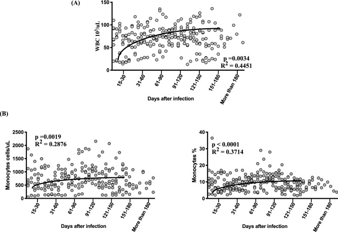
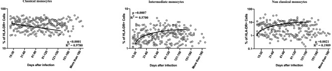
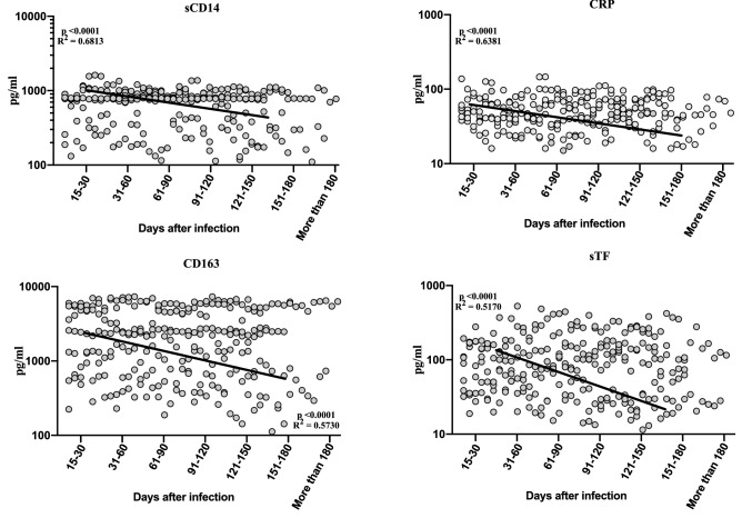
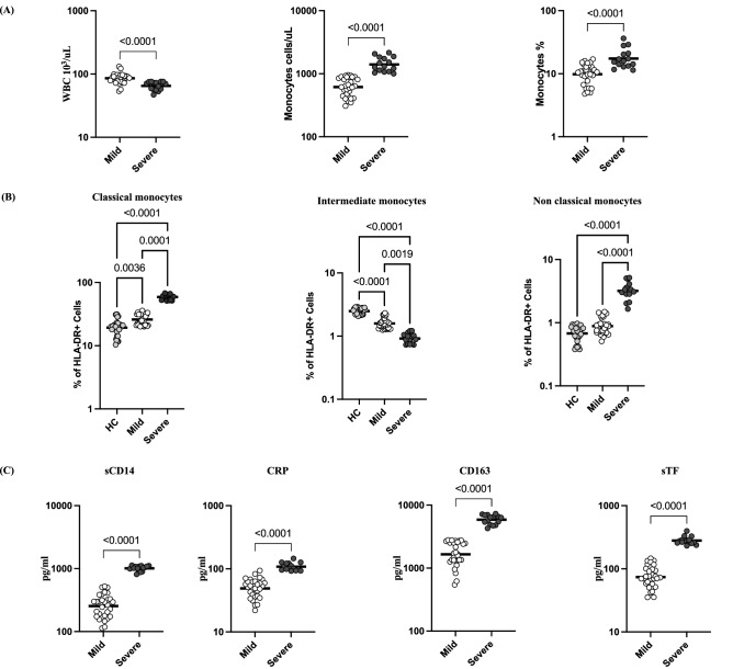
Monocytes are thought to play an important role in host defence and pathogenesis of COVID-19. However, a comprehensive examination of monocyte numbers and function has not been performed longitudinally in acute and convalescent COVID-19. We examined the absolute counts of monocytes, the frequency of monocyte subsets, the plasma levels of monocyte activation markers using flowcytometry and ELISA in seven groups of COVID-19 individuals, classified based on days since RT-PCR confirmation of SARS-CoV2 infection. Our data shows that the absolute counts of total monocytes and the frequencies of intermediate and non-classical monocytes increases from Days 15-30 to Days 61-90 and plateau thereafter. In contrast, the frequency of classical monocytes decreases from Days 15-30 till Days 121-150. The plasma levels of sCD14, CRP, sCD163 and sTissue Factor (sTF)-all decrease from Days 15-30 till Days 151-180. COVID-19 patients with severe disease exhibit higher levels of monocyte counts and higher frequencies of classical monocytes and lower frequencies of intermediate and non-classical monocytes and elevated plasma levels of sCD14, CRP, sCD163 and sTF in comparison with mild disease. Thus, our study provides evidence of dynamic alterations in monocyte counts, subset frequencies and activation status in acute and convalescent COVID-19 individuals.


Conflict of interest statement

The authors declare no competing interests.

Figures

Figure 1
Figure 1
Increase in WBC and monocyte absolute counts and percentages in convalescent COVID-19 individuals over time. (A) Analysis of WBC absolute counts were shown using best fit curve model from acute and convalescent COVID-19 individuals classified as groups based on days since RT-PCR confirmation (B) Analysis of absolute monocyte counts and percentages were shown using best fit curve model from acute and convalescent COVID-19 individuals classified as groups based on days since RT-PCR confirmation. Analysis was done by first order model polynomial fit curve, for absolute count of WBC (p = 0.0034, R2 = 0.4451) and monocyte absolute count (percentage p = 0.0019, R2 = 0.2876 and p < 0.0001, R = 0.3714) by Akaike’s Information Criterion and each dot represent single individuals. Thick black line represents best fit curve.
Figure 2
Figure 2
Alterations in frequencies of circulating monocyte subsets in convalescent COVID-19 individuals over time. Analysis of monocyte subsets from acute and convalescent COVID-19 individuals classified as groups based on days since RT-PCR confirmation. The frequencies of monocyte (classical, intermediate and non-classical) subsets are shown with first order model polynomial fit curve, classical monocyte p < 0.0001, R2 = 0.9780, intermediate monocytes p = 0.0007, R2 = 0.5700, non-classical monocytes p = 0.0021, R2 = 0.1909 by Akaike’s Information Criterion and each dot represent single individuals. Thick black line represents best fit curve.
Figure 3
Figure 3
Increased levels of monocyte activation markers in convalescent COVID-19 individuals over time. Circulating plasma levels of monocyte activation markers sCD14, CRP, sCD163 and sTF from acute and convalescent COVID-19 individuals classified as groups based on days since RT-PCR confirmation. The levels of monocyte activation markers were shown with second order model polynomial fit curve, sCD14, p < 0.0001, R2 = 0.6813; CRP, p < 0.0001, R2 = 0.6381; sCD163, p < 0.0001, R2 = 0.5730, sTF, p < 0.0001, R2 = 0.5170 by Akaike’s Information Criterion and each dot represent single individuals. Thick black line represents best fit curve.
Figure 4
Figure 4
Severe COVID-19 disease associated with altered frequencies of monocyte subsets and increased monocyte activation markers. (A) WBC absolute count and Monocyte absolute count and percentage were shown for mild (n = 30) and severe (n = 15) COVID-19 individuals sampled between days 15 to 60 following RT-PCR confirmation. The data are represented as scatter plots with each circle representing a single individual. (B) The frequencies of monocyte subsets in mild (n = 30) and severe (n = 15) COVID-19 individuals sampled between days 15 to 60 following RT-PCR confirmation and healthy control individuals (n = 30). The data are represented as scatter plots with each circle representing a single individual. p values were calculated using the Kruskal–Wallis test. (C) Circulating plasma levels of monocyte activation markers sCD14, CRP, sCD163 and sTF in mild (n = 30) and severe (n = 15) COVID-19 sampled between days 15 to 60 following RT-PCR confirmation. The data are represented as scatter plots with each circle representing a single individual. p values were calculated using the Mann–Whitney U-test.

References

    1. Shi C, Pamer EG. Monocyte recruitment during infection and inflammation. Nat. Rev. Immunol. 2011;11:762–774. doi: 10.1038/nri3070. - DOI - PMC - PubMed
    1. Prame Kumar K, Nicholls AJ, Wong CHY. Partners in crime: Neutrophils and monocytes/macrophages in inflammation and disease. Cell Tissue Res. 2018;371:551–565. doi: 10.1007/s00441-017-2753-2. - DOI - PMC - PubMed
    1. Kapellos TS, et al. Human monocyte subsets and phenotypes in major chronic inflammatory diseases. Front. Immunol. 2019;10:2035. doi: 10.3389/fimmu.2019.02035. - DOI - PMC - PubMed
    1. Martinez FO, Combes TW, Orsenigo F, Gordon S. Monocyte activation in systemic Covid-19 infection: Assay and rationale. EBioMedicine. 2020;59:102964. doi: 10.1016/j.ebiom.2020.102964. - DOI - PMC - PubMed
    1. Su GL, Simmons RL, Wang SC. Lipopolysaccharide binding protein participation in cellular activation by LPS. Crit. Rev. Immunol. 1995;15:201–214. doi: 10.1615/CritRevImmunol.v15.i3-4.10. - DOI - PubMed

Publication types