

Role of Mast Cells in the Pathogenesis of Pruritus in Mastocytosis
- PMID: 34642766
- PMCID: PMC9425624
- DOI: 10.2340/actadv.v101.350
Role of Mast Cells in the Pathogenesis of Pruritus in Mastocytosis
Abstract
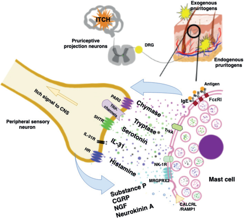
Pruritus can be defined as an unpleasant sensation that evokes a desire to scratch and significantly impairs patients' quality of life. Pruritus is widely observed in many dermatoses, including mastocytosis, a rare disease characterized by abnormal accumulation of mast cells, which can involve skin, bone marrow, and other organs. Increasing evidence highlights the role of mast cells in neurogenic inflammation and itching. Mast cells release various pruritogenic mediators, initiating subsequent mutual communication with specific nociceptors on sensory nerve fibres. Among important mediators released by mast cells that induce pruritus, one can distinguish histamine, serotonin, proteases, as well as various cytokines. During neuronal-induced inflammation, mast cells may respond to numerous mediators, including neuropeptides, such as substance P, neurokinin A, calcitonin gene-related peptide, endothelin 1, and nerve growth factor. Currently, treatment of pruritus in mastocytosis is focused on alleviating the effects of mediators secreted by mast cells. However, a deeper understanding of the intricacies of the neurobiology of this disease could help to provide better treatment options for patients.
Conflict of interest statement
DK has no conflicts of interest to declare.
AR has worked as a consultant or speaker for AbbVie, Bioderma, Celgene, Chema Elektromet, Eli Lilly, Galderma, Janssen, Leo Pharma, Medac, Menlo Therapeutics, Novartis, Pierre-Fabre, Sandoz, and Trevi, and participated as principal investigator or subinvestigator in clinical trials sponsored by AbbVie, Drug Delivery Solutions Ltd, Galderma, Genentech, Janssen, Kymab Limited, Leo Pharma, Menlo Therapeutics, MetrioPharm, MSD, Novartis, Pfizer, and Trevi.
Figures
Similar articles
-
Mast cell-neural interactions contribute to pain and itch.Immunol Rev. 2018 Mar;282(1):168-187. doi: 10.1111/imr.12622. Immunol Rev. 2018. PMID: 29431216 Free PMC article. Review.
-
Pathogenesis and treatment of pruritus.Curr Allergy Asthma Rep. 2010 Jul;10(4):236-42. doi: 10.1007/s11882-010-0117-z. Curr Allergy Asthma Rep. 2010. PMID: 20428977
-
Mastocytosis: current concepts in diagnosis and treatment.Ann Hematol. 2002 Dec;81(12):677-90. doi: 10.1007/s00277-002-0575-z. Epub 2002 Nov 29. Ann Hematol. 2002. PMID: 12483363 Review.
-
Methods for preclinical assessment of antipruritic agents and itch mechanisms independent of mast-cell histamine.Biol Pharm Bull. 2015;38(5):635-44. doi: 10.1248/bpb.b15-00090. Biol Pharm Bull. 2015. PMID: 25947907 Review.
-
Pruritogenic mediators in psoriasis vulgaris: comparative evaluation of itch-associated cutaneous factors.Br J Dermatol. 2003 Oct;149(4):718-30. doi: 10.1046/j.1365-2133.2003.05586.x. Br J Dermatol. 2003. PMID: 14616362
Cited by
-
Diospyros lotus leaf extract and its main component, myricitrin, inhibit both histamine‑dependent and histamine‑independent itching.Exp Ther Med. 2025 Apr 16;29(6):121. doi: 10.3892/etm.2025.12871. eCollection 2025 Jun. Exp Ther Med. 2025. PMID: 40297616 Free PMC article.
-
Raepenol™ Cream, a Complex of Natural Compounds, Promotes Wound Healing and Relieves Pruritus In Vivo.In Vivo. 2024 Sep-Oct;38(5):2318-2327. doi: 10.21873/invivo.13697. In Vivo. 2024. PMID: 39187315 Free PMC article.
-
Causal relationship between 41 inflammatory factors, circulating white blood cells, and pruritus: A 2-sample bidirectional Mendelian randomization study.Medicine (Baltimore). 2024 Dec 13;103(50):e40894. doi: 10.1097/MD.0000000000040894. Medicine (Baltimore). 2024. PMID: 39686490 Free PMC article.
-
The Role of Mast Cells in the Induction and Maintenance of Inflammation in Selected Skin Diseases.Int J Mol Sci. 2023 Apr 10;24(8):7021. doi: 10.3390/ijms24087021. Int J Mol Sci. 2023. PMID: 37108184 Free PMC article. Review.
-
Mastocytosis and Skin Cancer: The Current State of Knowledge.Int J Mol Sci. 2023 Jun 7;24(12):9840. doi: 10.3390/ijms24129840. Int J Mol Sci. 2023. PMID: 37372988 Free PMC article. Review.
References
-
- Reich A, Szepietowski JC. Diagnostic procedures of itch. Curr Probl Dermatol 2016; 50: 24–28. - PubMed
-
- Le M, Miedzybrodzki B, Olynych T, Chapdelaine H, Ben-Shoshan M. Natural history and treatment of cutaneous and systemic mastocytosis. Postgrad Med 2017; 129: 896–901. - PubMed
-
- Bulat V, Mihić LL, Šitum M, Buljan M, Blajić I, Pušić J. Most common clinical presentations of cutaneous mastocytosis. Acta Clin Croat Suppl 2009; 48: 59–64. - PubMed
MeSH terms
LinkOut - more resources
Full Text Sources
