. 2023 Feb;25(1):97-109.
doi: 10.1007/s11307-021-01642-9. Epub 2021 Oct 12.

Identification of a Suitable Untargeted Agent for the Clinical Translation of ABY-029 Paired-Agent Imaging in Fluorescence-Guided Surgery

Affiliations

Identification of a Suitable Untargeted Agent for the Clinical Translation of ABY-029 Paired-Agent Imaging in Fluorescence-Guided Surgery

Cheng Wang et al. Mol Imaging Biol. 2023 Feb.

Abstract

Purpose: Non-specific uptake and retention of molecular targeted agents and heterogeneous tissue optical properties diminish the ability to differentiate between tumor and normal tissues using molecular targeted fluorescent agents. Paired-agent imaging (PAI) can increase the diagnostic ability to detect tumor tissue by mitigating these non-specific effects and providing true molecular contrast by co-administration of an untargeted control imaging agent with a targeted agent. This study evaluates the suitability of available clinically translatable untargeted agents for the translation of PAI in fluorescence-guided surgery using an affibody-based targeted imaging agent (ABY-029).

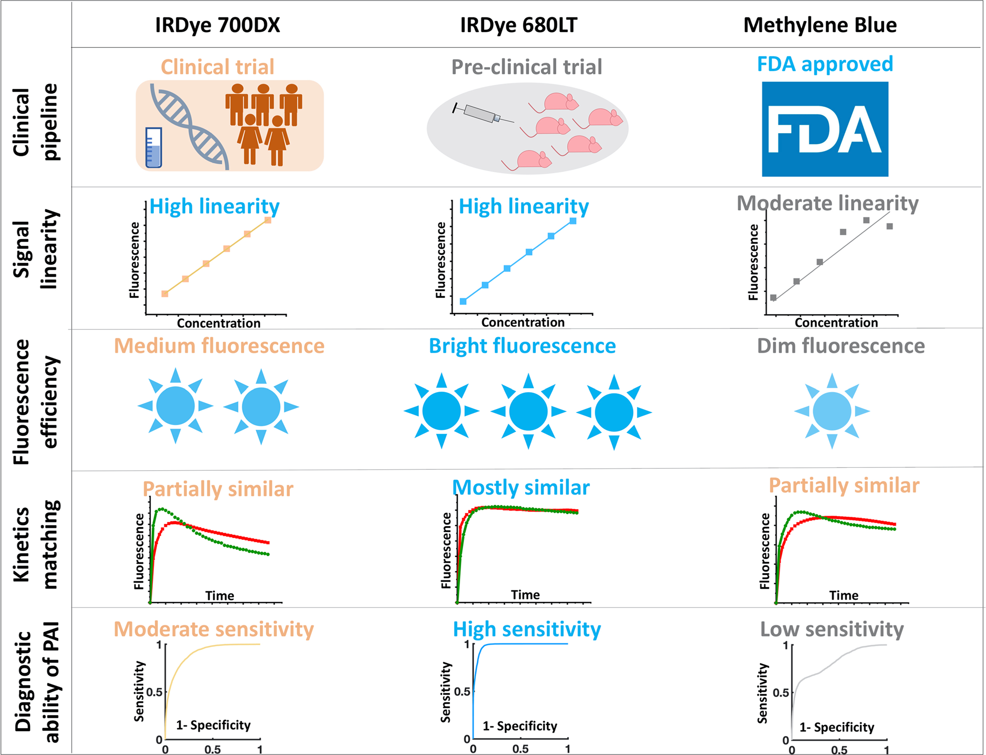
Experimental: DESIGN: Three untargeted agents that fluoresce near 700 nm and exhibit good clinical safety profiles (methylene blue, IRDye 700DX, and IRDye 680LT) were tested in combination with the clinically tested IRDye 800CW-labeled anti-epidermal growth factor receptor (EGFR) affibody molecule, ABY-029 (eIND 122,681). Properties of the untargeted agent important for human use and integrity of PAI were tested: (1) plasma protein binding; (2) fluorescence signal linearity in in vitro whole blood dilution; (3) in vivo pharmacokinetic matching to targeted agent in negative control tissue; and (4) in vivo diagnostic accuracy of PAI vs single agent imaging (SAI) of ABY-029 alone in orthotopic oral head and neck squamous cell carcinomas.

Results: IRDye 680LT outperformed IRDye 700DX and methylene blue with the highest signal linearity (R2 = 0.9998 ± 0.0002, 0.9995 ± 0.0004, 0.91 ± 0.02, respectively), the highest fluorescence yield in whole blood at 1 μM (104.42 ± 0.05, 103.68 ± 0.09, 101.9 ± 0.2, respectively), and the most closely matched ABY-029 pharmacokinetics in EGFR-negative tissues (binding potential error percentage = 0.31% ± 0.37%, 10.25% ± 1.30%, and 8.10% ± 5.37%, respectively). The diagnostic ability of PAI with ABY-029 and IRDye 680LT outperformed conventional SAI with an area-under-the-receiver-operating-characteristic curve (AUC) value of 0.964 vs. 0.854, and 0.978 vs. 0.925 in the Odyssey scanning system and Pearl wide field imaging system, respectively.

Conclusion: PAI is a highly promising methodology for increasing detection of tumors in fluorescence-guided surgery. Although not yet clinically approved, IRDye 680LT demonstrates promise as an untargeted agent when paired with ABY-029. The clinical translation of PAI to maximize tumor excision, while minimizing normal tissue removal, could improve both patient survival and life quality.

Keywords: ABY-029; Epidermal growth factor receptor; Fluorescence-guided surgery; Head and neck squamous cell carcinoma; IRDye 680LT; IRDye 700DX; Methylene blue; Molecular imaging.


Figures

Fig. 1.
Fig. 1.
Gel electrophoresis for BSA-dye binding test. The non-targeted control dyes were loaded in 4–20% Criterion™ Tris–HCl protein gel with or without BSA. Both 700-nm (a) and 800-nm (b) fluorescence images were acquired by Odyssey scanner. The portion of free dye bound with BSA and overall fluorescence enhancements are reported for each dye (c). Methylene blue was precluded from analysis because the unassociated dye runs in opposite direction to the BSA-associated dye.
Fig. 2.
Fig. 2.
Linearity of fluorescence emission of non-targeted control agents in whole blood. Paired agents premixed (+) or not premixed (−) with BSA were diluted in whole bovine blood. Fluorescence images in both channels were acquired by Odyssey CLx (a). For each condition, concentration vs. signal intensity was plotted in the log–log scale to visualize all concentrations tested since concentration was varied over several orders of magnitude (b). The coefficient of determination (R2) and intercept of the regression analyses are compared for each non-targeted agent tested (c).
Fig. 3.
Fig. 3.
Pharmacokinetics comparison between ABY-029 and untargeted control agents in receptor-negative muscle. The compartment model representing each imaging agent and BP (a) are shown with corresponding fluorescence images acquired over time on the Odyssey CLx in the muscle after paired-agent infusion (b). The optimal normalization ratio was determined by calculating the minimized mean squared error of each targeted-and untargeted imaging pair in receptor-negative muscle (c). The minimized mean-square error calculated for the paired-agents in each animal is summarized (d).
Fig. 4.
Fig. 4.
Diagnostic ability of PAI and SAI collected using the Odyssey CLx for xenograft tongue tumors. a Targeted and untargeted fluo-rescence images of xenograft tongue tumors in a murine model were taken on the Odyssey CLx, and BP maps were calculated. Histograms of pixel intensity for targeted, untargeted, BP in normal, and tumor tissue were plotted based on ROIs defined in pathological slices. A representative area under the curve (AUC) of the receiver operating characteristic (ROC) curve is reported for an individual animal in each imaging condition and was used to compare the diagnostic ability of three imaging methods. b ROCs are shown for each cohort of animals imaged with the same paired-agents and AUCs are reported.
Fig. 5.
Fig. 5.
Diagnostic ability of PAI and SAI collected using the Pearl Impulse for xenograft tongue tumors. a Targeted and untargeted fluorescence images were collected, and BP maps calculated for each imaging-agent pair. Histograms of pixel intensity in tumor and normal tissue are plotted based on ROIs defined in pathological slices. A representative AUC of the ROC is reported for an individual animal in each imaging condition. b ROCs are shown for each cohort of animals imaged with the same paired-agents, and AUCs are reported.
Fig. 6.
Fig. 6.
The performance of untargeted agents in paired agent imaging. Five perspectives, clinical pipeline, signal linearity, fluorescence efficiency, kinetics matching, and diagnostic ability of PAI were analyzed among three untargeted agents.

References

    1. Siegel RL, Miller KD, Fuchs HE, Jemal A (2021) Cancer statistics, 2021. CA: A cancer journal for clinicians 71:7–33. 10.3322/caac.21654 - DOI - PubMed
    1. Eldeeb H, Macmillan C, Elwell C, Hammod A (2012) The effect of the surgical margins on the outcome of patients with head and neck squamous cell carcinoma: single institution experience. Cancer Biol Med 9:29–33 - PMC - PubMed
    1. Functional outcomes after transoral robotic surgery for head and neck cancer – Iseli Tim A., Kulbersh Brian D., Iseli Claire E., Carroll William R., Rosenthal Eben L., J. Scott Magnuson, 2009. 10.1016/j.otohns.2009.05.014. Accessed 2 Dec 2020 - DOI - PubMed
    1. DiNardo LJ, Lin J, Karageorge LS, Powers CN (2000) Accuracy, utility, and cost of frozen section margins in head and neck cancer surgery. Laryngoscope 110:1773–1776. 10.1097/00005537-200010000-00039 - DOI - PubMed
    1. Rosenthal EL, Moore LS, Tipirneni K et al. (2017) Sensitivity and specificity of cetuximab-IRDye800CW to identify regional metastatic disease in head and neck cancer. Clin Cancer Res 23:4744–4752 - PMC - PubMed

Publication types