. 2021 Nov 15;10(11):bio058669.
doi: 10.1242/bio.058669. Epub 2021 Nov 29.

Regulation of NANOG and SOX2 expression by activin A and a canonical WNT agonist in bovine embryonic stem cells and blastocysts

Affiliations

Regulation of NANOG and SOX2 expression by activin A and a canonical WNT agonist in bovine embryonic stem cells and blastocysts

Yao Xiao et al. Biol Open. .

Abstract

Bovine embryonic stem cells (ESC) have features associated with the primed pluripotent state including low expression of one of the core pluripotency transcription factors, NANOG. It has been reported that NANOG expression can be upregulated in porcine ESC by treatment with activin A and the WNT agonist CHIR99021. Accordingly, it was tested whether expression of NANOG and another pluripotency factor SOX2 could be stimulated by activin A and the WNT agonist CHIR99021. Immunoreactive NANOG and SOX2 were analyzed for bovine ESC lines derived under conditions in which activin A and CHIR99021 were added singly or in combination. Activin A enhanced NANOG expression but also reduced SOX2 expression. CHIR99021 depressed expression of both NANOG and SOX2. In a second experiment, activin A enhanced blastocyst development while CHIR99021 treatment impaired blastocyst formation and reduced number of blastomeres. Activin A treatment decreased blastomeres in the blastocyst that were positive for either NANOG or SOX2 but increased those that were CDX2+ and that were GATA6+ outside the inner cell mass. CHIR99021 reduced SOX2+ and NANOG+ blastomeres without affecting the number or percent of blastomeres that were CDX2+ and GATA6+. Results indicate activation of activin A signaling stimulates NANOG expression during self-renewal of bovine ESC but suppresses cells expressing pluripotency markers in the blastocyst and increases cells expressing CDX2. Actions of activin A to promote blastocyst development may involve its role in promoting trophectoderm formation. Furthermore, results demonstrate the negative role of canonical WNT signaling in cattle for pluripotency marker expression in ESC and in formation of the inner cell mass and epiblast during embryonic development. This article has an associated First Person interview with the first author of the paper.

Keywords: Activin; Bovine; Embryonic stem cells; Pluripotency; WNT signaling.


Conflict of interest statement

Competing interests The authors declare no competing or financial interests.

Figures
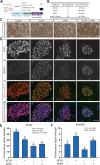
Fig. 1.
Fig. 1.
Treatment of embryonic stem cells with activin A enhanced NANOG expression while activation of canonical WNT signaling attenuated SOX2 and NANOG expression. (A) Experimental design. Zona-free blastocysts were seeded onto mouse embryonic fibroblasts (MEF) with the culture medium consisting of base medium mTeSR, FGF2, IWR-1 and additional treatments of 25 ng/ml activin A (ACTA), 1.5 μM CHIR99021 (CHIR), 25 ng/ml ACTA+1.5 μM CHIR or control. The total number of blastocysts analyzed is in panel B. Blastocysts were produced in two embryo production replicates. (B) Efficiency of derivation of cell lines. There was no significant difference among treatments. Subsequent observations were carried out on the number of cell lines indicated. (C) Morphological characteristics of cells derived under different conditions at week 5 of passage 5 or 6. Scale bar: 100 μm. (D) Dual immunolabeling of SOX2 and NANOG after 5–6 weeks of treatment. Scale bar: 50 μm. (E, F) Quantification of immunofluorescence for SOX2 (E) and NANOG (F). Data are least-squares means±SEM. The P-value for main effects and the interaction that were P<0.10 or less are shown in the upper left regions of each figure. Bars with different letters differ (P<0.05) as determined by mean-separation test.
Fig. 2.
Fig. 2.
Effects of activin A treatment and activation of canonical WNT on development of bovine embryos to the blastocyst stage. (A) Experimental design. In vitro produced bovine embryos were treated with 25 ng/ml activin A (ACTA), 1.5 μM CHIR99021 (CHIR), 25 ng/ml ACTA+1.5 μM CHIR or control from day 4–7.5 of development. Blastocysts were collected for immunolabeling of lineage markers including SOX2, CDX2, NANOG and GATA6. Effects of treatments on percent of presumptive zygotes (B) and percent of cleaved embryos (C) becoming blastocysts were observed on day 7.5 in six embryo production replicates. (D) Total cell numbers of blastocysts. Data in panels B–D represent least-squares means±SEM. The P-value for main effects and the interaction that were P<0.10 or less are shown in the upper left regions of each figure. Bars with different letters differ (P<0.05) as determined by mean-separation test.
Fig. 3.
Fig. 3.
Representative images of day 7.5 bovine blastocysts that were immunolabeled for SOX2 and CDX2 or NANOG and GATA6. Shown are images generated by maximum projection of z-stacks. Scale bar: 50 µm.
Fig. 4.
Fig. 4.
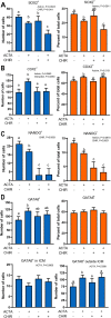
Effects of activin A treatment and activation of canonical WNT signaling from day 4 to 7.5 of development on lineage segregation in the resultant blastocysts. Treatments were 25 ng/ml activin A (ACTA), 1.5 μM CHIR99021 (CHIR), 25 ng/ml ACTA+1.5 μM CHIR or control from day 4–7.5 of development. Quantification of number and percent of cells that were SOX2+ (A), CDX2+ (B), NANOG+ (C), and GATA6+ (D). Data for numbers of GATA6+ cells were compiled for the entire blastocyst, the inner cell mass (ICM) only, and for cells outside the ICM. The P-values for main effects and the interaction that were P<0.10 or less are shown in the upper left regions of each figure. Bars with different letters differ (P<0.05) as determined by mean-separation test.

References

    1. Adachi, K., Suemori, H., Yasuda, S. Y., Nakatsuji, N. and Kawase, E. (2010). Role of SOX2 in maintaining pluripotency of human embryonic stem cells. Genes Cells 15, 455-470. 10.1111/j.1365-2443.2010.01400.x - DOI - PubMed
    1. Ai, Z., Shao, J., Wu, Y., Yu, M., Du, J., Shi, X., Shi, X., Zhang, Y. and Guo, Z. (2016). CHIR99021 enhances Klf4 expression through β-catenin signaling and miR-7a regulation in J1 mouse embryonic stem cells. PLoS ONE 11, e0150936. 10.1371/journal.pone.0150936 - DOI - PMC - PubMed
    1. Aparicio, I. M., Garcia-Herreros, M., Fair, T. and Lonergan, P. (2010). Identification and regulation of glycogen synthase kinase-3 during bovine embryo development. Reproduction 140, 83-92. 10.1530/REP-10-0040 - DOI - PubMed
    1. Baillie-Benson, P., Moris, N. and Martinez Arias, A. (2020). Pluripotent stem cell models of early mammalian development. Curr. Opin. Cell Biol. 66, 89-96. 10.1016/j.ceb.2020.05.010 - DOI - PubMed
    1. Bayerl, J., Ayyash, M., Shani, T., Manor, Y. S., Gafni, O., Massarwa, R., Kalma, Y., Aguilera-Castrejon, A., Zerbib, M., Amir, H.et al. (2021). Principles of signaling pathway modulation for enhancing human naive pluripotency induction. Cell Stem Cell 28, 1549-1565.e12. 10.1016/j.stem.2021.04.001 - DOI - PMC - PubMed

Publication types