

Striatal topographical organization: Bridging the gap between molecules, connectivity and behavior
- PMID: 34643358
- PMCID: PMC8524362
- DOI: 10.4081/ejh.2021.3284
Striatal topographical organization: Bridging the gap between molecules, connectivity and behavior
Abstract
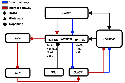
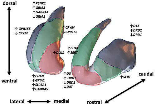
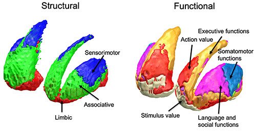
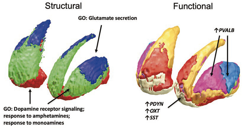
The striatum represents the major hub of the basal ganglia, receiving projections from the entire cerebral cortex and it is assumed to play a key role in a wide array of complex behavioral tasks. Despite being extensively investigated during the last decades, the topographical organization of the striatum is not well understood yet. Ongoing efforts in neuroscience are focused on analyzing striatal anatomy at different spatial scales, to understand how structure relates to function and how derangements of this organization are involved in various neuropsychiatric diseases. While being subdivided at the macroscale level into dorsal and ventral divisions, at a mesoscale level the striatum represents an anatomical continuum sharing the same cellular makeup. At the same time, it is now increasingly ascertained that different striatal compartments show subtle histochemical differences, and their neurons exhibit peculiar patterns of gene expression, supporting functional diversity across the whole basal ganglia circuitry. Such diversity is further supported by afferent connections which are heterogenous both anatomically, as they originate from distributed cortical areas and subcortical structures, and biochemically, as they involve a variety of neurotransmitters. Specifically, the cortico-striatal projection system is topographically organized delineating a functional organization which is maintained throughout the basal ganglia, subserving motor, cognitive and affective behavioral functions. While such functional heterogeneity has been firstly conceptualized as a tripartite organization, with sharply defined limbic, associative and sensorimotor territories within the striatum, it has been proposed that such territories are more likely to fade into one another, delineating a gradient-like organization along medio-lateral and ventro-dorsal axes. However, the molecular and cellular underpinnings of such organization are less understood, and their relations to behavior remains an open question, especially in humans. In this review we aimed at summarizing the available knowledge on striatal organization, especially focusing on how it links structure to function and its alterations in neuropsychiatric diseases. We examined studies conducted on different species, covering a wide array of different methodologies: from tract-tracing and immunohistochemistry to neuroimaging and transcriptomic experiments, aimed at bridging the gap between macroscopic and molecular levels.
Figures
References
-
- DiFiglia M, Carey J. Large neurons in the primate neostriatum examined with the combined Golgi-electron microscopic method. J Comp Neurol 1986;244:36–52. - PubMed
-
- Graveland GA, Difiglia M. The frequency and distribution of medium-sized neurons with indented nuclei in the primate and rodent neostriatum. Brain Res 1985;327:307–11. - PubMed
-
- Pasik P, Pasik T, Holstein GR, Hámori J. GABAergic elements in the neuronal circuits of the monkey neostriatum: a light and electron microscopic immunocytochemical study. J Comp Neurol 1988;270:157–70. - PubMed
-
- Alexander GE, Crutcher MD, DeLong MR. Basal gangliathalamocortical circuits: parallel substrates for motor, oculomotor, ‘prefrontal’ and ‘limbic’ functions. Prog Brain Res 1990;85:119–46. - PubMed
-
- Karachi C, Francois C, Parain K, Bardinet E, Tande D, Hirsch E, et al. Three-dimensional cartography of functional territories in the human striatopallidal complex by using calbindin immunoreactivity. J Comp Neurol 2002;450:122–34. - PubMed
Publication types
MeSH terms
LinkOut - more resources
Full Text Sources
Miscellaneous
