. 2021 Oct 28;125(42):11628-11636.
doi: 10.1021/acs.jpcb.1c06019. Epub 2021 Oct 13.

Tau Protein Binding Modes in Alzheimer's Disease for Cationic Luminescent Ligands

Affiliations

Tau Protein Binding Modes in Alzheimer's Disease for Cationic Luminescent Ligands

Yogesh Todarwal et al. J Phys Chem B. .

Abstract

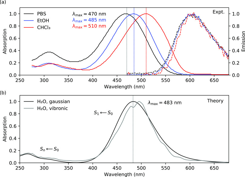
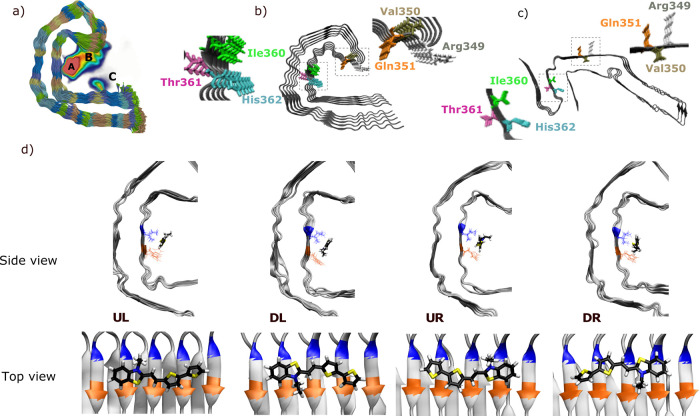
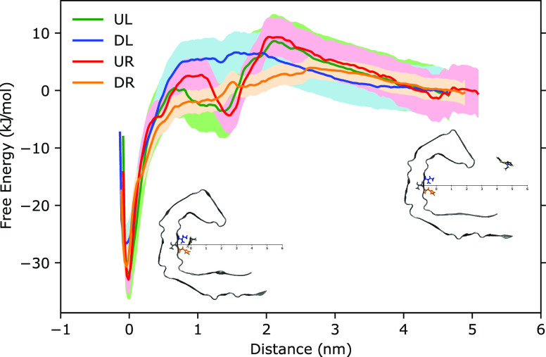
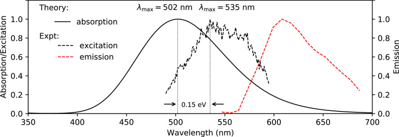
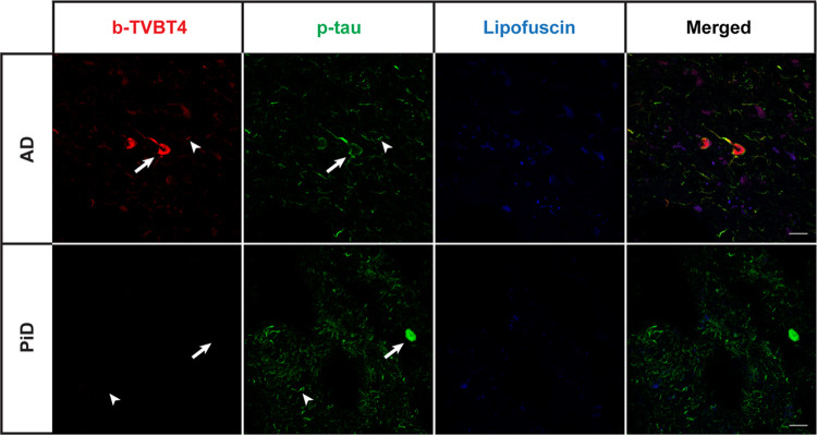
The bi-thiophene-vinylene-benzothiazole (bTVBT4) ligand developed for Alzheimer's disease (AD)-specific detection of amyloid tau has been studied by a combination of several theoretical methods and experimental spectroscopies. With reference to the cryo-EM tau structure of the tau protofilament ( Nature 2017, 547, 185), a periodic model system of the fibril was created, and the interactions between this fibril and bTVBT4 were studied with nonbiased molecular dynamics simulations. Several binding sites and binding modes were identified and analyzed, and the results for the most prevailing fibril site and ligand modes are presented. A key validation of the simulation work is provided by the favorable comparison of the theoretical and experimental absorption spectra of bTVBT4 in solution and bound to the protein. It is conclusively shown that the ligand-protein binding occurs at the hydrophobic pocket defined by the residues Ile360, Thr361, and His362. This binding site is not accessible in the Pick's disease (PiD) fold, and fluorescence imaging of bTVBT4-stained brain tissue samples from patients diagnosed with AD and PiD provides strong support for the proposed tau binding site.


Conflict of interest statement

The authors declare no competing financial interest.

Figures

Figure 1
Figure 1
Molecular structure of the bTVBT4 ligand. Selected ground and excited state (S0/S1) bond length (Å) parameters of the π-conjugated backbone are given. Natural population analysis (NPA) charges for the bi-thiophene and benzothiazole moieties (as separated by the dashed line) in the S0 and S1 states are given in red. The S0 and S1 states are described at the levels of DFT/B3LYP and TDDFT/CAM-B3LYP, respectively.
Figure 2
Figure 2
(a) Experimental absorption and emission spectra of bTVBT4 in different solvents (PBS is phosphate-buffered saline) obtained at room temperature. (b) Theoretical absorption spectra for bTVBT4 in water solution using Gaussian and vibronic line broadenings. The 10 lowest states are included in the calculations performed at the level of TDDFT/CAM-B3LYP(100%).
Figure 3
Figure 3
Summary of the MD simulations of the interactions between bTVBT4 and the amyloid fibrillar structure of tau. (a) Density map of the bTVBT4 interactions with the tau Alzheimer’s fold, where red indicates the highest, blue a lower, and white a zero ligand density, (b) Alzheimer’s fold (PDB ID: 5O3L), (c) Pick’s fold (PDB ID: 6GX5), and (d) identified binding modes for bTVBT4 in site A during MD simulation, with separation of down (D), up (U), left (L), and right (R) orientation of the ethyl group with respect to His362 (orange) and Ile360 (blue).
Figure 4
Figure 4
Free energy profiles for bTVBT4 in the UL (green), DL (blue), UR (red), and DR (orange) modes, obtained by the potential of mean force approach by pulling the ligand from binding site A to become free in solution.
Figure 5
Figure 5
Theoretical absorption and experimental excitation and emission spectra for bTVBT4 bound to the tau protein in the Alzheimer fold. The 10 lowest states are included in the calculations performed at the level of TDDFT/CAM-B3LYP(100%). The experimental excitation and emission spectra were recorded at room temperature from tau deposits in a bTVBT4-stained AD brain tissue section washed with PBS (see SI section 2.2 for further details).
Figure 6
Figure 6
Fluorescence images of the brain section from a patient diagnosed with Alzheimer’s disease (AD, top panel) or Pick’s disease (PiD, bottom panel) stained with 100 nM bTVBT4 (red) and phospho-tau antibody AT8 (p-tau, green). Arrow: neurofibrillary tangle (AD), Pick body (PiD). Arrowhead: neuropil thread. As the autofluorescence from lipofuscin granules can overlap with bTVBT4 emission, an additional channel in which the settings only allowed excitation of lipofuscin (blue) is also shown. Scale bars represent 20 μm.

References

    1. Nguyen P. H.; Ramamoorthy A.; Sahoo B. R.; Zheng J.; Faller P.; Straub J. E.; Dominguez L.; Shea J.-E.; Dokholyan N. V.; De Simone A.; et al. Amyloid Oligomers: A Joint Experimental/Computational Perspective on Alzheimer’s Disease, Parkinson’s Disease, Type II Diabetes, and Amyotrophic Lateral Sclerosis. Chem. Rev. 2021, 121, 2545–2647. 10.1021/acs.chemrev.0c01122. - DOI - PMC - PubMed
    1. Fitzpatrick A. W. P.; Falcon B.; He S.; Murzin A. G.; Murshudov G.; Garringer H. J.; Crowther R. A.; Ghetti B.; Goedert M.; Scheres S. H. W.; et al. Cryo-EM structures of tau filaments from Alzheimer’s disease. Nature 2017, 547, 185–190. 10.1038/nature23002. - DOI - PMC - PubMed
    1. van Eersel J.; Bi M.; Ke Y. D.; Hodges J. R.; Xuereb J. H.; Gregory G. C.; Halliday G. M.; Götz J.; Kril J. J.; Ittner L. M. Phosphorylation of soluble tau differs in Pick’s disease and Alzheimer’s disease brains. J. Neural. Transm. 2009, 116, 1243–1251. 10.1007/s00702-009-0293-y. - DOI - PubMed
    1. Hanger D. P.; Byers H. L.; Wray S.; Leung K.-Y.; Saxton M. J.; Seereeram A.; Reynolds C. H.; Ward M. A.; Anderton B. H. Novel Phosphorylation Sites in Tau from Alzheimer Brain Support a Role for Casein Kinase 1 in Disease Pathogenesis. J. Biol. Chem. 2007, 282, 23645–23654. 10.1074/jbc.M703269200. - DOI - PubMed
    1. Smet-Nocca C.Tau Protein: Methods and Protocols; Smet-Nocca C., Ed.; Methods in Molecular Biology; Humana Press, 2017; Vol. 1523. https://www.springer.com/gp/book/9781493965960

Publication types