. 2021 Oct 13;13(615):eabh1486.
doi: 10.1126/scitranslmed.abh1486. Epub 2021 Oct 13.

Ubiquitination and degradation of SUMO1 by small-molecule degraders extends survival of mice with patient-derived tumors

Affiliations

Ubiquitination and degradation of SUMO1 by small-molecule degraders extends survival of mice with patient-derived tumors

Anita C Bellail et al. Sci Transl Med. .

Abstract

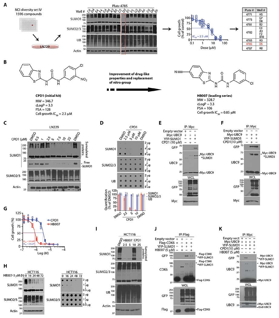
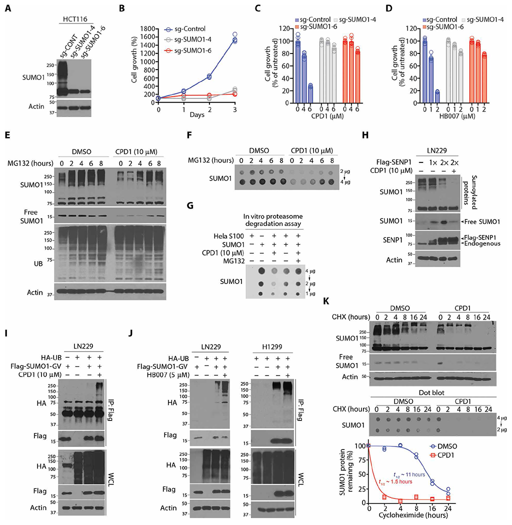
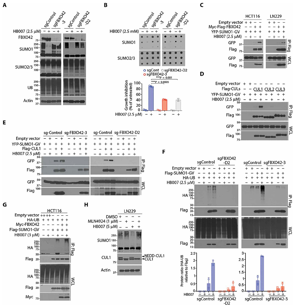
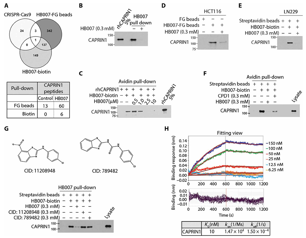
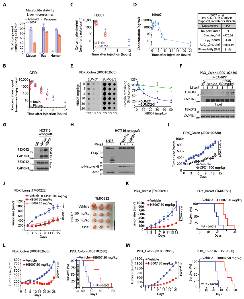
Discovery of small-molecule degraders that activate ubiquitin ligase–mediated ubiquitination and degradation of targeted oncoproteins in cancer cells has been an elusive therapeutic strategy. Here, we report a cancer cell–based drug screen of the NCI drug-like compounds library that enabled identification of small-molecule degraders of the small ubiquitin-related modifier 1 (SUMO1). Structure-activity relationship studies of analogs of the hit compound CPD1 led to identification of a lead compound HB007 with improved properties and anticancer potency in vitro and in vivo. A genome-scale CRISPR-Cas9 knockout screen identified the substrate receptor F-box protein 42 (FBXO42) of cullin 1 (CUL1) E3 ubiquitin ligase as required for HB007 activity. Using HB007 pull-down proteomics assays, we pinpointed HB007’s binding protein as the cytoplasmic activation/proliferation-associated protein 1 (CAPRIN1). Biolayer interferometry and compound competitive immunoblot assays confirmed the selectivity of HB007’s binding to CAPRIN1. When bound to CAPRIN1, HB007 induced the interaction of CAPRIN1 with FBXO42. FBXO42 then recruited SUMO1 to the CAPRIN1-CUL1-FBXO42 ubiquitin ligase complex, where SUMO1 was ubiquitinated in several of human cancer cells. HB007 selectively degraded SUMO1 in patient tumor–derived xenografts implanted into mice. Systemic administration of HB007 inhibited the progression of patient-derived brain, breast, colon, and lung cancers in mice and increased survival of the animals. This cancer cell–based screening approach enabled discovery of a small-molecule degrader of SUMO1 and may be useful for identifying other small-molecule degraders of oncoproteins.


Conflict of interest statement

Competing interests: A.C.B. and C. Hao are the cofounders of HB Therapeutics Inc. H.-Y.L. is an employee of Synovel Laboratory LLC. M.B., C.l.S, and B.C.S.C. are the employees of Horizon Discovery. A.S. is a cofounder of Anagin Corp. A.C.B., C. Hao, and H.-Y.L. are co-inventors on the U.S. provisional patent application 62/669,640 and the PCT/US2019/031245 patent application entitled “Compositions and methods for treating cancer.” The other authors declare that they have no competing interests.

Figures

Fig. 1.
Fig. 1.. Discovery of the hit compound CPD1 and chemical lead HB007.
(A) Workflow in LN229 cell–based drug screening of the NCI library through Western blots and cell viability assay with the identification of 11 active compounds with D5 characterized as the hit compound (highlighted in red). (B) The chemical structures and pharmacological properties of the hit compound CPD1 and the lead compound HB007. cLogP, partition coefficient log P; PSA, polar surface area. (C) LN229 cells were treated for 72 hours with indicated doses of CPD1 and analyzed by Western blots for conjugated and unconjugated/free SUMO1 as indicated (right). SUMO2/3 and β-actin were used, respectively, as the selectivity and the loading control. (D) LN229 cells were treated with CDP1 for 72 hours and examined by dot blots for total SUMO1 concentrations with the amounts of loading proteins indicated (right). Dot intensity was evaluated using ImageJ (bottom). (E and F) LN229 cells were cotransfected with Myc-UBC9 and YFP-SUMO1 or YFP-SUMO3 or empty vector as control and subjected to myc IP and Western blotting for UBC9-SUMO1 (E) and UBC9-SUMO3 conjugates (F) as indicated (right) with whole-cell lysate (WCL) as the loading control. (G) LN229 cells were treated with a series of dilutions of CPD1 or HB007 for 5 days and examined by cell viability for cell growth inhibition with the IC50 values indicated (points: n = 6). (H) HCT116 cells were treated with HB007 for the time indicated and analyzed by Western (left) and dot blots (right) for conjugated and total SUMO1 concentrations. (I) HCT116 cells were treated with DMSO (control), CPD1, or HB007 for 72 hours with the indicated doses (micromolar) and analyzed by Western blotting using the indicate antibodies (left). (J) LN229 cells were cotransfected with Flag-CDK6 and YFP-SUMO1, treated with HB007 for 24 hours, and subjected to Flag IP and Western blotting using CDK6 and green fluorescent protein (GFP) antibody (that recognizes YFP) for SUMO1-CDK6 conjugates as indicated (right). (K) Myc IP and Western blotting for SUMO1-UBC9 conjugates as indicated (right) in myc-UBC9– and YFP-SUMO1–transfected LN229 cells after CPD1 and HB007 treatment for 24 hours.
Fig. 2.
Fig. 2.. CPD1 and HB007 induce SUMO1 ubiquitination and degradation.
(A) Western blots for SUMO1 protein concentrations in SUMO1 knockout HCT116 sgSUMO1-4, sgSUMO1-6, and control (sg-CONT) clones. (B) Cell growth analysis of SUMO1 knockout HCT116 sgS1-4, sgS1-6, and sg-CONT clones (n = 3). (C and D) Cell viability assay of SUMO1 knockout HCT116 and sg-CONT clones after being treated with CPD1 (C) or HB007 (D) with the indicated doses for 3 days (points: n = 6). (E and F) LN229 cells were treated with CPD1 for 24 hours, followed by MG132 for indicated times (top), and then analyzed by Western blots for conjugated and unconjugated/free forms (E) and dot blots for the total amounts of SUMO1 protein (F). (G) HeLa cell S100 fraction was added with SUMO1, CPD1, and/or MG132 and analyzed by dot blotting for the total amount of SUMO1. (H) Flag-SENP1 was overexpressed in LN229 cells, and the cells were treated with CDP1 for 48 hours and examined by Western blot for conjugated and unconjugated/free SUMO1 as indicated (right). (I) Flag-SUMO1-GV– and HA-UB–transfected LN229 cells were treated or untreated with CDP1 for 24 hours; Flag IP was analyzed by Western blots for SUMO1 polyubiquitination. (J) Flag-SUMO1-GV and HA-UB were cotransfected in LN229 (left) or H1299 (right), and the cells were treated or untreated with HB007 for 48 hours and subjected to Flag IP and Western blots for SUMO1 polyubiquitination. (K) LN229 cells were treated with CPD1 in the presence or absence of CHX for indicated times and examined by Western and dot blots for conjugated and unconjugated/free (top) and total amounts of SUMO1 protein (middle). The total amounts of SUMO1 protein were normalized with actin using the ImageJ and plotted for the half-life of SUMO1 protein (bottom) (n = 2 technical replicates). t1/2, half-time.
Fig. 3.
Fig. 3.. Discovery of a HB007-targeted E3 ligase pathway using a genome-scale CRISPR-Cas9 knockout screen.
(A and B) CRISPR-Cas9 FBXO42 knockout (sgFBXO42-3 and sgFBXO42-D2) and sgRNA control (sgControl) HCT116 clones were treated with HB007 for 72 hours and analyzed by Western blot for conjugated SUMO1 (A), dot blots for SUMO1 total amounts [(B) top], and cell viability assay for cell growth inhibition [(B) bottom)] (means ± SD; n = 3; ***P < 0.001 by unpaired t test). (C) Flag-FBXO42 and YFP-SUMO1-GV were cotransfected in HCT116 or LN229 cells. After 24 hours of treatment with HB007, the cells were subjected to Flag IP and Western blots using a GFP/YFP antibody for the interaction of FBXO42 and SUMO1. (D) Flag-CUL1, CUL2, or CUL3 was cotransfected with YFP-SUMO1-GV in HCT116 cells, and after treatment with HB007 for 48 hours, the cells were subjected to Flag IP and Western blots using GFP/YFP antibodies for the interaction of SUMO1 and CUL1, CUL2, or CUL3. (E) The FBXO42 knockout sg-FBXO42-3, sg-FBXO42-D2, and sgRNA control HCT116 clone were transfected with YFP-SUMO-GV and Flag-CUL1; treated with HB007 for 24 hours; and subjected to Flag IP and Western blot. (F) The sgRNA control and sg-FBXO42-D2 (left) or sg-FBXO42-3 HCT116 clone (right) was transfected with Flag-SUMO1-GV and HA-UB, treated with HB007 for 24 hours, and subjected to Flag IP and Western blot for SUMO1 polyubiquitination (top) and densitometry analysis of the HA-UB blots for the poly-UB amounts (bottom) (n = 2). (G) HCT116 cells were cotransfected with Flag-SUMO1-GV, Myc-FBXO42, and/or HA-UB; treated or untreated with HB007 for 24 hours; and subjected to Flag IP and Western blotting for SUMO1 polyubiquitination. (H) LN229 cells were treated with MLN4924 for 24 hours, alone or in combination with HB007, and analyzed by Western blots for conjugated SUMO1 and neddylated or unneddylated CUL1 as indicated (right).
Fig. 4.
Fig. 4.. Identification of the HB007 binding protein CAPRIN1.
(A) Venn diagram of the data from genome-scale CRISPR-Cas9 knockout screen, HB007-FG bead and HB007-biotin/streptavidin-coated bead pull-down (top), and spectrometric total peptides counts of CAPRIN1 from LC-MS/MS analysis (bottom). (B) HB007-biotin was incubated with rhCAPRIN1 in the presence or absence of free HB007 and pulled down by streptavidin-coated beads and tested by immunoblotting for the binding of rhCAPRIN1 to HB007-biotin, with rhCAPRIN1 (5%) used as the loading control. (C) rhCAPRIN1 was premixed with 1 μM biotin, followed by HB007-biotin/streptavidin-coated bead pull-down in the presence of various doses of HB007. CAPRIN1 binding was identified by immunoblotting using CAPRIN1 antibodies. (D and E) HB007-FG beads and HB007-biotin were incubated with HCT116 lysate (D) and LN229 lysate (E) added or not with free HB007, and the pull-downs were tested by immunoblotting for the binding of cellular CAPRIN1 to HB007. (F) Immunoblot of HB007-biotin/streptavidin pull-down of HCT116 lysate added or not with the indicated concentrations of CPD1 or HB007. (G) The CPD1 similar but inactive compounds CID: 11208948 or CID: 789482 (top) or HB007 was added to HCT116 lysate that was then incubated with HB007-biotin. HB007-biotin/streptavidin pull-down was analyzed by immunoblotting. (H) Representative BLI sensorgrams of the interactions between rhCAPRIN1 and biotinylated HB007. Plots of the binding response during the association (0 to 600 s) and dissociation (600 to 1200 s) periods of the BLI assay at varying concentrations of CAPRIN1 (top) when HB007-biotin–loaded biosensors (quenched with biocytin) were dipped in CAPRIN1 wells. The plots have been processed with the double referencing technique and aligning of x and y axes. The red curves indicate the fit data. Residual binding for each plot at the varying concentrations (bottom). Binding curves were fit globally to a 1:1 binding model to calculate the binding constant (KD) from kinetic analysis as the ratio of the association (koff) and dissociation (kon) rate constants.
Fig. 5.
Fig. 5.. HB007-induced CAPRIN1-FBXO42 interaction and SUMO1 recruitment to CUL1 E3 Ligase.
(A) The CAPRIN1 knockout sgCAPRIN1-3, sgCAPRIN1-12, and sgRNA control clones of HCT116 cells were treated or untreated with HB007 (3 μM) for 72 hours and analyzed by dot blotting for total amounts of SUMO1 and SUMO2/3. (B) CAPRIN1 knockout and control HCT116 clones were treated with DMSO or HB007 (1 or 2 μM) for 10 days and tested by colony formation assays with colony numbers calculated and presented (means ± SD; n = 6; **P < 0.01 and ***P < 0.001 by unpaired t test). (C) CAPRIN1 knockout and control HCT116 clones were cotransfected with myc-FBXO42 and YFP-SUMO1-GV, treated or untreated with HB007 for 24 hours, and subjected to myc IP and Western blotting for FBXO42 and SUMO1 interaction. (D) CAPRIN1 knockout HCT116 clones were cotransfected with Flag-SUMO1-GV and HA-UB, treated or untreated with HB007 for 24 hours, and subjected to Flag IP and Western blotting for SUMO1 polyubiquitination. Whole-cell lysate was included as the loading control. (E) HCT116 cells were transfected with Myc-Flag-FBXO42 and treated with HB007 for 8 hours with the doses indicated, followed by endogenous CAPRIN1-IP using CAPRIN1 antibodies. Western blots revealed the interaction of Myc-Flag-FBXO42 and endogenous CUL1. (F) HCT116 cells were transfected with Flag-CAPRIN1, treated with MLN4924 for 4 hours, and submitted to Flag IP and Western blotting for CAPRIN1-CUL1 interaction. (G) HCT116 were treated with HB007 for 8 hours and subjected to endogenous CAPRIN1-IP, followed by Western blot for CUL1 and FBXO42 interaction. Immunoglobulin G (IgG)–IP was used as a negative control. (H) Schematic representation of CAPRIN1 and its deletion mutants (left). Interaction of CAPRIN1 with HB007 and CUL1 was analyzed by HB007-biotin pull-down and Flag IP of CAPRIN1 and its mutants.
Fig. 6.
Fig. 6.. The selective activity of CPD1 and HB007 against different cancer cell types.
(A) NSCLC (lung), colon, breast carcinoma, and brain glioblastoma cell lines and matched normal lung Nuli-1, colon HIEC6, and breast MCF10A epithelial cells, and brain normal human astrocytes (NHAs) were analyzed by Western blotting for SUMO1 conjugation, CAPRIN1, and FBXO42 expression. (B) The IC50 values were compared between CPD1 and HB007 5 days treatment of 25 cancer cell lines (n = 2 calculated from two independent experiments totaling n = 12). (C to E) The normal lung epithelial cell Nuli-1 and NSCLC cell lines were treated or untreated with HB007 for 72 hours and analyzed by cell viability assay for growth inhibition (n = 6 biological replicate) (C), Western blot for conjugated SUMO1 (D), and dot blot for total SUMO1 amounts (E). SUMO2/3 was used as the selectivity control. (F) The human colon carcinoma HCT116, normal colon epithelial HIEC6, normal human astrocytes NHA, and BALB/c mouse fibroblasts were treated with HB007 for 48 hours and analyzed by Western blot for G3BP1 amounts. (G) HCT116, HEK293, and BALB/c cells were treated with HB007 for 8 hours and subjected to IP by a CAPRIN1 antibody (CAP), followed by Western blot for CAPRIN1 and G3BP1 interaction. IgG was used as the negative control. (H) HCT116, HIEC6, and NHA were treated with HB007 and subjected to IP using a G3BP1 antibody, followed by Western blot for its interaction with CAPRIN1 and CUL1. (I) Enzymatic analysis of the inhibition of CYP enzymes by HB007 at 10 μM in human liver microsomes. (J) The selectivity profile of HB007 (10 μM) against 67 diverse key human proteins as indicated.
Fig. 7.
Fig. 7.. The PKs and in vivo anticancer activity of CPD1 and HB007.
(A) HB007 was incubated for 45 min with mouse, rat, and human microsomes, and the amount of HB007 remaining was quantified by LC-MS (n = 3). Atenolol and verapamil were used as controls. (B and C) The PK assessment of CPD1 (B) and HB007 (C) by analyzing mouse plasma and brain tissue was carried out following intraperitoneal injection of a compound (20 mg/kg) (n = 3 mice per time point) in NOD/SCID mice. (D) The PKs of HB007 were determined by analyzing the compound in rat plasma samples after administrated orally in a solution formulation (n = 3). (E) Colon cancer PDX mice were treated with the indicated doses of HB007 through intraperitoneal injection once per day for 3 days beginning after a week of tumor inoculation; xenograft tissues were analyzed by dot blotting for total amounts of SUMO1 with SUMO2/3 as the control (left) with the densities quantified (right). (F) Colon cancer PDX mice [as in (E)] were subjected to IP by a CAPRIN1 antibody followed by Western blot for FBXO42 and CAPRIN1 interaction. (G) HCT116 xenograft mice were treated with vehicle or HB007 once per day for 3 days beginning after 10 days tumor inoculation; xenograft tissue and subjected to IP using CAPRIN1 antibody followed by Western blot for FBXO42 and CAPRIN1 interaction. (H) HCT116 xenografts [as in (G)] were subjected to Western blotting for caspase-3 cleavage for apoptotic cell death and phosphorylated histone (p-histone) H3 as a proliferation marker. (I) Colon cancer PDX mice were treated with CPD1 (100 mg/kg) for 15 days beginning after a week of tumor inoculation (means ± SEM, n = 10 per group, ***P=0.0001 by Wilcoxon tests). (J and K) PDX mice of lung NSCLC (J) and breast carcinoma (K) were treated with the indicated doses of CPD1 and/or HB007 for 15 days beginning after a week of tumor inoculation, and tumor sizes indicated that the treatment suppressed xenografts. (G) Right: Representative images of lung xenografts at the end of the treatment were presented. Data represent as means ± SEM. PDX mice of breast: n = 6 per group for vehicle and n = 7 per HB007, **P = 0.0078 at day 15 by Wilcoxon tests; PDX mice of lung: n = 7 per vehicle and n = 8 to 9 per treatment group, ****P < 0.0001 by Friedman test. (L and M) Mice bearing primary (L) and metastatic colon PDX xenografts (M) were treated with HB007; effect of the treatment on xenograft growth (left) and survival of mice as indicated by Kaplan-Meier survival curves (right). Data represent means ± SEM. PDX primary colon (J000102630): n = 5 per vehicle, n = 6 per treatment group, ***P < 0.0002 by Wilcoxon test; PDX metastatic colon (NCI #519858): n = 8 per group, **P = 0.002 by Wilcoxon test.

Similar articles

Cited by

References

    1. Salami J, Crews CM, Waste disposal-An attractive strategy for cancer therapy. Science 355, 1163–1167 (2017). - PubMed
    1. Schapira M, Calabrese MF, Bullock AN, Crews CM, Targeted protein degradation: Expanding the toolbox. Nat. Rev. Drug Discov 18, 949–963 (2019). - PubMed
    1. Baek K, Schulman BA, Molecular glue concept solidifies. Nat. Chem. Biol 16, 2–3 (2020). - PubMed
    1. Lu G, Middleton RE, Sun H, Naniong M, Ott CJ, Mitsiades CS, Wong KK, Bradner JE, Kaelin WG Jr., The myeloma drug lenalidomide promotes the cereblon-dependent destruction of Ikaros proteins. Science 343, 305–309 (2014). - PMC - PubMed
    1. Kronke J, Udeshi ND, Narla A, Grauman P, Hurst SN, McConkey M, Svinkina T, Heckl D, Comer E, Li X, Ciarlo C, Hartman E, Munshi N, Schenone M, Schreiber SL, Carr SA, Ebert BL, Lenalidomide causes selective degradation of IKZF1 and IKZF3 in multiple myeloma cells. Science 343, 301–305 (2014). - PMC - PubMed

Publication types