. 2021 Sep 24;12(9):e00403.
doi: 10.14309/ctg.0000000000000403.

Liquid Biopsy in Gastric Cancer: Analysis of Somatic Cancer Tissue Mutations in Plasma Cell-Free DNA for Predicting Disease State and Patient Survival

Affiliations

Liquid Biopsy in Gastric Cancer: Analysis of Somatic Cancer Tissue Mutations in Plasma Cell-Free DNA for Predicting Disease State and Patient Survival

Greta Varkalaite et al. Clin Transl Gastroenterol. .

Abstract

Introduction: Gastric cancer (GC) diagnosis in late stages and high mortality rates are the main issues that require new noninvasive molecular tools. We aimed to assess somatic mutational profiles in GC tissue and plasma cell-free DNA (cfDNA), evaluate their concordance rate, and analyze the role of multilayer molecular profiling to predict disease state and prognosis.

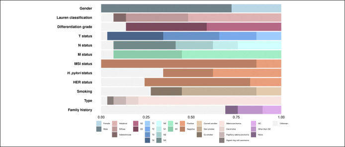
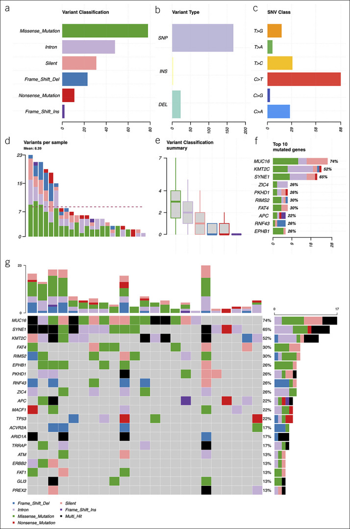
Methods: Treatment-naive GC patient group (n = 29) was selected. Whole exome sequencing (WES) of GC tissue was performed, and a unique 38-gene panel for deep targeted sequencing of plasma cfDNA was developed. Oncoproteins were measured by enzyme-linked immunosorbent assay, and other variables such as tumor mutational burden and microsatellite instability were evaluated using WES data.

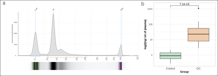
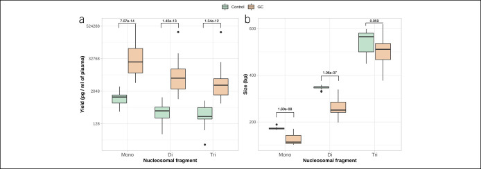
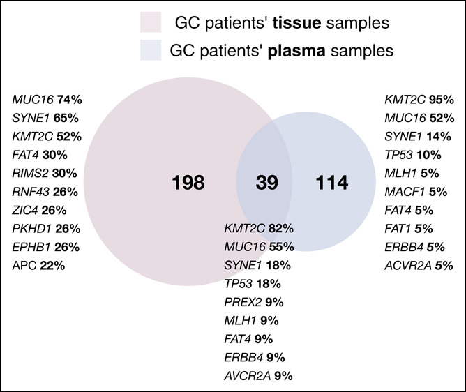
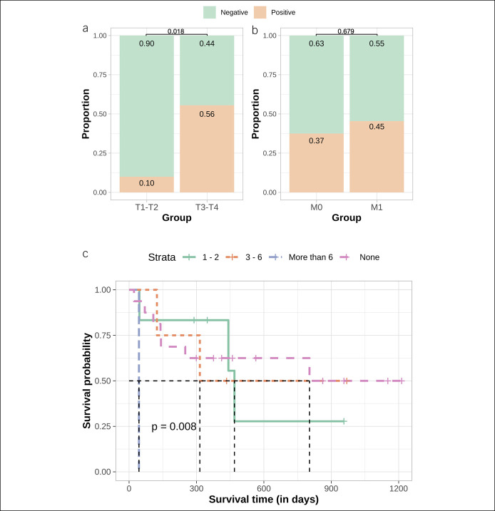
Results: The yield of cfDNA was increased 43.6-fold; the integrity of fragments was decreased in GC compared with controls. WES analysis of cancerous tissue and plasma cfDNA (targeted sequencing) mutational profiles revealed 47.8% concordance. The increased quantity of GC tissue-derived alterations detected in cfDNA was associated with worse patients' survival. Analysis of importance of multilayer variables and receiver operating characteristic curve showed that combination of 2 analytes: (i) quantity of tissue matching alterations and (ii) presence of any somatic alteration in plasma cfDNA resulted in area under curve 0.744 when discriminating patients with or without distant metastasis. Furthermore, cfDNA sequence alterations derived from tumor tissue were detected in patients who had even relatively small GC tumors (T1-T2).

Discussion: Our results indicate that quantitative and qualitative cfDNA mutational profile analysis is a promising tool for evaluating GC disease status or poorer prognosis.


Conflict of interest statement

Guarantor of the article: Jurgita Skieceviciene, PhD.

Specific author contributions: Juozas Kupcinskas, MD, PhD, and Jurgita Skieceviciene, PhD, contributed equally to this work. J.S., J.K., and A.F.: supervision and conceptualization. M.F. and G.V.: data collection, data analysis, interpretation, and visualization. G.V. and J.S.: writing original draft. M.F., J.K., A.F., and J.S.: manuscript review and editing. All authors approved the final manuscript version for submission.

Financial support: The study is a part of the MULTIOMICS project that has received funding from European Social Fund (project No. 09.3.3-LMT-K-712-01-0130) under grant agreement with the Research Council of Lithuania (LMTLT). M.F. is supported by the Deutsche Forschungsgemeinschaft (DFG).

Potential competing interests: The authors declare that they have no conflict of interest.

Ethics statement: Informed consent was obtained from all patients. Research was approved by the Kaunas Regional Biomedical Research Ethics Committee (No. BE-2-10, May 8, 2011 and No. BE-2-31, June 5, 2018, Kaunas, Lithuania).

Figures

Figure 1.
Figure 1.
Characteristics of patients with GC (n = 29). A bar graph representing the proportion of patients in each section. GC, gastric cancer; HER, human epidermal growth factor receptor; MSI, microsatellite instability.
Figure 2.
Figure 2.
(a) Representative electropherogram of cfDNA sample. Fragment size of cfDNA ranges from 100 to 1,000 bp, with the main peak around 170 bp; (b) yield of plasma total cfDNA (ng per ml of blood plasma) in control (n = 20) and GC (n = 29) samples. A 43.6-fold increase of cfDNA yield was determined in the GC patient group (P = 7.07 × 10⁻¹⁴). Results are shown on logarithmic scale. cfDNA, cell-free DNA; GC, gastric cancer.
Figure 3.
Figure 3.
(a) Yield of mononucleosomal, dinucleosomal, and trinucleosomal fragments (pg per ml of plasma) in control and GC patients' groups. Statistically significant increase of all cfDNA peaks was observed for patients with GC (P = 7.07 × 10⁻¹⁴, P = 1.42 × 10⁻¹³, and P = 1.34 × 10⁻¹², mononucleosomal, dinucleosomal, and trinucleosomal peaks, respectively); (b) size of mononucleosomal, dinucleosomal, and trinucleosomal fragments of cfDNA in control and GC patients' groups. Mononucleosomal and dinucleosomal cfDNA peaks of the patients with GC were significantly shorter compared with control cfDNA samples (P = 1.60 × 10⁻⁸ and P = 1.06 × 10⁻⁷, respectively). cfDNA, cell-free DNA; GC, gastric cancer.
Figure 4.
Figure 4.
Summary of the mutational spectra (only genes from custom gene panel) in gastric cancer tissue samples: (a) absolute variant class values, the most common variant class was missense mutations; (b) absolute variant type values, the most common variant type detected was SNPs; (c) distribution of various SNV substitutions, C > T substitutions were detected the most frequently; (d) absolute numbers of variants per sample, the dashed line shows the mean quantity of somatic variants per sample (8.39); (e) mean distribution of variant classes per sample, on average, missense mutations were most frequent; (f) top 10 mutated genes, x axis: absolute numbers (samples), percentages calculated from all somatic variants detected; and (g) oncoplot of the mutated genes in gastric cancer tissue samples, showing mutated genes and distribution of variant classes per sample. Color codes in (d-g) graphs are the same as in (a). DEL, deletion; INS, insertion; SNP, single nucleotide polymorphism; SNV, single nucleotide variant.
Figure 5.
Figure 5.
Venn diagram shows the quantity of unique and shared somatic alterations detected in GC patient tissue and plasma samples. Genes listed represents top 10 mutated genes in each case (tissue only, plasma only, and shared) and their frequency. GC, gastric cancer.
Figure 6.
Figure 6.
(a) Proportion of positive and negative samples with matching tissue and plasma cfDNA alterations comparing groups of T1-T2 and T3-T4. Approximately 10.0% of T1-T2 GC patients and 56.0% of T3-T4 GC patients had a detectable amount of circulating tumor DNA; difference was statistically significant (P = 0.018); (b) proportion of positive and negative samples with matching tissue and plasma cfDNA alterations comparing groups of M0 and M1. Approximately 37.0% of the GC without distant metastasis and 45.0% of GC with distant metastasis had a detectable amount of tumor cfDNA; difference was not significant (P = 0.679); (c) Kaplan-Meier survival analysis of patients with GC with 0 (pink line), 1–2 (green line), 3–6 (orange line), or more than 6 (blue line) tissue matching alterations detected in plasma cfDNA. Average survival in days decreases gradually when comparing patients with increasing quantity of mutations (P = 0.008). cfDNA, cell-free DNA; GC, gastric cancer.

References

    1. Bray F, Ferlay J, Soerjomataram I, et al. . Global cancer statistics 2018: GLOBOCAN estimates of incidence and mortality worldwide for 36 cancers in 185 countries. CA Cancer J Clin 2018;68:394–424. - PubMed
    1. Rajilic-Stojanovic M, Figueiredo C, Smet A, et al. Systematic review: Gastric microbiota in health and disease. Aliment Pharmacol Ther 2020;51:582–602. - PubMed
    1. Zhao Y, Zhang J, Cheng ASL, et al. . Gastric cancer: Genome damaged by bugs. Oncogene 2020;39:3427–42. - PMC - PubMed
    1. Bornschein J, Leja M, Kupcinskas J, et al. . Molecular diagnostics in gastric cancer. Front Biosci 2014;19:312–38. - PubMed
    1. Link A, Kupcinskas J. MicroRNAs as non-invasive diagnostic biomarkers for gastric cancer: Current insights and future perspectives. World J Gastroenterol 2018;24:3313–29. - PMC - PubMed

Publication types

Substances