. 2021 Nov 11;184(23):5728-5739.e16.
doi: 10.1016/j.cell.2021.09.031. Epub 2021 Oct 12.

Cyclic CMP and cyclic UMP mediate bacterial immunity against phages

Affiliations

Cyclic CMP and cyclic UMP mediate bacterial immunity against phages

Nitzan Tal et al. Cell. .

Abstract

The cyclic pyrimidines 3',5'-cyclic cytidine monophosphate (cCMP) and 3',5'-cyclic uridine monophosphate (cUMP) have been reported in multiple organisms and cell types. As opposed to the cyclic nucleotides 3',5'-cyclic adenosine monophosphate (cAMP) and 3',5'-cyclic guanosine monophosphate (cGMP), which are second messenger molecules with well-established regulatory roles across all domains of life, the biological role of cyclic pyrimidines has remained unclear. Here we report that cCMP and cUMP are second messengers functioning in bacterial immunity against viruses. We discovered a family of bacterial pyrimidine cyclase enzymes that specifically synthesize cCMP and cUMP following phage infection and demonstrate that these molecules activate immune effectors that execute an antiviral response. A crystal structure of a uridylate cyclase enzyme from this family explains the molecular mechanism of selectivity for pyrimidines as cyclization substrates. Defense systems encoding pyrimidine cyclases, denoted here Pycsar (pyrimidine cyclase system for antiphage resistance), are widespread in prokaryotes. Our results assign clear biological function to cCMP and cUMP as immunity signaling molecules in bacteria.

Keywords: Pycsar; anti-phage; bacteria; cCMP; cUMP; cyclase; defense; pb8; phage; pyrimidine.


Conflict of interest statement

Declaration of interests R.S. is a scientific cofounder and advisor of BiomX, Pantheon Bioscience, and Ecophage.

Figures

Figure 1.
Figure 1.. The Pycsar defense system encodes a cytidylate cyclase.
(A) A two-gene system encoding a protein with a nucleotide cyclase domain and a protein with transmembrane (TM) helices from Escherichia coli E831. The gene IDs in the IMG database (Chen et al., 2019) are shown above the genes. (B) Pycsar systems defend against phages. Systems from two E. coli strains were cloned, together with the native promoter regions, and transformed into E. coli MG1655. Fold defense was measured using serial dilution plaque assays, comparing the efficiency of plating of phages on the Pycsar-containing strain to that of the control strain that lacks the system. Data represent an average of three replicates (see detailed data in Figure S1B). (C) Mutation in the active site of the predicted cyclase domain (D102A, predicted to abolish metal binding), or deletion of the TM-domain gene, abolishes the defensive activity of Pycsar from E. coli E831. Data represent plaque-forming units per ml (PFU/ml) of T5 phages infecting control cells, cells expressing the WT Pycsar from E. coli E831, and cells expressing mutated Pycsar. Shown is the average of three replicates, with individual data points overlaid. (D) Thin-layer chromatography analysis of nucleotide second messenger synthesis by the E. coli E831 cyclase (EcPycC). N, all four NTPs; Pi, inorganic phosphate. Data are representative of 3 independent experiments. (E) HPLC analysis of chemical standards compared to the product of EcPycC. (F) Chemical structure of 3′,5′ cyclic cytidine monophosphate (cCMP). (G) MSMS fragmentation spectra of a cCMP standard (bottom) and the cyclase product (top). (H) Concentrations of cCMP in lysates extracted from EcPycC-expressing cells or control GFP-expressing cells, that were infected by T5 phage, as measured by LC-MSMS with synthesized cCMP standard. X-axis represents minutes post infection, with zero representing non-infected cells. Cells were infected by phage T5 at an MOI of 2 at 37°C. EcPycC expression was induced by 0.2% arabinose. Bar graphs represent the average of two biological replicates, with individual data points overlaid.
Figure 2.
Figure 2.. Diversity of pyrimidine cyclases in prokaryotic genomes.
(A) Phylogenetic tree of proteins with pyrimidine cyclase domains. Only non-redundant sequences were used to build the tree (Table S1). The percentage of genes that are located near known defense genes is indicated for each clade. Bootstrap values are indicated for major clades. The outermost ring indicates the type of effector gene that is associated with the cyclase gene; Upper left, color code for the effector types. TM stands for transmembrane domain. Pie charts represent the phylum distribution of cyclase-containing prokaryotes in each of the clades on the tree. The common domain organization of the Pycsar system in each clade is presented to the right of the pie chart. Genes for which cyclase activity was demonstrated in vitro are marked by grey wedges, with U marking cUMP production and C marking cCMP. (B) Thin-layer chromatography analysis of cyclase proteins from two representatives of each clade. Pi, inorganic phosphate; Data are representative of 3 independent experiments. (C) HPLC analysis of chemical standards compared to the enzymatic product of the BcPycC cyclase. (D) Chemical structure of 3′,5′ cyclic uridine monophosphate (cUMP). (E) MSMS fragmentation spectra of a cUMP standard (bottom) and the BcPycC cyclase product (top). (F) A Pycsar system from Xanthomonas perforans GEV1001, when cloned into E. coli MG1655, defends against phage T7. Data represent plaque-forming units per ml (PFU/ml) of T7 phage infecting control cells and system-expressing cells. Shown is the average of three replicates, with individual data points overlaid.
Figure 3.
Figure 3.. Structural and functional analysis of pyrimidine cyclases.
(A) Crystal structure of a uridylate cyclase from Burkholderia cepacia LK29. Individual protomers of the homodimer are depicted in light and dark blue. Active site locations are denoted by purple circles. (B) Zoom-in cutaway of the nucleotide binding pocket in the cUMP cyclase structure. UTP modeled into the apo protein active site highlights potential amino acid interactions (subscript b denotes opposing monomer). D1018 and K938 are residues essential for adenylate selectivity in mammalian adenylate cyclases, and are modeled here from superposition of BcPycC with a mammalian soluble adenylate cyclase (PDB 1CJK). (C) Zoom-in cutaway of predicted phosphate and ribose interactions in the cUMP cyclase structure. (D) Conserved motifs in cUMP and cCMP cyclase of clade B and clade E, respectively. The residues mutated in B. cepacia LK29 and E. coli E831 PycC cyclases are indicated above. (E) Thin-layer chromatography analysis of pyrimidine cyclase mutants of Burkholderia cepacia LK29 (left) and Escherichia coli E831 (right). The mutations in the cyclase proteins are indicated above. Marked in colors are mutations that completely abolished cUMP/cCMP production. (F) Plating efficiency of phage T5 on control cells, E. coli E831 Pycsar-expressing cells, and strains mutated in the cyclase protein. Data represent plaque-forming units per ml (PFU/ml), average of three replicates with individual data points overlaid.
Figure 4.
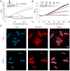
Figure 4.. cCMP mediates abortive infection in Pycsar.
(A) Growth curves of cells expressing the Pycsar system from E. coli E831 (purple) and control cells (black) with and without infection by phage T5 at multiplicity of infection (MOI) of 2 or 0.02. Results of three experiments are presented as individual curves. (B) Growth curves of Pycsar-expressing cells (purple) and control cells (black) with and without the addition of different concentrations of cCMP to the medium, or double distilled water (DDW) control. Results of three experiments are presented as individual curves. (C) Fluorescence microscopy images of E. coli MG1655 cells expressing the Pycsar system from E. coli E831 (lower panel), or control cells lacking the system (upper panel). Shown are DNA (blue) and membrane (red) stains. Images were captured at 60 min after the addition of 250 μM cCMP to the medium. Arrows point to abnormal membrane protrusions. Representative images from a single replicate out of three independent replicates are shown.
Figure 5.
Figure 5.. cUMP activates Pycsar TIR effectors that execute abortive infection.
(A) Growth curves of cells expressing the cyclase-TIR Pycsar system from Xanthomonas perforans GEV1001 (green) and control cells (black) with and without infection by phage T7 at multiplicity of infection (MOI) of 2 or 0.02. Results of three experiments are presented as individual curves. (B) Growth curves of XpPycsar-expressing cells (green) and control cells (black) with and without the addition of cUMP to the medium. Results of three experiments are presented as individual curves. (C) Concentrations of NAD+ in lysates extracted from XpPycsar expressing cells and control cells, infected by phage T7, as measured by LC-MSMS with synthesized NAD+ standard. X-axis represents minutes post infection, with zero representing non-infected cells. Cells were infected by phage T7 at an MOI of 2 at 37°C. Bar graphs represent the average of three biological replicates, with individual data points overlaid. (D) HPLC analysis of effector-mediated NAD+ cleavage by BcPycTIR. The TIR-domain effector cleaves NAD+ into the products ADP-ribose (ADPR) and nicotinamide (NAM), and the activity is strictly dependent on the presence of the cUMP molecule. cUMP was added at a concentration of 250 nM and incubated for 1 hr with the TIR effector from B. cepacia LK29 at 500 nM in the presence of 500 μM NAD+. Data are representative of 3 independent experiments. (E) Analysis of NAD+ cleavage activity of BcPycTIR using the fluorescent substrate ε-NAD+ at varying concentration of cNMPs. Data are average of three biological replicates with individual data points overlaid. (F) NAD+ cleavage activity of BcPycTIR in response to cUMP at finer resolution of concentrations span as compared to panel E. (G) Electron microscopy analysis of BcPycTIR filament formation in the presence of 50 μM cCMP, cUMP, or without a ligand. The 2D class average of the effector when activated by cUMP is presented at the bottom right. Scale bar for 2D class average image is 100 Å.
Figure 6.
Figure 6.. Phage escape from Pycsar-mediated defense.
(A) Representative phage mutants capable of escaping defense by the Pycsar system of E. coli E831. Shown are 10-fold serial dilution plaque assays, comparing the plating efficiency of WT and mutant phages on E. coli MG1655 cells that contain the system and a control strain that lacks the system and contains an empty vector instead. Mutations of all escape mutant phages are detailed in Table S2. (B) Positions of mutations within the T5 Pb8 protein. (C) Concentrations of cCMP in cell lysates extracted from cells infected by WT or mutant T5 phages, as measured by LC-MSMS with synthesized cCMP standard. X-axis represents minutes post infection, with zero representing non-infected cells. Cells were infected at an MOI of 2 at 37°C. Bar graphs represent the average of three biological replicates, with individual data points overlaid. EcPycC expression was induced by 0.02% arabinose. (D) A competition fitness assay between WT and mutant T5 phage on both EcPycsar-expressing and control cells. Y-axis represents the fraction of each strain out of the total phage mixture based on sequenced DNA reads. X-axis represents the infection passage, with 0 representing the mixture prior to first passage. In each passage, phage lysate was taken and used for infection of a fresh bacterial culture.

Comment in

References

    1. Bähre H, Danker KY, Stasch JP, Kaever V, and Seifert R (2014). Nucleotidyl cyclase activity of soluble guanylyl cyclase in intact cells. Biochem. Biophys. Res. Commun 443, 1195–1199. - PubMed
    1. Bähre H, Hartwig C, Munder A, Wolter S, Stelzer T, Schirmer B, Beckert U, Frank DW, Tümmler B, Kaever V, et al. (2015). cCMP and cUMP occur in vivo. Biochem. Biophys. Res. Commun 460, 909–914. - PMC - PubMed
    1. Baym M, Kryazhimskiy S, Lieberman TD, Chung H, Desai MM, and Kishony R (2015). Inexpensive Multiplexed Library Preparation for Megabase-Sized Genomes. PLoS One 10, e0128036. - PMC - PubMed
    1. Beckert U, Grundmann M, Wolter S, Schwede F, Rehmann H, Kaever V, Kostenis E, and Seifert R (2014). cNMP-AMs mimic and dissect bacterial nucleotidyl cyclase toxin effects. Biochem. Biophys. Res. Commun 451, 497–502. - PubMed
    1. Bernheim A, and Sorek R (2020). The pan-immune system of bacteria: antiviral defence as a community resource. Nat. Rev. Microbiol 18, 113–119. - PubMed

Publication types

MeSH terms