Comparative Study
. 2021 Oct 12;37(2):109813.
doi: 10.1016/j.celrep.2021.109813.

Sex-biased islet β cell dysfunction is caused by the MODY MAFA S64F variant by inducing premature aging and senescence in males

Affiliations
Comparative Study

Sex-biased islet β cell dysfunction is caused by the MODY MAFA S64F variant by inducing premature aging and senescence in males

Emily M Walker et al. Cell Rep. .

Abstract

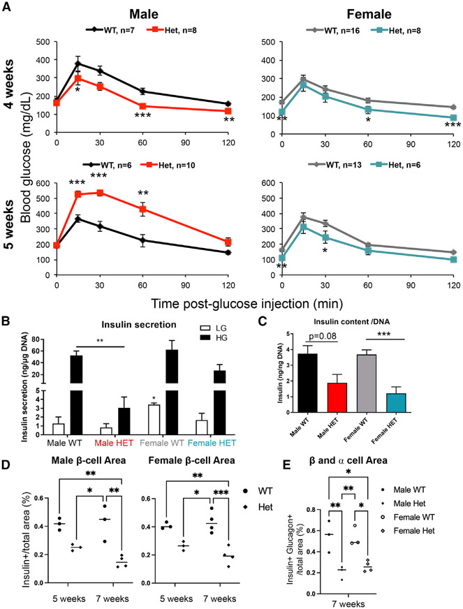
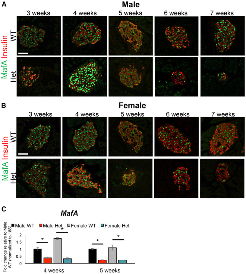
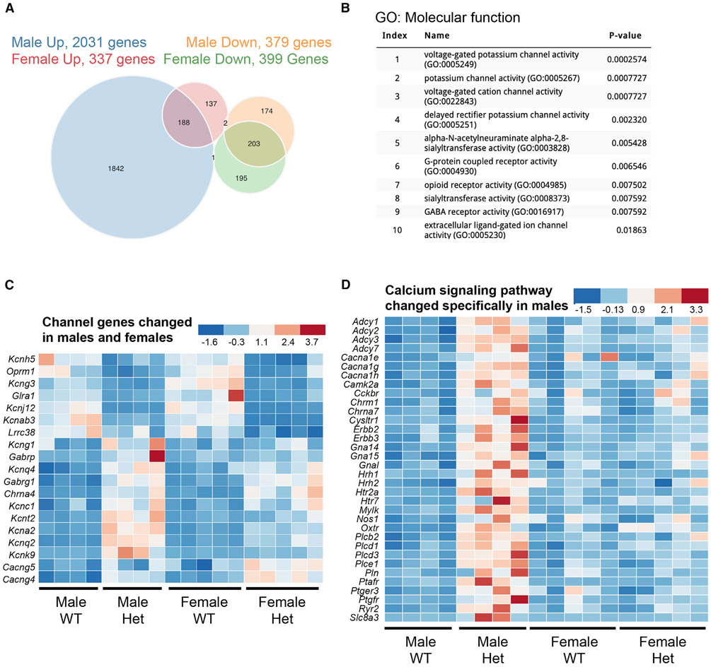
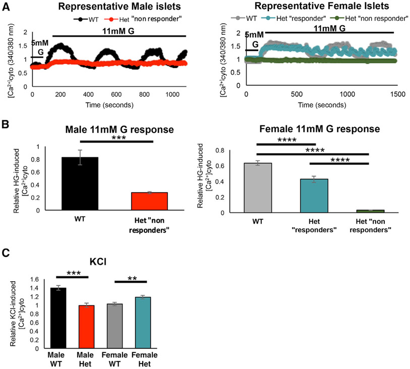
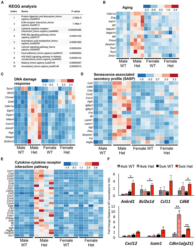
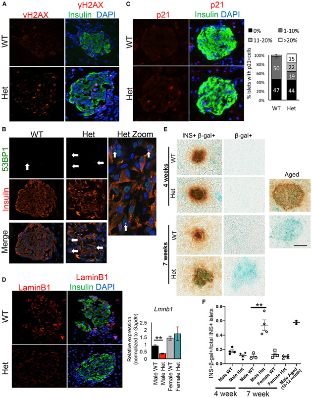
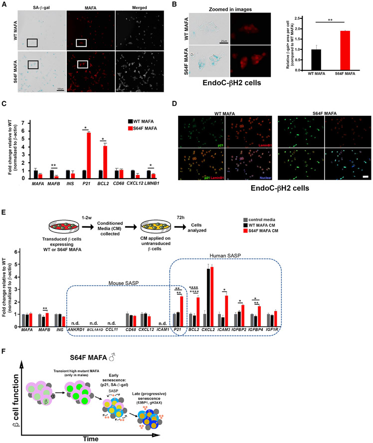
A heterozygous missense mutation of the islet β cell-enriched MAFA transcription factor (p.Ser64Phe [S64F]) is found in patients with adult-onset β cell dysfunction (diabetes or insulinomatosis), with men more prone to diabetes than women. This mutation engenders increased stability to the unstable MAFA protein. Here, we develop a S64F MafA mouse model to determine how β cell function is affected and find sex-dependent phenotypes. Heterozygous mutant males (MafAS64F/+) display impaired glucose tolerance, while females are slightly hypoglycemic with improved blood glucose clearance. Only MafAS64F/+ males show transiently higher MafA protein levels preceding glucose intolerance and sex-dependent changes to genes involved in Ca2+ signaling, DNA damage, aging, and senescence. MAFAS64F production in male human β cells also accelerate cellular senescence and increase senescence-associated secretory proteins compared to cells expressing MAFAWT. These results implicate a conserved mechanism of accelerated islet aging and senescence in promoting diabetes in MAFAS64F carriers in a sex-biased manner.

Keywords: MAFA; beta cell; cellular senescence; diabetes; islet biology; sexual dimorphism.


Conflict of interest statement

Declaration of interests The authors declare no competing interests.

Figures

Figure 1.
Figure 1.. Male but not female MafAS64F/+ mice become glucose intolerant between 4 and 5 weeks of age
(A) Fasted male and female animals underwent intraperitoneal glucose tolerance tests at 4 and 5 weeks of age. Male heterozygous (termed Het) MafAS64F/+ mice (red line) had improved glucose clearance at 4 weeks but become glucose intolerant by 5 weeks. Female MafAS64F/+ mice (teal line) had significantly lower fasting blood glucose levels and improved glucose clearance at both time points. (B) High glucose-stimulated insulin secretion in isolated islets was impaired in male MafAS64F/+ samples at 5 weeks. Islets were incubated with 4.6 mM (low, LG) or 16.8 mM (high, HG) glucose for 1 h. Secreted insulin was normalized to DNA content. (C) Islet insulin content trended lower in MafAS64F/+ males and was significantly decreased in females. Levels were normalized to DNA content. (D) Male (left panel) and female (right panel) islet β cell area was reduced at 7 weeks in MafAS64F/+ mice. The area was calculated by dividing the total insulin+ area by the total pancreatic area (eosin staining) in pancreas sections obtained every 50 μm multiplied by 100 to obtain percentage (%). (E) Indicative of reduced islet area, the combined islet β and α cell area was significantly reduced in male and female MafAS64F/+ mice at 7 weeks. (A–C) Two-tailed Student t test; *p < 0.05; **p < 0.01; ***p < 0.001. (D and E) Two-way ANOVA; *p < 0.05; **p < 0.01; ***p < 0.005.
Figure 2.
Figure 2.. MAFA protein levels were transiently upregulated at 4 weeks in male MafAS64F/+ islets
(A) Immunostaining over the course of 3, 4, 5, 6, and 7 weeks revealed that MAFA protein staining intensity only increased at 4 weeks of age in male MafAS64F/+ (Het) islets. (B) In contrast, MAFA staining intensity was unchanged between female MafAS64F/+ and WT islets. (C) MafA mRNA levels were significantly reduced at 4 and 5 weeks in both male and female MafAS64F/+ islet samples. Fold change shown relative to male WT islets. Two-tailed Student t test; *p < 0.05; scale bars, 50 μm.
Figure 3.
Figure 3.. Male and female MafAS64F/+ islet cells regulate a common and distinct set of genes associated with Ca2+ and K+ channel activity
(A) The Venn diagram illustrates the total number of RNA-seq-identified genes differentially up- or down-regulated between 5-week-old WT and MafAS64F/+ islets. (B) Gene Ontology (GO): molecular function analysis (p < 0.05) of the 391 genes commonly up- or down-regulated in MafAS64F/+ (Het) islets revealed alterations in multiple ion channel activity pathways. (C) Heatmaps showing channel gene expression changes common between male and female MafAS64F/+ islets. (D) Heatmap of Ca2+ signaling pathway genes uniquely increased in male MafAS64F/+ islets identified by Kyoto Encyclopedia of Genes and Genomes (KEGG) analysis (also see Figure 5A). False discovery rate (FDR) < 0.05.
Figure 4.
Figure 4.. Glucose-induced Ca2+ oscillations and KCl-induced Ca2+ responses are altered in MafAS64F/+ islets
(A) Representative Fura2 traces show loss of the β cell glucose-induced Ca2+ oscillations (11 mM glucose [G]) in 5-week-old male MafAS64F/+ (Het) islets (red line, “non-responders”). Female MafAS64F/+ islets had 2 different functionally responsive islet populations (“responders,” teal line; non-responders, green line). (B) Quantitation of male (left; 2-tailed Student t test; ***p < 0.001) and female (right; 1-way ANOVA; ****p < 0.0001) islets showed reduced cytoplasmic Ca2+ following stimulation with 11 mM glucose. The average peak amplitude was quantitated by dividing the first peak ΔCa2+ after 11 mM glucose application by the baseline response at 5 mM glucose. (C) The Ca2+ response to 30 mM KCl is reduced in male MafAS64F/+ islets but increased in female MafAS64F/+ islets. Representative traces for this experiment are shown in Figure S8. Two-tailed Student t test; **p < 0.01; ***p < 0.001.
Figure 5.
Figure 5.. Male MafAS64F/+ islets display increased expression of aging, DDR, SASP, and cytokine pathway gene signatures
(A) KEGG analysis of the 1,842 genes specifically upregulated in 5-week-old male MafAS64F/+ (Het) islets. (B–E) Heatmaps reveal male MafAS64F/+ islets have increased expression of pathway genes associated with (B) aging, (C) DDR, (D) SASP, and (E) cytokine-cytokine receptor interactions. FDR < 0.05. (F) qRT-PCR confirmation of pathway gene changes in 5-week-old male MafAS64F/+ islets (solid bars). Gene expression was unchanged at 4-week-old male MafAS64F/+ islets, with the exception of Cdkn1a (p21) upregulation (hash-marked bars). Two-tailed Student t test; *p < 0.05; **p < 0.01.
Figure 6.
Figure 6.. DNA damage and senescence markers are increased in dysfunctional male MafAS64F/+ islets
(A–C) gH2AX staining (A) and 53BP1 (B), markers of DNA double-strand breaks, and p21, a cell-cycle inhibitor (C), were present in male MafAS64F/+ (Het) islets. Male MafAS64F/+ islets had a significant increase in the proportion of islets with >10% p21+ cells. (D) Male MafAS64F/+ islets showed reduced LaminB1 protein (left) and mRNA (right). (E and F) SA-β-gal was not produced in 4-week-old male MafAS64F/+ islets, but was detected in MafAS64F/+ males by 7 weeks. (E) The SA-β-gal in 7-week-old MafAS64F/+ islets was of similar intensity to 10- to 12-month-old WT male mouse islets. (F) The proportion of SA-β-gal+ islets was quantitated for each sample (n = 3–4). Two-tailed Student t test; **p < 0.01; scale bar, 50 μm.
Figure 7.
Figure 7.. Human β cells expressing MAFAS64F demonstrate accelerated senescence and a species-specific SASP signature
(A) SA-β-gal staining on EndoC-βH2 cells transduced to express WT MAFA or MAFAS64F and stained for MAFA (red). Scale bar, 100 μm. (B) Inset areas outlined in boxes in (A) are magnified in the left panel. Scale bar, 25 μm. β-gal+ area was significantly increased in MAFAS64F cells (right panel). (C) Human expression of β cell-enriched proteins and senescence associated proteins identified in male MafAS64F/+ mice. Two-tailed Student t test; *p < 0.05; **p < 0.01. (D) Immunostaining for senescence markers p21 (green) was increased and LaminB1 (red) decreased in MAFAS64F-expressing EndoC-βH2 cells. (E) CM from WT MAFA or MAFAS64F-expressing EndoC-βH2 cells were collected, purified and added to EndoC-βH2 cells cultured for 72 h. SASP genes upregulated in male MafAS64F/+ mice were not identified in EndoC-βH2 cells. However, novel, species-specific secretory senescence-associated factors were identified in this context. N.D., not detectable. One-way ANOVA; *p < 0.05; **p < 0.01; ****p < 0.0001. (F) Schematic of temporal senescence and aging responses in MafAS64F-expressing male β cells. In male MafAS64F/+, transiently high MafA protein triggers molecular insults to induce premature senescence marked by cell-cycle arrest, senescence staining (blue cytoplasmic shade), and initiation of a senescence-associated secretory phenotype (SASP, pink secretory factors), resulting in impaired GSIS. Late (progressive) senescence propagates this phenotype with SASP amplification and diversification.

References

    1. Aguayo-Mazzucato C, Koh A, El Khattabi I, Li WC, Toschi E, Jermendy A, Juhl K, Mao K, Weir GC, Sharma A, and Bonner-Weir S (2011). Mafa expression enhances glucose-responsive insulin secretion in neonatal rat β cells. Diabetologia 54, 583–593. - PMC - PubMed
    1. Aguayo-Mazzucato C, Andle J, Lee TB Jr., Midha A, Talemal L, Chipashvili V, Hollister-Lock J, van Deursen J, Weir G, and Bonner-Weir S (2019). Acceleration of β cell aging determines diabetes and senolysis improves disease outcomes. Cell Metab. 30, 129–142.e4. - PMC - PubMed
    1. Ahlgren U, Jonsson J, Jonsson L, Simu K, and Edlund H (1998). β-cell-specific inactivation of the mouse Ipf1/Pdx1 gene results in loss of the β-cell phenotype and maturity onset diabetes. Genes Dev. 12, 1763–1768. - PMC - PubMed
    1. Arda HE, Li L, Tsai J, Torre EA, Rosli Y, Peiris H, Spitale RC, Dai C, Gu X, Qu K, et al. (2016). Age-Dependent Pancreatic Gene Regulation Reveals Mechanisms Governing Human β Cell Function. Cell Metab. 23, 909–920. - PMC - PubMed
    1. Arrojo E Drigo R, Lev-Ram V, Tyagi S, Ramachandra R, Deerinck T, Bushong E, Phan S, Orphan V, Lechene C, Ellisman MH, and Hetzer MW (2019). Age mosaicism across multiple scales in adult tissues. Cell Metab. 30, 343–351.e3. - PMC - PubMed

Publication types

MeSH terms