. 2021 Oct 8;36(8):e360801.
doi: 10.1590/ACB360801. eCollection 2021.

Effects of dipotassium glycyrrhizinate on wound healing

Affiliations

Effects of dipotassium glycyrrhizinate on wound healing

Camila Dos Santos Leite et al. Acta Cir Bras. .

Abstract

Purpose: Dipotassium glycyrrhizinate (DPG) has anti-inflammatory properties, besides promoting the regeneration of skeletal muscle. However, it has not been reported on skin wound healing/regeneration. This research aimed to characterize the effects of DPG in the treatment of excisional wounds by second intention.

Methods: Male adults (n=10) and elderly (n=10) Wistar rats were used. Two circular wounds were excised on the dorsal skin. The excised normal skins were considered adult (GAN) and elderly (GIN) naïve. For seven days, 2% DPG was applied on the proximal excision: treated adult (GADPG) and elderly (GIDPG), whereas distal excisions were untreated adult (GANT) and elderly (GINT). Wound healing areas were daily measured and removed for morphological analyses after the 14th and the 21st postoperative day. Slides were stained with hematoxylin-eosin, Masson's trichrome, and picrosirius red.

Results: Histological analysis revealed intact (GAN/GIN) and regenerated(GANT/GINT/GADPG/GIDPG) skins. No differences of wounds' size were found among treated groups. Epidermis was thicker after 14 days and thinner after 21 days of DPG administration. Higher collagen I density was found in GIDPG (14th day) and GADPG (21st day).

Conclusions: DPG induced woundhealing/skin regeneration, with collagen I, being more effective in the first 14 days after injury.


Conflict of interest statement

Conflict of interest: Nothing to declare

Author’s contribution: Conception and design of the study: Rocha T; Acquisition of data: Leite CS and Pires OC; Interpretation of data: Priolli DG; Analysis and interpretation of data: Rocha T; Technical procedures: Leite CS, Tenis DG and Ziegler JVN; Statistics analysis: Priolli DG; Manuscript preparation and writing: Leite CS; Critical revision: Pires OC.

Figures

Figure 1
Figure 1. Fluxogram of experimental groups: adults (GA); elderly (GI); non-treated–GANT14, GANT21, GINT14, and GINT21; treated–GADPG14, GADPG21, GIDPG14, and GIDPG21.
Figure 2
Figure 2. Graphical representation (groups GANT, GADPG, GINT and GIDPG; area in mm3) and macroscopical analysis (a-f) of wound healing overtime of skin resection [after 7 (a, d), 14 (b, e), 21 (c, f) days]. Observe the macroscopic aspect in adults (n=10)(a-c) and elderly (n=10, Bonferroni post-hoc test) (d-f) animals, untreated (arrow) and treated (double-arrow) with DPG (bar=1.3 cm).
Figure 3
Figure 3. Graphical representation and histological analysis of epidermis thickness (groups GAN - a, GANT14 - b, GADPG14 - c, GANT21 - d, GADPG21 - e; GIN - f, GINT14 - g, GIDPG14 - h, GINT21 - i and GIDPG21 - j; bar=100μm). Observe the normal aspects in naïve (a, f) and regenerated (b-e; g-j) skins. Note the increase of epidermis thickness after DPG treatment in adult (n=10), Bonferroni post-hoc test (c) and elderly (n=10) (h, j) animals.
Figure 4
Figure 4. Graphical representation and histological analysis of dermis thickness (groups GAN - a, GANT14 - b, GADPG14 - c, GANT21 - d, GADPG21 - e; GIN - f, GINT14 - g,GIDPG14 - h, GINT21 - i and GIDPG21 - j; bar=100 μm). Observe the normal aspects and collagen distribution in naïve (a, f) and regenerated (b-e; g-j) skins. Cutaneous appendages were not observed in regenerated dermis (b-e; g-j). (n=10, Dunnett post-hoc test).
Figure 5
Figure 5. Graphical representation of collagens I (a, c) and III (b, d) distribution in the dermis of groups GAN, GANT14, GADPG14, GANT21, GADPG21, GIN, GINT14, GIDPG14, GINT21 and GIDPG21. Observe that the amount of collagen I is higher than the one of collagen III in adult (a) and elderly (c) animals. Note that the collagen III significantly reduced in (b) adult and elderly (d) animals after skin resection (n=10, Dunnett post-hoc test).
Figure 6
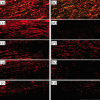
Figure 6. Histological analysis of collagen I (red) and collagen III (green) distribution in the dermis of groups GAN (a), GIN (b), GANT14 (c), GINT14 (d), GADPG14 (e), GIDPG14 (f), GANT21 (g) GINT21 (h), GADPG21 (i) and GIDPG21 (j) (bar=100 μm). (n=10, Dunnett post-hoc test).

References

    1. Rodrigues M, Kosaric N, Bonham CA, Gurtner GC. Wound healing: a cellular perspective. Physiol Rev. 2019;99(1):665–706. doi: 10.1152/physrev.00067.2017. - DOI - PMC - PubMed
    1. Sorg H, Tilkorn DJ, Hager S, Hauser J, Mirastschijski U. Skin wound healing: an update on the current knowledge and concepts. Eur Surg Res. 2017;58(1-2):81–94. doi: 10.1159/000454919. - DOI - PubMed
    1. Anderson K, Hamm RL. Factors that impair wound healing. J Am Coll Clin Wound Spec. 2014;4(4):84–91. doi: 10.1016/j.jccw.2014.03.001. - DOI - PMC - PubMed
    1. Guo S, Dipietro LA. Factors affecting wound healing. J Dent Res. 2010;89(3):219–229. doi: 10.1177/0022034509359125. - DOI - PMC - PubMed
    1. Oriá RB, Ferreira FVA, Santana EN, Fernandes MR, Brito GAC. Study of age-related changes in human skin using histomorphometric and autofluorescence approaches. An Bras Dermatol. 2003;78(4):425–434. doi: 10.1590/S0365-05962003000400004. - DOI