. 2022 Feb;7(1):62-70.
doi: 10.1136/svn-2021-001028. Epub 2021 Oct 13.

Novel role of STAT3 in microglia-dependent neuroinflammation after experimental subarachnoid haemorrhage

Affiliations

Novel role of STAT3 in microglia-dependent neuroinflammation after experimental subarachnoid haemorrhage

Zhiyuan Vera Zheng et al. Stroke Vasc Neurol. 2022 Feb.

Abstract

Background and purpose: Signal transducer and activator of transcription 3 (STAT3) may contribute to the proinflammation in the central nervous system diseases by modulating the microglial responses. Thus, this study was intended to investigate the effect of STAT3 on microglia-dependent neuroinflammation and functional outcome after experimental subarachnoid haemorrhage (SAH).

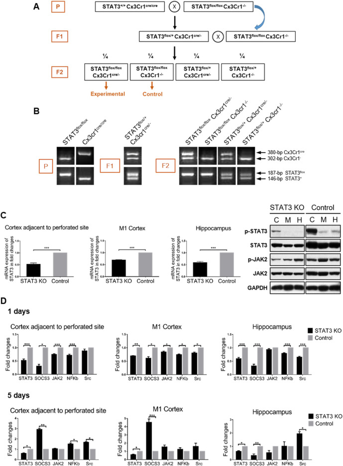
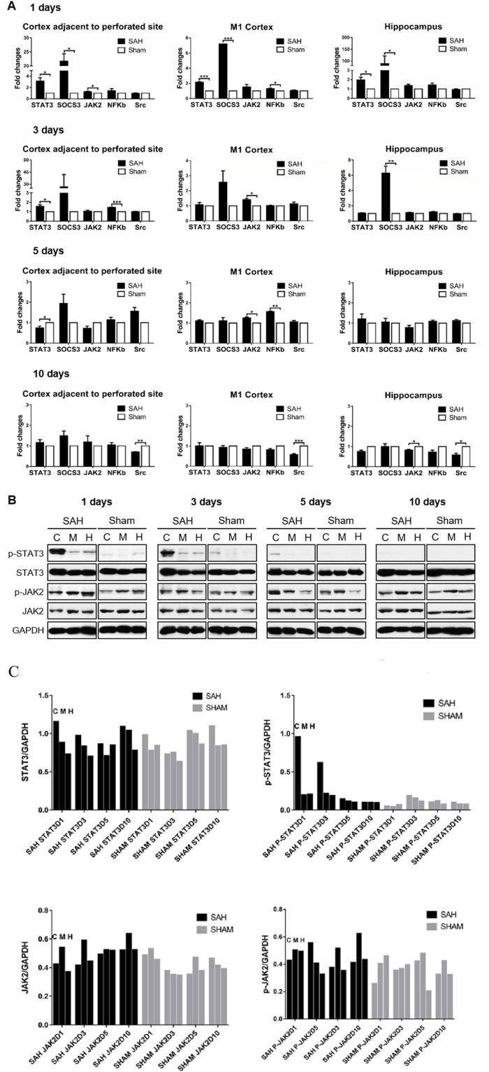
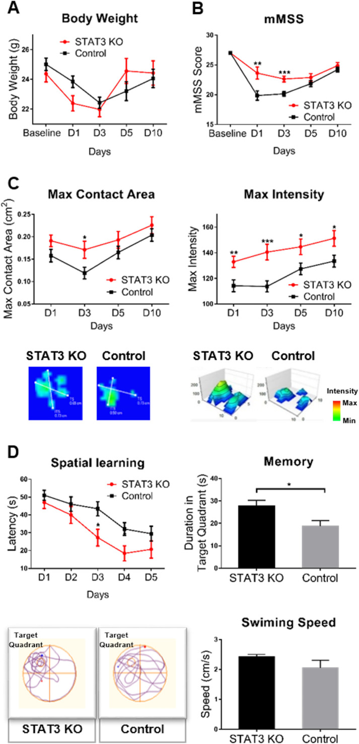
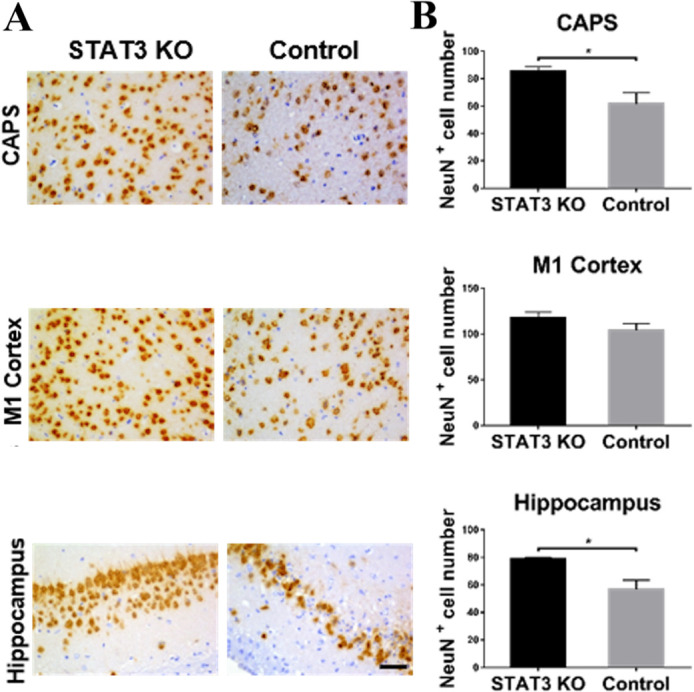
Methods: The SAH model was established by endovascular perforation in the mouse. Real-time PCR (RtPCR) and western blot were used to examine the dynamic STAT3 signalling pathway responses after SAH. To clarify the role of the STAT3 signalling pathway in the microglia-dependent neuroinflammation after SAH, the microglia-specific STAT3 knockout (KO) mice were generated by the Cre-LoxP system. The neurological functions were assessed by Catwalk and Morris water maze tests. Neuronal loss after SAH was determined by immunohistochemistry staining. Microglial polarisation status after STAT3 KO was then examined by RtPCR and immunofluorescence.

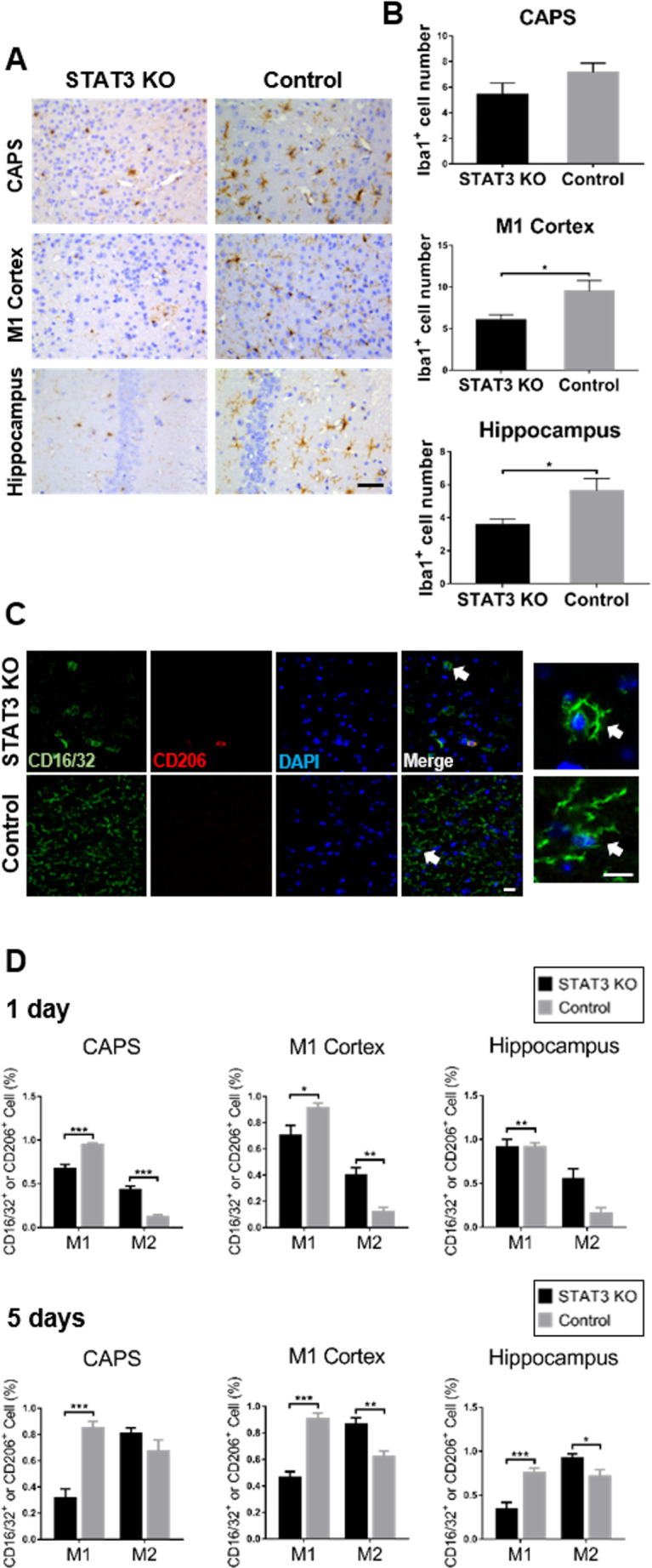
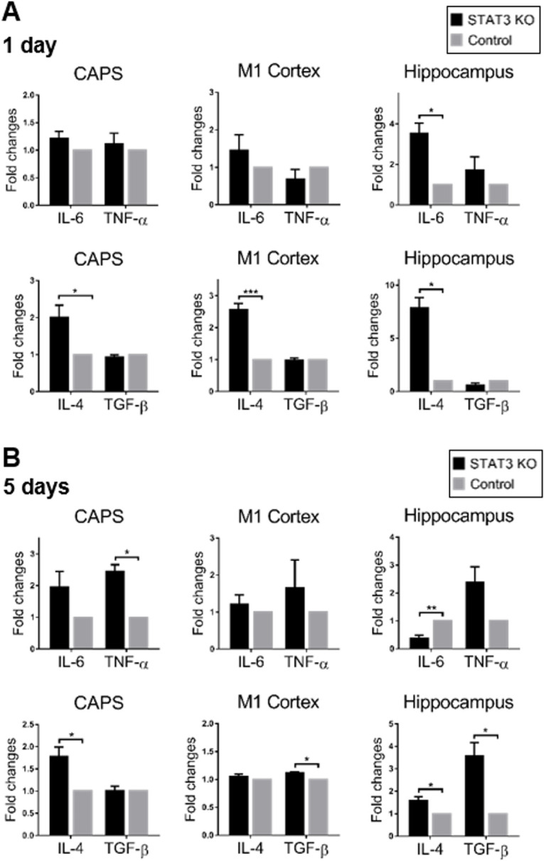
Results: The STAT3 and Janus kinase-signal transducer 2 activated immediately with the upregulation and phosphorylation after SAH. Downstream factors and related mediators altered dynamically and accordingly. Microglial STAT3 deletion ameliorated the neurological impairment and alleviated the early neuronal loss after SAH. To investigate the underlying mechanism, we examined the microglial reaction after STAT3 KO. STAT3 deletion reversed the increase of microglia after SAH. Loss of STAT3 triggered the early morphological changes of microglia and primed microglia from M1 to M2 polarisation. Functionally, microglial STAT3 deletion suppressed the SAH-induced proinflammation and promoted the anti-inflammation in the early phase.

Conclusions: STAT3 is closely related to the microglial polarisation transition and modulation of microglia-dependent neuroinflammation. Microglial STAT3 deletion improved neurological function and neuronal survival probably through promoting M2 polarisation and anti-inflammatory responses after SAH. STAT3 may serve as a promising therapeutic target to alleviate early brain injury after SAH.

Keywords: hemorrhage; inflammation; subarachnoid.


Conflict of interest statement

Competing interests: None declared.

Figures

Figure 1
Figure 1
Characterisation of microglia-specific STAT3 KO mice. (A) The schematic description of the breeding strategy. (B) PCR analysis of genotypes. STAT3flox allele was electrophoretically separated and demonstrated as a 187 bp fragment. Wild-type STAT3 allele was 146 bp. Cx3Cr1cre fragment was 380 bp and Cx3Cr1 was 302 bp. (C) Biochemical determination of STAT3 KO at the gene and protein levels. RNA and protein were extracted from the CAPS (C), M1 cortex (M) and hippocampus (H) of the SAH brain. RtPCR examination was performed to determine the mRNA expression of STAT3 in STAT3 KO mice compared with the control. WB was performed to determine the STAT3/JAK2 phosphorylation status of STAT3 KO mice. (D) The STAT3 signalling pathway activation status of STAT3 KO mice at 1 and 5 days after SAH. The mRNA expression of the mediators involved in the STAT3 pathway is shown in the fold change in the CAPS, M1 cortex and hippocampus of STAT3 KO mice compared with the control. n=3 per group. Values are mean±SEM. *P≤0.05, **P0.01, ***P≤0.001. CAPS, cortex adjacent to the perforated site; JAK2, Janus kinase-signal transducer 2; KO, knockout; RtPCR, real-time PCR; SAH, subarachnoid haemorrhage; STAT3, signal transducer and activator of transcription 3; WB, western blot.
Figure 2
Figure 2
STAT3 signalling pathway activated after experimental SAH. (A) RNA extract was prepared from CAPS, M1 cortex and hippocampus of SAH brain at 1, 3, 5 and 10 days. The mRNA expression of mediators involved in the STAT3 pathway after SAH was determined by RtPCR and shown in the fold change compared with the sham-operated group. n=3–4/group/time point. Values were mean±SEM. *P≤0.05, **P≤0.01, ***P≤0.001. (B) STAT3/JAK2 phosphorylation status was detected by WB at 1, 3, 5 and 10 days. The levels of phosphorylated and total STAT3/JAK2 relative to internal control GAPDH are shown in the cortex adjacent to the perforated site (C), M1 cortex (M) and hippocampus (H), respectively, of SAH and sham-operated mice. n=3/group/time point. (C) The quantitative analysis of the pSTAT3/STAT3 and pJAK2/JAK of (B) to show the trend of pSTAT3/STAT3 and pJAK2/JAK in the SAH and sham groups. CAPS, cortex adjacent to the perforated site; JAK2, Janus kinase-signal transducer 2; NF-κB, nuclear factor-kappa B; RtPCR, real-time PCR; SAH, subarachnoid haemorrhage; STAT3, signal transducer and activator of transcription 3; WB, western blot.
Figure 3
Figure 3
Microglial STAT3 deletion ameliorated the neurological impairment after SAH. (A) The time-course body weight changes were examined in STAT3 KO and control groups of mice. (B)The sensorimotor functions were assessed by the mMSS (5–27 scores). (C) Gait analysis was performed by the Catwalk system. The max contact area (CM2) and max intensity of paw were detected. STAT3 KO mice showed the alleviated paw contraction and increased paw intensity compared with the control. (D) Cognitive functions including learning ability and memory were assessed by the MWM test. Spatial learning ability was defined as the latency to escape the platform. Memory was defined as the time mice spent in the target quadrant where the platform was placed previously. The bottom figure shows the typical swim path and average swimming speed of STAT3 KO and control groups of mice. n=8–16 per group. Values are mean±SEM. *P≤0.05, **P≤0.01, ***P≤0.001. KO, knockout; mMSS, Mouse Motor and Sensory Scale; MWM, Morris water maze; SAH, subarachnoid haemorrhage; STAT3, signal transducer and activator of transcription 3.
Figure 4
Figure 4
Microglial STAT3 deletion alleviated the neuronal loss after SAH. (A) Neuronal loss was detected by NeuN immunohistochemistry in the CAPS, M1 cortex and hippocampus of the brain. The representative NeuN labelled coronal brain sections were shown on day 5 after SAH. (B) NeuN-positive neurons were quantified in the aforementioned brain areas of STAT3 KO and control groups of mice. Bar=50 µm. STAT3 KO n=7, control n=8. Values are ean±SEM. *P≤0.05, **P≤0.01, ***P≤0.001. CAPS, cortex adjacent to the perforated site; KO, knockout; SAH, subarachnoid haemorrhage; STAT3, signal transducer and activator of transcription 3.
Figure 5
Figure 5
Stat3 deletion primed microglia to M2 polarisation. (A) Microglia was examined by iba1 immunohistochemistry in CAPS, M1 cortex and hippocampus of the brain. The representative iba1 labelled coronal brain sections were shown on day 1 after SAH. Bar=50 µm. (B) Iba1-positive microglia were quantified in the aforementioned brain areas of STAT3 KO and control groups of mice. (C) Microglial polarisation status was determined by immunofluorescence. The representative confocal microscopic images showed the visualisation of CD16/32 (M1, green), CD206 (M2, red) and DAPI (nuclei, blue) coexpression in CAPS on day 1 after SAH. The white arrows indicate the highly magnified view of the typical microglial processes. Bar=20 µm for all magnifications. (D) CD16/32-positive (M1) or CD206-positive (M2) cells were quantified in the aforementioned brain areas of STAT3 KO and control groups of mice at 1 and 5 days after SAH. n=5–8 per group. Values were the mean±SEM. *P≤0.05, **P≤0.01, ***P≤0.001. CAPS, cortex adjacent to the perforated site; KO, knockout; SAH, subarachnoid haemorrhage; STAT3, signal transducer and activator of transcription 3.
Figure 6
Figure 6
Microglial STAT3 deletion promoted anti-inflammation after SAH. (A, B) The expression of M1/M2 microglia-related cytokines were detected by RtPCR at 1 and 5 days after SAH. The mRNA expression of M1-phenotype related pro-inflammatory factors (IL-6 and TNF-α) and M2-phenotype related anti-inflammatory factors (IL-4 and TGF-β) was shown in fold change in STAT3 KO mice compared with the control. Values were the mean±SEM *p≤0.05, **p≤0.01, ***p≤0.001. CAPS, cortex adjacent to the perforated site; IL, interleukin; KO, knockout; SAH, subarachnoid haemorrhage; STAT3, signal transducer and activator of transcription 3; TGF-β, transforming growth factor-β.

References

    1. Cahill J, Zhang JH. Subarachnoid hemorrhage: is it time for a new direction? Stroke 2009;40:S86–7. 10.1161/STROKEAHA.108.533315 - DOI - PMC - PubMed
    1. de Rooij NK, Linn FHH, van der Plas JA, et al. . Incidence of subarachnoid haemorrhage: a systematic review with emphasis on region, age, gender and time trends. J Neurol Neurosurg Psychiatry 2007;78:1365–72. 10.1136/jnnp.2007.117655 - DOI - PMC - PubMed
    1. Zheng VZ, Wong GKC. Neuroinflammation responses after subarachnoid hemorrhage: a review. J Clin Neurosci 2017;42:7–11. 10.1016/j.jocn.2017.02.001 - DOI - PubMed
    1. van Dijk BJ, Vergouwen MDI, Kelfkens MM, et al. . Glial cell response after aneurysmal subarachnoid hemorrhage - functional consequences and clinical implications. Biochim Biophys Acta 2016;1862:492–505. 10.1016/j.bbadis.2015.10.013 - DOI - PubMed
    1. Saijo K, Glass CK. Microglial cell origin and phenotypes in health and disease. Nat Rev Immunol 2011;11:775–87. 10.1038/nri3086 - DOI - PubMed

Publication types

MeSH terms

Substances