Clinical Trial
. 2021 Oct 13;11(1):20357.
doi: 10.1038/s41598-021-99749-0.

Interindividual variability of electric fields during transcranial temporal interference stimulation (tTIS)

Affiliations
Clinical Trial

Interindividual variability of electric fields during transcranial temporal interference stimulation (tTIS)

Jill von Conta et al. Sci Rep. .

Abstract

Transcranial temporal interference stimulation (tTIS) is a novel non-invasive brain stimulation technique for electrical stimulation of neurons at depth. Deep brain regions are generally small in size, making precise targeting a necessity. The variability of electric fields across individual subjects resulting from the same tTIS montages is unknown so far and may be of major concern for precise tTIS targeting. Therefore, the aim of the current study is to investigate the variability of the electric fields due to tTIS across 25 subjects. To this end, the electric fields of different electrode montages consisting of two electrode pairs with different center frequencies were simulated in order to target selected regions-of-interest (ROIs) with tTIS. Moreover, we set out to compare the electric fields of tTIS with the electric fields of conventional tACS. The latter were also based on two electrode pairs, which, however, were driven in phase at a common frequency. Our results showed that the electric field strengths inside the ROIs (left hippocampus, left motor area and thalamus) during tTIS are variable on single subject level. In addition, tTIS stimulates more focally as compared to tACS with much weaker co-stimulation of cortical areas close to the stimulation electrodes. Electric fields inside the ROI were, however, comparable for both methods. Overall, our results emphasize the potential benefits of tTIS for the stimulation of deep targets, over conventional tACS. However, they also indicate a need for individualized stimulation montages to leverage the method to its fullest potential.


Conflict of interest statement

CSH holds a patent on brain stimulation. JC, FHK, BCB, AA, AT declare no competing interests.

Figures
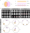
Figure 1
Figure 1
Concept of temporal interference stimulation (tTIS) and selected electrode montages to target three different regions of interest (ROIs). (a) For tTIS, two pairs of stimulation electrodes are attached to the scalp. Between the electrodes of each electrode pair, a high frequency alternating current is applied at e.g., 1 kHz and 1.01 kHz. The superposition of the signals causes an amplitude modulation oscillating at the difference of both frequencies (in this case 10 Hz). (b) Anatomical locations (in red) of the defined ROIs on the MNI brain: left hippocampus, left motor cortex and thalamus. (c) The electrode montages targeting the left hippocampus and the left motor cortex were adapted from Rampersad et al. for tTIS and for tACS. The electrode montage targeting the thalamus was defined with F7-PO7, and F8-PO8 for tTIS and for tACS.
Figure 2
Figure 2
Electric field simulation results for transcranial temporal interference stimulation (tTIS) and transcranial alternating current stimulation (tACS), targeting the left hippocampus. (a) Electric field distribution on the MNI brain for tTIS and tACS. (b) Electric field distribution for two representative subjects targeting the left hippocampus for tTIS (left). Spatial correlation of the electric fields over the whole brain across all subjects (right). (c) Parameter that characterize the electric field simulations for tTIS and tACS (mean electric field inside the ROIs—bottom left, the maximum of the electric fields inside vs. outside the ROIs—bottom middle, proportion of voxel that exposed to field strength greater than 0.1 V/m inside the ROIs and for the whole gray matter—bottom right). Violin plots indicate the distribution of the underlying data. Note that the violin plots include a boxplot (in the center of the violin with the median (white dots), 25% quartile and 75% quartile, upper- and lower adjacent value. Additionally, individual data points are overlayed (black dots).
Figure 3
Figure 3
Electric field simulation results for transcranial temporal interference stimulation (tTIS) and transcranial alternating current stimulation (tACS), targeting the left motor area. (a) Electric field distribution on the MNI brain for tTIS and tACS. (b) Electric field distribution for two representative subjects targeting the left motor area for tTIS (left). Spatial correlation of the electric fields over the whole brain across all subjects (right). (c) Parameters that characterize the electric field simulations for tTIS and tACS (mean electric field inside the ROIs—bottom left, the maximum of the electric fields inside vs. outside the ROIs—bottom middle, proportion of voxel that exposed to field strength greater than 0.1 V/m inside the ROIs and for the whole gray matter—bottom right).
Figure 4
Figure 4
Electric field simulation results for transcranial temporal interference stimulation (tTIS) and transcranial alternating current stimulation (tACS), targeting the thalamus. (a) Electric field distribution on the MNI brain for tTIS and tACS. (b) Electric field distribution for two representative subjects targeting thalamus for tTIS (left). Spatial correlation of the electric fields over the whole brain across all subjects (right). (c) Parameter that characterize the electric field simulations for tTIS and tACS (mean electric field inside the ROIs—bottom left, the maximum of the electric fields inside vs. outside the ROIs—bottom middle, proportion of voxel that exposed to field strength greater than 0.1 V/m inside the ROIs and for the whole gray matter—bottom right).

References

    1. Miniussi C, Harris JA, Ruzzoli M. Modelling non-invasive brain stimulation in cognitive neuroscience. Neurosci. Biobehav. Rev. 2013;37:1702–1712. doi: 10.1016/j.neubiorev.2013.06.014. - DOI - PubMed
    1. Paulus W. Transcranial electrical stimulation (tES-tDCS; tRNS, tACS) methods. Neuropsychol. Rehabil. 2011;21:602–617. doi: 10.1080/09602011.2011.557292. - DOI - PubMed
    1. Nitsche MA, et al. Transcranial direct current stimulation: State of the art 2008. Brain Stimul. 2008;1:206–223. doi: 10.1016/j.brs.2008.06.004. - DOI - PubMed
    1. Benabid AL, Chabardes S, Mitrofanis J, Pollak P. Deep brain stimulation of the subthalamic nucleus for the treatment of Parkinson’s disease. Lancet Neurol. 2009;8:67–81. doi: 10.1016/S1474-4422(08)70291-6. - DOI - PubMed
    1. Rodriguez-Oroz MC, et al. Bilateral deep brain stimulation in Parkinson’s disease: A multicentre study with 4 years follow-up. Brain. 2005;128:2240–2249. doi: 10.1093/brain/awh571. - DOI - PubMed

Publication types