

Development of prefrontal cortex
- PMID: 34645980
- PMCID: PMC8511863
- DOI: 10.1038/s41386-021-01137-9
Development of prefrontal cortex
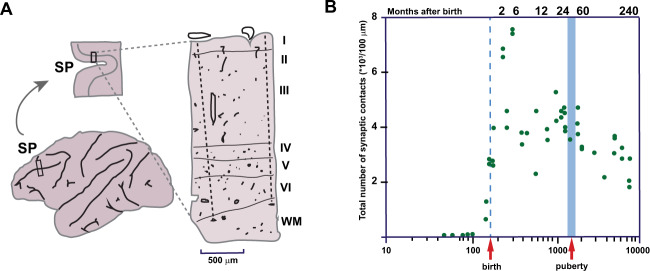
Abstract
During evolution, the cerebral cortex advances by increasing in surface and the introduction of new cytoarchitectonic areas among which the prefrontal cortex (PFC) is considered to be the substrate of highest cognitive functions. Although neurons of the PFC are generated before birth, the differentiation of its neurons and development of synaptic connections in humans extend to the 3rd decade of life. During this period, synapses as well as neurotransmitter systems including their receptors and transporters, are initially overproduced followed by selective elimination. Advanced methods applied to human and animal models, enable investigation of the cellular mechanisms and role of specific genes, non-coding regulatory elements and signaling molecules in control of prefrontal neuronal production and phenotypic fate, as well as neuronal migration to establish layering of the PFC. Likewise, various genetic approaches in combination with functional assays and immunohistochemical and imaging methods reveal roles of neurotransmitter systems during maturation of the PFC. Disruption, or even a slight slowing of the rate of neuronal production, migration and synaptogenesis by genetic or environmental factors, can induce gross as well as subtle changes that eventually can lead to cognitive impairment. An understanding of the development and evolution of the PFC provide insight into the pathogenesis and treatment of congenital neuropsychiatric diseases as well as idiopathic developmental disorders that cause intellectual disabilities.
© 2021. The Author(s).
Conflict of interest statement
The authors declare no competing interests.
Figures


References
-
- Fuster JM. Prefrontal neurons in networks of executive memory. Brain Res Bull. 2000;52:331–6. - PubMed
-
- Fuster JM. Memory networks in the prefrontal cortex. Prog brain Res. 2000;122:309–16. - PubMed
-
- Goldman-Rakic PS. Circuitry of the primate prefrontal cortex and the regulation of behavior by representational memory. Handbook of Physiology, The Nervous System, Higher Functions of the Brain. In: Plum F, editor. Handbook of physiology, the nervous system, higher functions of the brain. Bethesda: American Physiological Society; 1987. pp. 373–417.
-
- Li H, Yan G, Luo W, Liu T, Wang Y, Liu R, et al. Mapping fetal brain development based on automated segmentation and 4D brain atlasing. Brain Struct Funct. 2021;226:1961–72. - PubMed
Publication types
MeSH terms
Substances
Grants and funding
LinkOut - more resources
Full Text Sources
Miscellaneous
