. 2021 Oct;598(7882):652-656.
doi: 10.1038/s41586-021-03968-4. Epub 2021 Oct 13.

Leprosy in wild chimpanzees

Affiliations

Leprosy in wild chimpanzees

Kimberley J Hockings et al. Nature. 2021 Oct.

Abstract

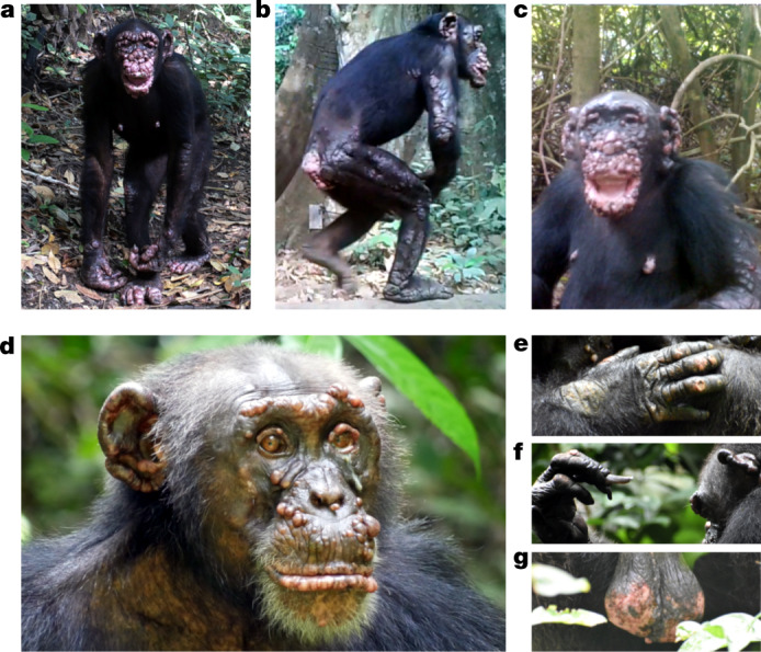
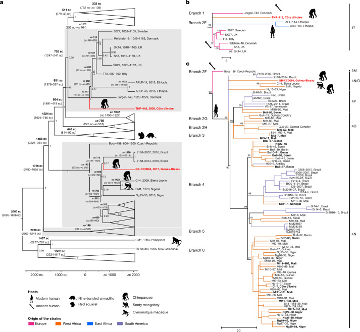
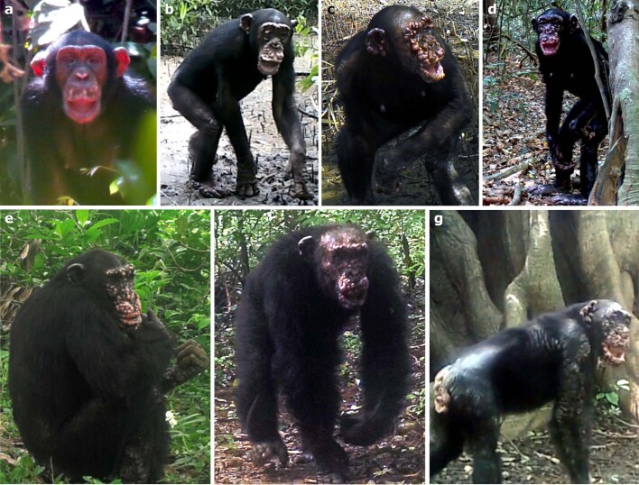
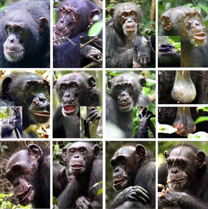
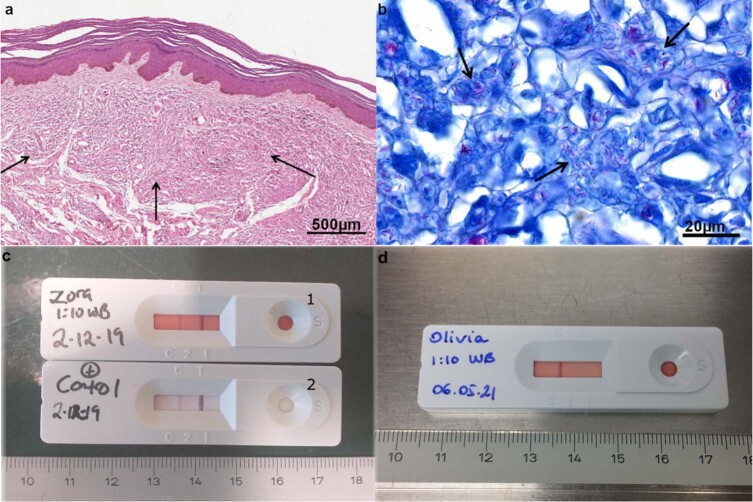
Humans are considered as the main host for Mycobacterium leprae1, the aetiological agent of leprosy, but spillover has occurred to other mammals that are now maintenance hosts, such as nine-banded armadillos and red squirrels2,3. Although naturally acquired leprosy has also been described in captive nonhuman primates4-7, the exact origins of infection remain unclear. Here we describe leprosy-like lesions in two wild populations of western chimpanzees (Pan troglodytes verus) in Cantanhez National Park, Guinea-Bissau and Taï National Park, Côte d'Ivoire, West Africa. Longitudinal monitoring of both populations revealed the progression of disease symptoms compatible with advanced leprosy. Screening of faecal and necropsy samples confirmed the presence of M. leprae as the causative agent at each site and phylogenomic comparisons with other strains from humans and other animals show that the chimpanzee strains belong to different and rare genotypes (4N/O and 2F). These findings suggest that M. leprae may be circulating in more wild animals than suspected, either as a result of exposure to humans or other unknown environmental sources.


Conflict of interest statement

The authors declare no competing interests.

Figures

Fig. 1
Fig. 1. Clinical manifestations of leprosy in three chimpanzees at CNP, Guinea-Bissau and TNP, Côte d’Ivoire.
a–c, Clinical signs of leprosy in two adult female chimpanzees in CNP (images extracted from camera traps). a, Rita has large hypopigmented nodules covering the entire body; disfigurement of the face, ears, hands and feet (ulcerated lesions and swelling). b, Rita has extensive plaques covering all limbs, with hair loss. c, Brinkos has large hypopigmented nodules covering the entire face, with extreme disfigurement of the face and ears, and ulcerated plaques on the arms and the nipples. dg, Clinical signs of leprosy in an adult male chimpanzee, Woodstock, at TNP. d, Multiple hypopigmented nodules on the ears, brow ridges, eyelid margins, nostrils, lips and the area between the upper lip and the nose. e, Hypopigmentation and swelling of the hands with ulcerations and hair loss on the dorsal side of the joints. f, Claw hand with nail loss and abnormal overgrowth of fingernails. g, Scrotal reddening and ulceration with fresh blood.
Fig. 2
Fig. 2. Phylogeny of M. leprae strains from human and animal hosts.
a, Bayesian dated phylogenetic tree of 278 M. leprae genomes including the two new chimpanzee strains (in bold red). Hypermutated samples with mutations in the nth gene were excluded from the analysis. The tree is drawn to scale, with branch lengths representing years of age. Median estimates of node ages are shown in black above branches; 95% HPD intervals are shown in grey. Some M. leprae branches are collapsed to increase readability. b, Maximum parsimony tree of branch 2F. c, Maximum parsimony tree of the branch 4. The tree was initially constructed using 286 genomes (Supplementary Table 6), including 2 new chimpanzee strains (in bold red) and 21 new genomes from West Africa (in bold), 500 bootstrap replicates and M. lepromatosis as outgroup. Sites with missing data were partially deleted (80% genome coverage cutoff), resulting in 4,470 variable sites used for the tree calculation. Subtrees corresponding to branches were retrieved in MEGA7. Corresponding genotypes are indicated on the side of each subtree. Samples are binned according to geographical origin as given in the legend. Scale bars (b, c), number of nucleotide substitutions. Animal silhouettes are available under Public Domain licence at PhyloPic (http://PhyloPic.org/).
Extended Data Fig. 1
Extended Data Fig. 1. Maps of the chimpanzee study sites and chimpanzee communities.
a, Map of the CNP, Guinea-Bissau and the TNP, Côte d’Ivoire, West Africa. b, Location of the chimpanzee communities at CNP that were monitored between 2015 and 2019 (1, Caiquene-Cadique; 2, Lautchande; 3, Cambeque; 4, Cabante; 5, Canamine; 6, Madina; 7, Amindara; 8, Guiledje). Estimated home ranges of chimpanzee communities at CNP are shown by 100% minimum convex polygons of direct chimpanzee observations and indirect chimpanzee traces and nests during the study period. Red outline represents chimpanzee communities with at least one individual with clinical manifestations of leprosy, confirmed using molecular analysis; orange outline represents chimpanzee communities with at least one individual with clinical manifestations of leprosy; yellow colour represents monitored communities where clinical manifestations of leprosy have not been observed nor confirmed through molecular analysis. c, Location of the three habituated chimpanzee communities monitored at TNP (N, north; S, south; E, east). Estimated home ranges of chimpanzee communities at TNP are shown by 100% minimum convex polygons of direct chimpanzee follows from December 2013 to October 2016. Red outline represents the community with individuals with clinical manifestations of leprosy, confirmed using molecular analysis and serological tests; blue colour represents communities where leprosy has not been recorded.
Extended Data Fig. 2
Extended Data Fig. 2. Disease progression of leprosy in chimpanzees at CNP.
Adult female chimpanzee Rita over the course of 5 years (ad) and disease manifestations in three additional adult chimpanzees (eg). a, 2013/05 – Hypopigmentation of skin around the mouth and nose, small nodule on the lower lip and left ear (opportunistically recorded with a video camera before the start of longitudinal health monitoring with camera traps). b, 2015/12 – Large nodules between the upper lip and nose, with multiple small nodules on the eyelids, cheek, ears margins, lower lip and brow ridge. Small dry patches with hair loss on the wrists, knees and elbows. c, 2017/12 – Nodules increase in number, with apparent swelling and reddening, facial disfigurement and claw hand. Plaques appear on the wrist, knee and elbow joints, with an increase in hair thinning. d, 2018/05 – Face and ears completely covered by large nodules, with facial disfigurement and generalized hair loss on limbs and lower back. Nodule formation and swelling of fingers and toes, with disfigurement of hands and feet, and more severe claw hand. Some plaques on the body are ulcerated, and the individual has clear weight loss. e, Jimi (Lautchande in 2018/06) – First observation of lesions in 2015. The head is completely covered with multiple nodules of reddish colour, some of which are ulcerated. Ear margins are thickened. Hands and feet present nodules and plaques, and the scrotum is affected (not visible on picture). f, Baaba (Cambeque in 2017/08) – First observation of lesions in 2017. Multiple hypopigmented nodules on the brow ridge, cheek and upper and lower lips. Ears have thickened margins and nodules. There is hair thinning, with multiple small plaques present on the upper and lower limbs, back, abdomen and shoulders. g, Brinkos (Caiquene-Cadique in 2018/10) – First observation of lesions in 2015. Facial disfigurement, with the ulceration of nodules and a hanging lower lip. Hands and feet are ulcerated, and fingers are swollen. There are nodules on the nipples, and plaques covering the lower back, shoulders and arm are ulcerated, with hair loss.
Extended Data Fig. 3
Extended Data Fig. 3. Disease progression of leprosy in an adult male chimpanzee at TNP (Woodstock) over the course of 2 years (2018–2020) (ai) and an adult female chimpanzee at TNP (Zora) over the course of 2008–2009 (jm).
a, 2017/01 – Woodstock before the appearance of clinical signs. b, 2018/06 – First hypopigmented nodules appear on the face (arrows), with swelling and hypopigmentation on both hands, and ulceration on the right hand. c, 2018/10 – Existing nodules increase in size and new smaller ones appear (arrows). Development of mucopurulent discharge from the left eye, and lower eyelid is turned outward. Hair loss and ulceration on dorsal part of right wrist and hand. d, 2019/04 – Most existing nodules increase in size and become pedunculated, and the nodule under the eye shrinks, and several new nodules appear (see arrows). Suspected start of nasal involvement, and right ear starts to become disfigured. Both hands are slightly swollen and hypopigmented, with the loss of nail plate on the fourth finger of the left hand, and the third and fifth fingers show early stage of abnormal nail overgrowth. e, 2019/10 – Facial lesions increase in size, and some become darkly pigmented. New lesions appear on the brow ridge, with nodules above the lips and between the lips, and the nose becomes pedunculated. The loss of nail plate, and nail bed becomes exposed on the first and second fingers of the left hand. f, 2020/04 – In general, facial nodules seem smaller than before, and the nodule under the left eye disappears. On the left hand, the nail of the fourth finger shows an advanced stage of abnormal nail overgrowth, and the third and fifth fingernails show early stage of abnormal nail overgrowth. g, 2020/07 – Facial nodules seem larger with many hypopigmented, and both ears are swollen and disfigured. Nasal involvement becomes apparent. Both hands are swollen and hypopigmented. Skin ulcerations present on the right hand, with possible claw hand on the left hand. h, 2019/04 – Slight hypopigmentation of scrotum. i, 2020/07 – Reddening and ulceration of scrotum; fresh blood observed. j, 2007/12 – Zora before the appearance of clinical signs of leprosy. k, 2008/01 – Appearance of nodules on the right ear and both eyebrow ridges. l, 2008/12 – Appearance of nodules on the left ear, and ulceration of the skin at the second, third and fourth proximal interphalangeal joint level of the right hand. m, 2009/04 – Nodular lesions on both ears and brow ridge seem aggravated, with nodular lesions on the lips, and above the mouth (four months before death).
Extended Data Fig. 4
Extended Data Fig. 4. Confirmation of leprosy infection in Zora through histopathology of skin sample and lateral flow test.
a, Lepromatous leprosy, skin with diffuse histiocytic infiltrate in the dermis. The haematoxylin and eosin stain was conducted once; scale bar, 500 μm. b, Lepromatous leprosy, skin, acid-fast bacilli in histiocytes. The inflammatory infiltrate consists predominantly of histiocytes admixed with fewer lymphocytes. Histiocytes show foamy or vacuolated cytoplasm and containing bacteria surrounded by a clear zone. Fite-Faraco stain; scale bar, 20 μm. Fite-Faraco stain was conducted once and was controlled by a positive control slide containing mycobacteria. c, whole blood from Zora (1) and the positive control (2). d, whole blood from a chimpanzee at TNP (Olivia) not infected with M. leprae, used as negative control. C, control lane; T, test lane.
Extended Data Fig. 5
Extended Data Fig. 5. Geographical distribution of M. leprae genotypes in Africa based on genome data.
The genotype 2F has never been reported in West Africa and is the least identified in Ethiopia. The genotype 4N/O was only reported in one human sample from West Africa. Data included only M. leprae genomes (Supplementary Note 5 and Supplementary Table 7). The map was downloaded from https://www.amcharts.com/svg-maps/ under a free licence and modified for the current figure in Inkscape, an open source digital illustration software package (https://inkscape.org).
Extended Data Fig. 6
Best-fitting root analysis using TempEst.
Extended Data Fig. 7
Extended Data Fig. 7. Maximum Likelihood tree to confirm the topological placement of GB-CC064 and TNP-418.
286 genomes (Supplementary Table 6) were used, including the two new chimpanzee strains (in bold red), 500 bootstrap replicates (value in black with the Tamura Nei model and in blue for the general time model) and M. lepromatosis as outgroup. Sites with missing data were partially deleted (80% genome coverage cutoff). a, Maximum Likelihood tree of the branch 2F. b, Maximum Likelihood tree of the branch 4.

Comment in

  • Circulating leprosy in the wild.
    Du Toit A. Du Toit A. Nat Rev Microbiol. 2022 Jan;20(1):2. doi: 10.1038/s41579-021-00653-1. Nat Rev Microbiol. 2022. PMID: 34697496 No abstract available.

Similar articles

Cited by

References

    1. Walker, S. L. et al. in Manson’s Tropical Infectious Diseases 23rd edn, 506–518 (Elsevier, 2014).
    1. Truman R. Leprosy in wild armadillos. Lepr. Rev. 2005;76:198–208. doi: 10.47276/lr.76.3.198. - DOI - PubMed
    1. Avanzi C, et al. Red squirrels in the British Isles are infected with leprosy bacilli. Science. 2016;354:744–747. doi: 10.1126/science.aah3783. - DOI - PubMed
    1. Suzuki K, Tanigawa K, Kawashima A, Miyamura T, Ishii N. Chimpanzees used for medical research shed light on the pathoetiology of leprosy. Future Microbiol. 2011;6:1151–1157. doi: 10.2217/fmb.11.97. - DOI - PubMed
    1. Meyers WM, et al. Leprosy in a mangabey monkey–naturally acquired infection. Int. J. Lepr. Mycobact. Other Mycobact. Dis. 1985;53:1–14. - PubMed

Publication types