. 2021 Nov;599(7883):131-135.
doi: 10.1038/s41586-021-04010-3. Epub 2021 Oct 13.

Oestrogen engages brain MC4R signalling to drive physical activity in female mice

Affiliations

Oestrogen engages brain MC4R signalling to drive physical activity in female mice

William C Krause et al. Nature. 2021 Nov.

Abstract

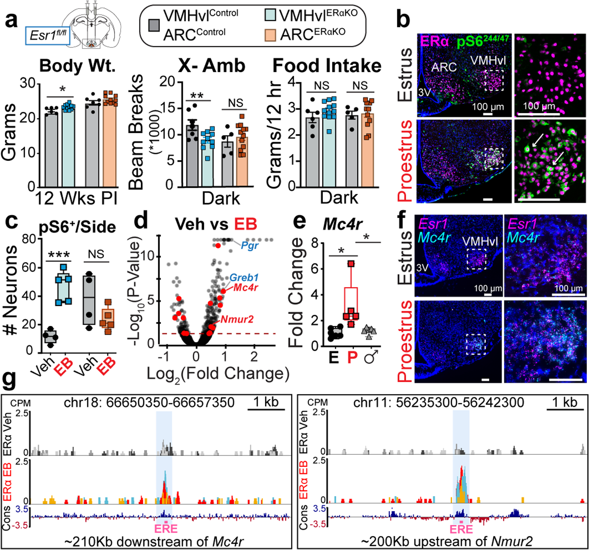
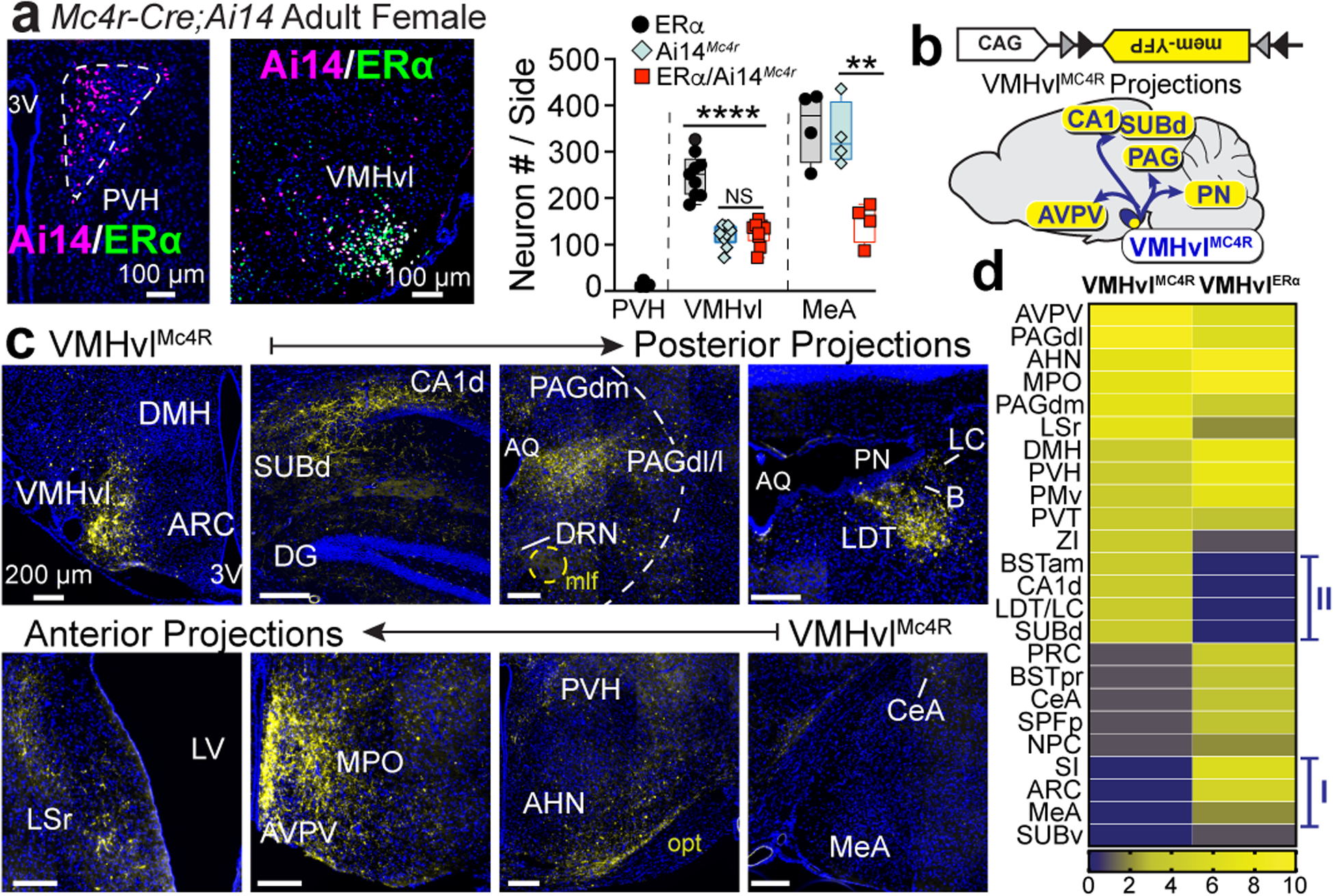
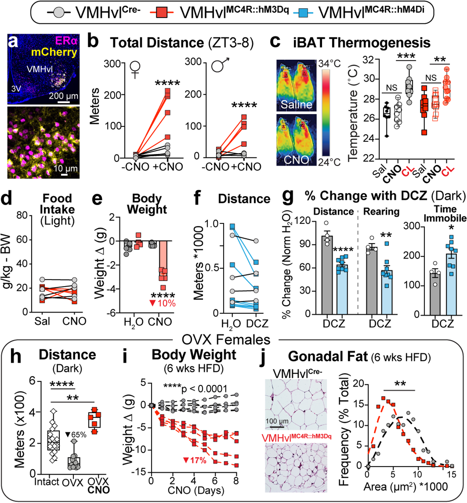
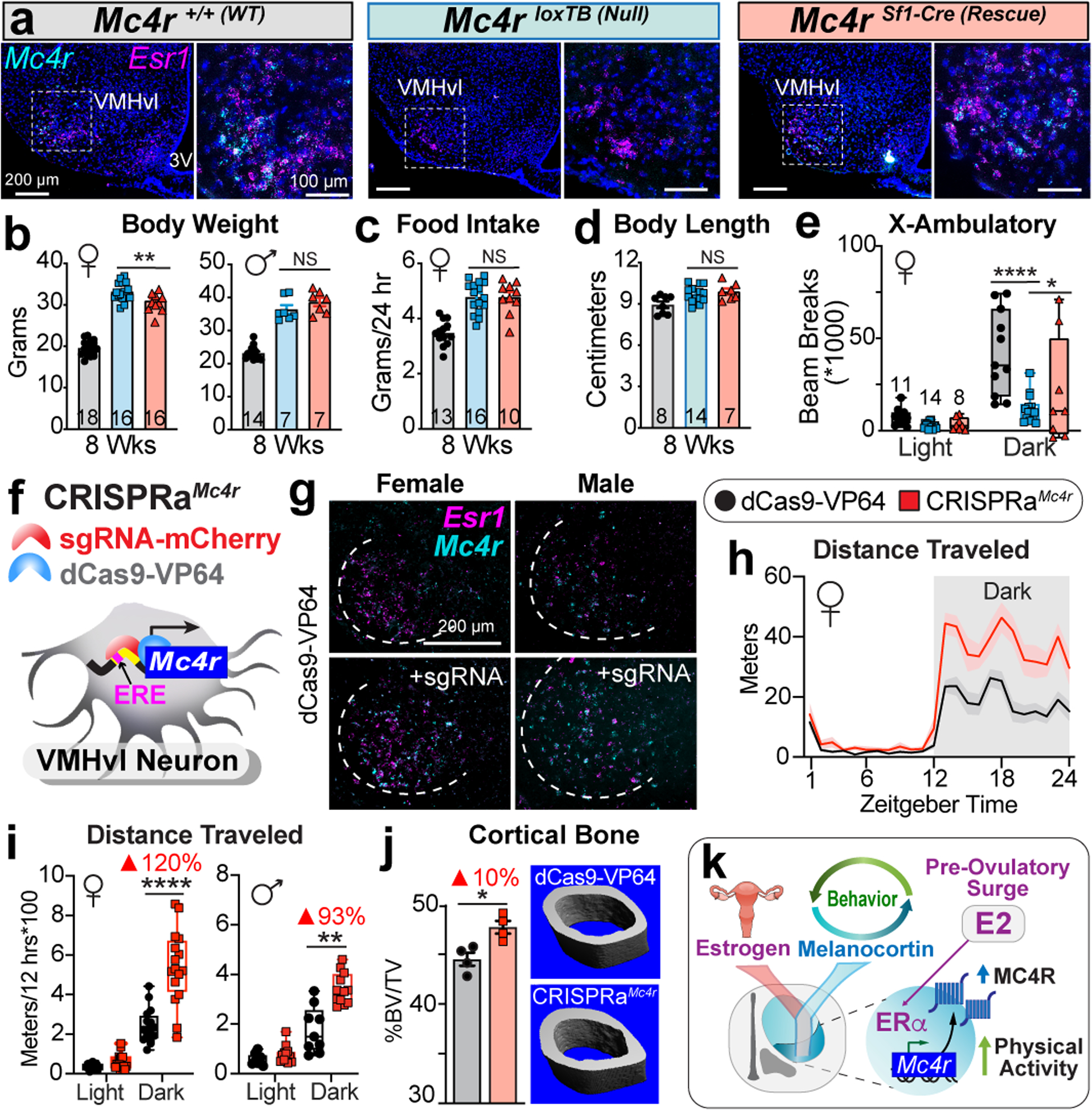
Oestrogen depletion in rodents and humans leads to inactivity, fat accumulation and diabetes1,2, underscoring the conserved metabolic benefits of oestrogen that inevitably decrease with age. In rodents, the preovulatory surge in 17β-oestradiol (E2) temporarily increases energy expenditure to coordinate increased physical activity with peak sexual receptivity. Here we report that a subset of oestrogen-sensitive neurons in the ventrolateral ventromedial hypothalamic nucleus (VMHvl)3-7 projects to arousal centres in the hippocampus and hindbrain, and enables oestrogen to rebalance energy allocation in female mice. Surges in E2 increase melanocortin-4 receptor (MC4R) signalling in these VMHvl neurons by directly recruiting oestrogen receptor-α (ERα) to the Mc4r gene. Sedentary behaviour and obesity in oestrogen-depleted female mice were reversed after chemogenetic stimulation of VMHvl neurons expressing both MC4R and ERα. Similarly, a long-term increase in physical activity is observed after CRISPR-mediated activation of this node. These data extend the effect of MC4R signalling - the most common cause of monogenic human obesity8 - beyond the regulation of food intake and rationalize reported sex differences in melanocortin signalling, including greater disease severity of MC4R insufficiency in women9. This hormone-dependent node illuminates the power of oestrogen during the reproductive cycle in motivating behaviour and maintaining an active lifestyle in women.


Figures

Fig. 1.
Fig. 1.. VMHvl neurons are sensitive to estrogen and maintain energy expenditure in adult females.
a, Body weight (*P=0.0310), ambulatory activity (**P=0.0014), and food intake in VMHvlERαKO (n=10), ARCERαKO (n=12), and control (grey, n=7/5) females. b, ERa and pS6244/47 co-expression (arrows) in proestrus and estrus (representative of 5 mice). c, Number of pS6244/47-labeled VMHvl (***P=0.0005) and ARC cells in vehicle (n=4) or EB (n=5) treated females. d, Enrichment of peptide ligand-binding receptors (red) (Benjamini-Hochburg adjusted P<0.05, dashed line). e, VMHvl Mc4r expression in proestrus (n=5), male (♂, n=6, *P=0.0189) and estrus (n=5, *P=0.0163) mice. f, Mc4r and Esr1 expression in estrus and proestrus (representative of 5 mice). g, CPM-normalized coverage tracks of ERE-containing ERα binding sites (pink boxes) within Mc4r (2/3 biological replicates) and Nmur2 (2/3 biological replicates) loci (MACS2, q < 0.01) in sub-cortical nuclei from vehicle and EB treated gonadectomized mice. Data are mean ± SEM, scatter, or box plots (whiskers indicate minimum and maximum values, edges of box are 25th and 75th percentiles, and center line indicates mean). a, c, unpaired 2-tailed t Test; a, RM 2-way ANOVA; e, 1-way ANOVA. Holm-Šidák multiple comparisons.
Figure 2.
Figure 2.. VMHvlMC4R neurons are molecularly and anatomically distinct subset of VMHvlERα neurons.
a, ERα and Ai14 expression and quantification in the PVH (n=4), VMHvl (n=9, ****P<0.0001), and MeA (n=4, **P=0.0057) of Ai14Mc4r female mice. b, Labeling vector and map of major VMHvlMC4R projections. c, VMHvl projections to anterior (upper row) and posterior (lower row) regions. Images representative of bilateral VMHvl targeting (n=3 mice, scale bars=200μm). d, Semi-quantitative comparison of VMHvlMC4R and VMHvlERα projection intensities. Anatomical abbreviations in Extended Data Table 1. See Fig. 1c legend for box plot description. a, 1-way ANOVA Holm-Šidák multiple comparisons.
Fig. 3.
Fig. 3.. VMHvlMC4R neurons control physical activity levels and when stimulated reverse inactivity and hypometabolism in obese OVX females.
a, ERα and mCherry in VMHvlMC4R::hM3Dq female. b, Spontaneous activity in VMHvlMC4R::hM3Dq (female=5, ****P<0.0001, male=5, ****P<0.0001) and VMHvlCre- (female=5, male=4) mice ± CNO injection. c, Thermography of VMHvlCre- (left) and VMHvlMC4R::hM3Dq (right) females and iBAT surface temperatures 30 and 45 min after Sal, CNO, or CL injection (n=4/5 VMHvlCre- P<0.0001, VMHvlMC4R::hM3Dq P=0.0003). d, Body weight normalized food consumption in females (n=5/5) following Sal/CNO injection during light period (ZT4–9). e, 24-hour weight change in females (n=5/5) administered drinking water (H2O) or CNO-water (CNO, ****P<0.0001). f, g, Dark period (ZT12–24) activity in VMHvlMC4R::hM4Di (n=8) and VMHvlCre- (n=4) females administered water or DCZ-water. ****P<0.0001, **P=0.0068, *P=0.0213. h, Activity levels in intact (n=16), OVX (n=12, ****P<0.0001), and OVX+CNO VMHvlMC4R::hM3Dq (n=5, **P=0.0023) mice. i, j, Body weight and gonadal white adipocyte area following 8-day CNO treatment of OVX/HFD mice (n=5/5). Data are mean ± SEM, scatter, or box plots (see Fig. 1c legend). b, c, d, e, f, h, RM 2-way ANOVA; g, unpaired 2-tailed t Tests; h, 1-way ANOVA; i, nested t Test. Holm-Šidák multiple comparisons as appropriate.
Fig. 4.
Fig. 4.. Sex-specific role for MC4R signaling in the VMHvl can be bypassed using CRISPR-mediated activation.
a, Esr1 and Mc4r expression in Mc4r+/+, Mc4rloxTB, and Mc4rSf1-Cre females. b, Body weights in 8-week-old female (**P=0.0026) and male Mc4r+/+, Mc4rloxTB, and Mc4rSf1-Cre mice. c, Food intake in female cohorts. d, Body length in female cohorts. e, Light and dark period activity in females (****P<0.0001, *P=0.0153). e, CRISPRaMc4r targets Mc4r promotor. f, Esr1 and Mc4r expression in control and CRISPRaMC4R female and male mice. g, Home-cage activity in CRISPRaMc4r (n=6) and control (n=5) female mice. h, Distances for three most active runs from CRISPRaMc4r and control female (n=6/5, ****P<0.0001) and male (n=4/3) mice. i, Cortical bone volume fraction for female mice 4 months post-infection (P=0.0129). j, VMHvlERα/MC4R neurons integrates estrogen and melanocortin signaling to generate a specialized hormone-dependent activity node in females. Data are mean ± SEM or box plots (see Fig. 1c legend). b, c, 1-way ANOVA; d, h, RM 2-way ANOVA; Holm-Šidák multiple comparisons. i, unpaired 2-tailed t Tests.

Comment in

References

    1. Mauvais-Jarvis F, Clegg DJ & Hevener AL The role of estrogens in control of energy balance and glucose homeostasis. Endocr Rev 34, 309–338 (2013). - PMC - PubMed
    1. Carr MC The emergence of the metabolic syndrome with menopause. J Clin Endocrinol Metab 88, 2404–2411 (2003). - PubMed
    1. Correa SM, et al. An estrogen-responsive module in the ventromedial hypothalamus selectively drives sex-specific activity in females. Cell Rep 10, 62–74 (2015). - PMC - PubMed
    1. Herber CB, et al. Estrogen signaling in arcuate Kiss1 neurons suppresses a sex-dependent female circuit promoting dense strong bones. Nat Commun 10, 163 (2019). - PMC - PubMed
    1. Martinez de Morentin PB, et al. Estradiol regulates brown adipose tissue thermogenesis via hypothalamic AMPK. Cell Metab 20, 41–53 (2014). - PMC - PubMed

Publication types

MeSH terms