Review
. 2022 Jun;22(6):339-352.
doi: 10.1038/s41577-021-00626-8. Epub 2021 Oct 13.

Lessons in self-defence: inhibition of virus entry by intrinsic immunity

Affiliations
Review

Lessons in self-defence: inhibition of virus entry by intrinsic immunity

Saliha Majdoul et al. Nat Rev Immunol. 2022 Jun.

Abstract

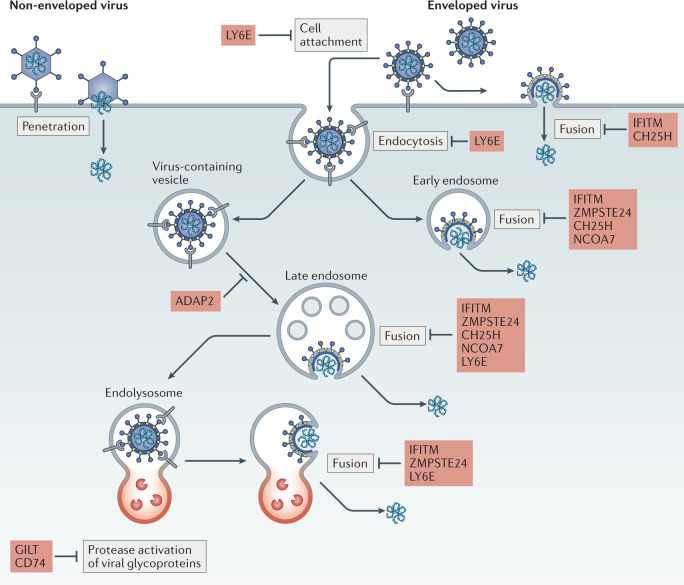
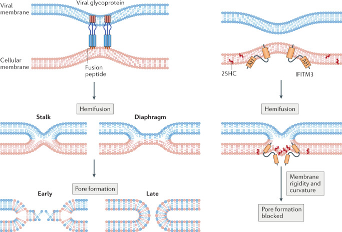
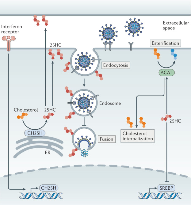
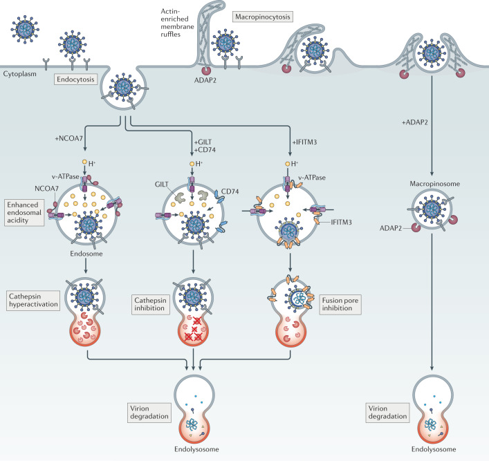
Virus entry, consisting of attachment to and penetration into the host target cell, is the first step of the virus life cycle and is a critical 'do or die' event that governs virus emergence in host populations. Most antiviral vaccines induce neutralizing antibodies that prevent virus entry into cells. However, while the prevention of virus invasion by humoral immunity is well appreciated, considerably less is known about the immune defences present within cells (known as intrinsic immunity) that interfere with virus entry. The interferon-induced transmembrane (IFITM) proteins, known for inhibiting fusion between viral and cellular membranes, were once the only factors known to restrict virus entry. However, the progressive development of genetic and pharmacological screening platforms and the onset of the COVID-19 pandemic have galvanized interest in how viruses infiltrate cells and how cells defend against it. Several host factors with antiviral potential are now implicated in the regulation of virus entry, including cholesterol 25-hydroxylase (CH25H), lymphocyte antigen 6E (LY6E), nuclear receptor co-activator protein 7 (NCOA7), interferon-γ-inducible lysosomal thiol reductase (GILT), CD74 and ARFGAP with dual pleckstrin homology domain-containing protein 2 (ADAP2). This Review summarizes what is known and what remains to be understood about the intrinsic factors that form the first line of defence against virus infection.


Conflict of interest statement

The authors declare no competing interests.

Figures

Fig. 1
Fig. 1. Pathways for enveloped virus entry into cells and its restriction.
Viral envelope glycoproteins on the surface of the virion mediate cell attachment by interacting directly with a host receptor on the cell surface. Receptor interaction drives conformational changes allowing exposure of the fusion peptide, which inserts itself into host membranes to drive virus–cell fusion. Fusion occurs at the plasma membrane or, in the case of viral glycoproteins that exhibit pH-dependent fusogenic activity and/or activation by cellular proteases, at endosomal or lysosomal (referred to as endolysosomal) membranes. Completion of membrane fusion allows passage of the viral ribonucleoprotein complex, including viral nucleic acid, into the host cell cytoplasm — a prerequisite for subsequent stages of the virus life cycle. Host factors discussed in this Review are listed in red beside the entry step they inhibit. For comparison, entry by non-enveloped viruses usually involves membrane fusion-independent penetration into the host cell, as depicted on the left. ADAP2, ARFGAP with dual pleckstrin homology domain-containing protein 2; CH25H, cholesterol 25-hydroxylase; GILT, interferon-γ-inducible lysosomal thiol reductase; IFITM, interferon-induced transmembrane protein; LY6E, lymphocyte antigen 6E; NCOA7, nuclear receptor co-activator protein 7; ZMPSTE24, zinc metalloproteinase STE24.
Fig. 2
Fig. 2. Stages of virus–cell membrane fusion and the antifusion activities of IFITM3 and 25-hydroxycholesterol.
Close apposition of viral and cellular membranes is induced by viral envelope glycoproteins (shown at the upper left and not drawn thereafter), followed by the formation of a hemifusion stalk. Hemifusion is characterized by lipid mixing between the outer leaflets and alignment of the inner leaflets of each bilayer and may progress from a stalk to a diaphragm-like structure. Finally, further lipid mixing leads to partial opening of the fusion pore, and the pore is further dilated to complete membrane fusion. Interferon-induced transmembrane protein 3 (IFITM3) promotes hemifusion while inhibiting pore formation in a process that requires its amphipathic helix (AH) and protein oligomerization. IFITM3 promotes membrane rigidity and curvature in a manner that may disfavour formation of the fusion pore, and 25-hydroxycholesterol (25HC) produced by cholesterol 25-hydroxylase (CH25H) may function similarly.
Fig. 3
Fig. 3. The impacts of 25-hydroxycholesterol on virus entry and cholesterol homeostasis.
Cholesterol 25-hydroxylase (CH25H) is localized to the endoplasmic reticulum (ER), where it catalyses the oxidation of cholesterol to produce the oxysterol 25-hydroxycholesterol (25HC). 25HC acts in an autocrine and paracrine manner to inhibit virus entry at the level of fusion. 25HC promotes acyl-CoA cholesterol acyltransferase (ACAT) activity to increase cholesterol esterification, which regulates cholesterol availability in membrane compartments. Furthermore, 25HC provides negative feedback in cholesterol metabolism by repressing sterol regulatory element-binding protein (SREBP)-induced genes that promote cholesterol biosynthesis.
Fig. 4
Fig. 4. Intrinsic inhibitors of virus entry promote virion degradation in endolysosomes via distinct mechanisms.
The antiviral activities exhibited by nuclear receptor co-activator protein 7 (NCOA7), interferon-induced transmembrane (IFITM) proteins, interferon-γ-inducible lysosomal thiol reductase (GILT), CD74 and ARFGAP with dual pleckstrin homology domain-containing protein 2 (ADAP2) act against viruses entering cells through pH-dependent fusion in endosomes. Whereas NCOA7 and IFITM3 have been reported to interact with vacuolar ATPase (v-ATPase), only NCOA7 may increase the acidity (lower the pH) of the endosomal lumen. NCOA7-mediated acidification of endosomes is associated with enhanced cathepsin activity in endolysosomes, and this elevated level of proteolytic activity may promote virion degradation before virus–cell fusion occurs. IFITM3, on the other hand, inhibits membrane fusion itself, resulting in endosomal sequestration of virions that are eventually degraded in endolysosomes. This latter effect results from the ability of IFITM3 to promote endolysosomal delivery of viral and cellular cargo, a function that is not yet mechanistically understood. GILT and CD74 are believed to enforce endosomal sequestration of viruses as well, but not by inhibiting membrane fusion — they inhibit the activity of endolysosomal cathepsins, proteases that cleave some viral glycoproteins and render them competent for fusion. ADAP2 promotes internalization of virions by macropinocytosis, bypassing their preferred sites for pH-dependent fusion and resulting in their accelerated disposal in endolysosomes.

References

    1. Parvez MK, Parveen S. Evolution and emergence of pathogenic viruses: past, present, and future. Intervirology. 2017;60:1–7. doi: 10.1159/000478729. - DOI - PMC - PubMed
    1. Dimitrov DS. Virus entry: molecular mechanisms and biomedical applications. Nat. Rev. Microbiol. 2004;2:109–122. doi: 10.1038/nrmicro817. - DOI - PMC - PubMed
    1. White JM, Delos SE, Brecher M, Schornberg K. Structures and mechanisms of viral membrane fusion proteins: multiple variations on a common theme. Crit. Rev. Biochem. Mol. Biol. 2008;43:189–219. doi: 10.1080/10409230802058320. - DOI - PMC - PubMed
    1. Marsh M, Helenius A. Virus entry: open sesame. Cell. 2006;124:729–740. doi: 10.1016/j.cell.2006.02.007. - DOI - PMC - PubMed
    1. Bieniasz PD. Intrinsic immunity: a front-line defense against viral attack. Nat. Immunol. 2004;5:1109–1115. doi: 10.1038/ni1125. - DOI - PubMed

Publication types