

Median arcuate ligament syndrome diagnosis on Computed Tomography: what a radiologist needs to know
- PMID: 34646405
- PMCID: PMC8500833
- DOI: 10.1016/j.radcr.2021.06.093
Median arcuate ligament syndrome diagnosis on Computed Tomography: what a radiologist needs to know
Abstract
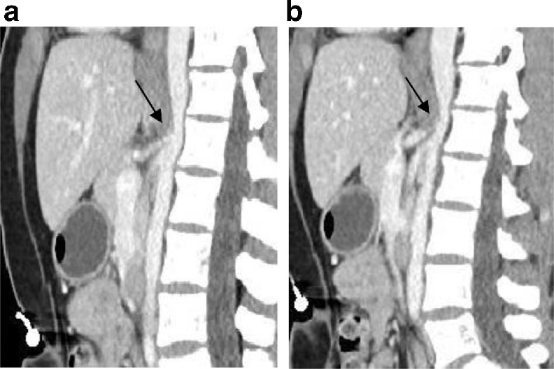
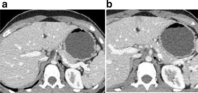
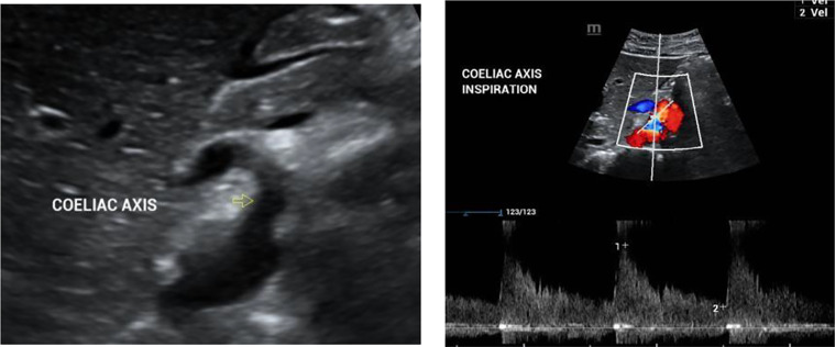
Median arcuate ligament syndrome or celiac artery compression syndrome is one of the abdominal vascular compression syndromes due to compression of proximal celiac artery by the median arcuate ligament. The median arcuate ligament unites diaphragmatic crura on either side at the level of aortic hiatus. The ligament has a low insertion causing compression of the celiac artery resulting in clinical symptoms of postprandial pain and weight loss. It is a rare syndrome, detected incidentally on routine Computed Tomography abdomen and pelvis studies. We present a rare case of a 35-year-old female who presented with abdominal pain. She was evaluated by Computed Tomography scan of the abdomen and pelvis. Ultrasound Doppler of mesenteric vasculature helped detect celiac artery stenosis. A referral to the vascular surgery department was made; however, the patient was managed conservatively.
Keywords: Celiac artery compression; Celiac artery stenosis; Computed Tomography (CT); Diagnostic Radiology; Median arcuate ligament syndrome (MALS); Ultrasound Doppler; Vascular compression syndrome.
Crown Copyright © 2021 Published by Elsevier Inc. on behalf of University of Washington.
Figures
References
-
- Harjola PT. A rare obstruction of the coeliac artery. Ann Chir Gynaecol Fenn. 1963;52:547–550. Report of a case. - PubMed
-
- Linder HH, Kemprud E. A clinicoanatomic study of the arcuate ligament of the diaphragm. Arch Surg. 1971;103:600–605. - PubMed
-
- Weber JM, Boules M, Fong K, Abraham B, Bena J, El-Hayek K. Median arcuate ligament syndrome is not a vascular disease. Ann Vasc Surg. 2016;30:22. 7.20. - PubMed
Publication types
LinkOut - more resources
Full Text Sources
