. 2021 Oct 1:29:100803.
doi: 10.1016/j.ymgmr.2021.100803. eCollection 2021 Dec.

Chitotriosidase as a biomarker for gangliosidoses

Affiliations

Chitotriosidase as a biomarker for gangliosidoses

Sarah Kim et al. Mol Genet Metab Rep. .

Abstract

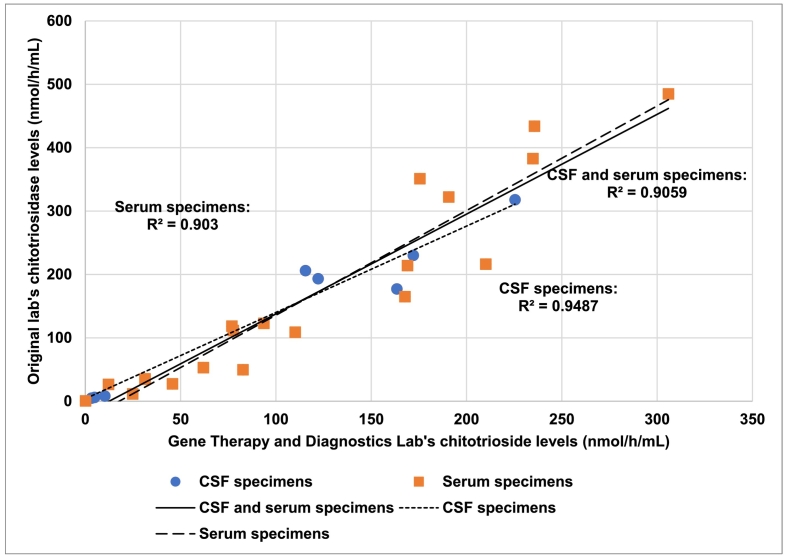
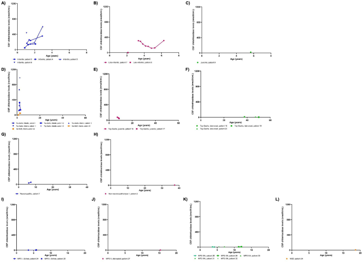
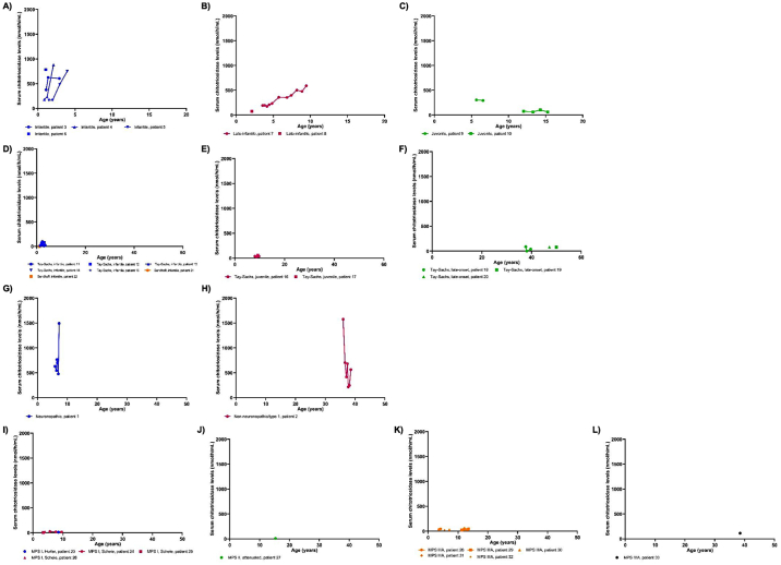
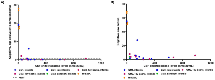
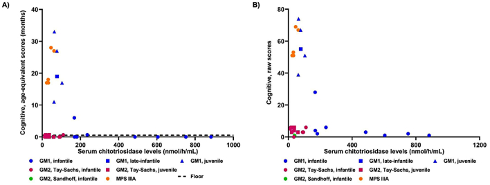
Elevated serum chitotriosidase (CHITO) is an indication of macrophage activation, and its capacity have been explored as a marker of inflammation in a number of disease states. For over a decade, CHITO plasma levels have been used by clinicians as a biomarker of inflammation in the lysosomal disease, Gaucher disease, including monitoring response to therapies in patients with Gaucher disease type I. Although it is becoming increasingly recognized that inflammation is a prominent component of many lysosomal diseases, the relation of CHITO levels to disease burden has not been well-characterized in the large majority of lysosomal diseases. Moreover, the role of CHITO in lysosomal diseases that affect the central nervous system (CNS) has not been systematically studied. In this study, one hundred and thirty-four specimens of CSF and serum were collected from 34 patients with lysosomal diseases affecting the CNS. This study included patients with GM1-gangliosidosis, GM2-gangliosidosis, mucopolysaccharidoses (MPS), multiple sulfatase deficiency and Gaucher disease. CHITO levels in the CSF were significantly higher in patients with more rapidly progressing severe neurological impairment: GM1-gangliosidosis vs MPS (p < 0.0001); GM2-gangliosidosis vs MPS (p < 0.0001). CHITO levels were higher in patients with the more severe phenotypes compared to milder phenotypes in GM1-gangliosidosis and GM2-gangliosidosis (serum CHITO in GM1-gangliosidosis infantile vs juvenile p = 0.025; CSF CHITO in Tay-Sachs infantile vs Tay-Sachs late-onset p < 0.0001). Moreover, higher CHITO levels in the CSF were significantly associated with lower cognitive test scores in patients with GM1-gangliosidosis, GM2-gangliosidosis, and MPS (p = 1.12*10-5, R2 = 0.72). Patients with infantile GM1-gangliosidosis showed increasing CSF CHITO over time, suggesting that CSF CHITO reflects disease progression and a possible surrogate endpoint for future clinical trials with infantile GM1-gangliosidosis. In summary, these results support the use of CSF CHITO to diagnose between different disease phenotypes and as a valuable tool for monitoring disease progression in patients. These results necessitate the inclusion of CHITO as an exploratory biomarker for clinical trials.

Keywords: Chitotriosidase; GM1-gangliosidosis; GM2-gangliosidosis; Gaucher; Lysosomal diseases; Mucopolysaccharidosis.


Figures

Fig. 1
Fig. 1
Comparison of chitotriosidase levels between labs. Eight cerebrospinal fluid (CSF) specimens and 20 serum specimens were assayed at both the Gene Therapy and Diagnostics Lab and the original lab. Three Pearson's correlation tests were performed to see if there was a correlation between chitotriosidase levels reported at the Gene Therapy and Diagnostics Lab and the original lab. Correlation tests were performed for serum specimens alone, CSF specimens alone, and CSF and serum specimens together.
Fig. 2
Fig. 2
Chitotriosidase levels in the CSF in multiple lysosomal diseases. Chitotriosidase levels in the cerebrospinal fluid (CSF) are shown for patients with lysosomal diseases. Sequential points from patients are connected by lines. A-C) GM1-gangliosidoses. Infantile: n = 11 CSF specimens, late-infantile: n = 9, juvenile: n = 1. D-F) GM2-gangliosidoses. Tay-Sachs infantile: n = 7 CSF specimens, Tay-Sachs juvenile: n = 3, Tay-Sachs adult: n = 3, and Sandhoff infantile: n = 3. G-H) Gaucher disease. Non-neuronopathic/type 1: n = 1 CSF specimen, neuronopathic phenotype: n = 2. I-L) Mucopolysaccharidoses (MPS) and multiple sulfatase deficiency (MSD). MPS IS: n = 3 CSF specimens, MPS II attenuated: n = 1, MPS IIIA: n = 7, and MSD: n = 1.
Fig. 3
Fig. 3
Chitotriosidase levels in the serum in multiple lysosomal diseases. Chitotriosidase levels in the serum are shown for patients with lysosomal diseases. Sequential points from patients are connected by lines. A-C) GM1-gangliosidoses. Infantile: n = 11 serum specimens, late-infantile: n = 12, juvenile: n = 6. D-F) GM2-gangliosidoses. Tay-Sachs infantile: n = 10 serum specimens, Tay-Sachs juvenile: n = 4, Tay-Sachs adult: n = 5, and Sandhoff infantile: n = 3. G-H) Gaucher disease. Non-neuronopathic/type 1: n = 7 serum specimens, neuronopathic phenotype: n = 6. I-L) Mucopolysaccharidoses (MPS). MPS IH: n = 1 serum specimen, MPS IS: n = 5, MPS II attenuated: n = 1, MPS IIIA: n = 9, and MPS IVA: n = 1.
Fig. 4
Fig. 4
Bayley cognitive domain and chitotriosidase levels in the CSF. Chitotriosidase levels in the CSF were plotted against the cognitive domain in the Bayley-III test measured as either age-equivalent scores (3A) or raw scores (3B). A) The floor (dotted line) designates the lowest possible score of the age-equivalent score, which is <16 days or 0.53 month.
Fig. 5
Fig. 5
Bayley cognitive domain and chitotriosidase levels in the serum. Chitotriosidase levels in the serum were plotted against the cognitive domain in the Bayley-III test measured as either age-equivalent scores (4A) or raw scores (4B). A) The floor (dotted line) designates the lowest possible score of the age-equivalent score, which is <16 days or 0.53 month.

Similar articles

Cited by

References

    1. Hollak C.E., van Weely S., van Oers M.H., Aerts J.M. Marked elevation of plasma chitotriosidase activity. A novel hallmark of Gaucher disease. J. Clin. Invest. 1994;93:1288–1292. doi: 10.1172/JCI117084. - DOI - PMC - PubMed
    1. Renkema G.H., Boot R.G., Strijland A., Donker-Koopman W.E., van den Berg M., Muijsers A.O., Aerts J.M. Synthesis, sorting, and processing into distinct isoforms of human macrophage chitotriosidase. Eur. J. Biochem. 1997;244:279–285. doi: 10.1111/j.1432-1033.1997.00279.x. - DOI - PubMed
    1. van Eijk M., van Roomen C.P., Renkema G.H., Bussink A.P., Andrews L., Blommaart E.F., Sugar A., Verhoeven A.J., Boot R.G., Aerts J.M. Characterization of human phagocyte-derived chitotriosidase, a component of innate immunity. Int. Immunol. 2005;17:1505–1512. doi: 10.1093/intimm/dxh328. - DOI - PubMed
    1. Guo Y., He W., Boer A.M., Wevers R.A., de Bruijn A.M., Groener J.E., Hollak C.E., Aerts J.M., Galjaard H., van Diggelen O.P. Elevated plasma chitotriosidase activity in various lysosomal storage disorders. J. Inherit. Metab. Dis. 1995;18:717–722. doi: 10.1007/BF02436762. - DOI - PubMed
    1. Michelakakis H., Dimitriou E., Labadaridis I. The expanding spectrum of disorders with elevated plasma chitotriosidase activity: an update. J. Inherit. Metab. Dis. 2004;27:705–706. doi: 10.1023/b:boli.0000043025.17721.fc. - DOI - PubMed

LinkOut - more resources