Review
. 2021 Sep 27:8:745279.
doi: 10.3389/fmed.2021.745279. eCollection 2021.

Mitochondrial Regulation of Diabetic Kidney Disease

Affiliations
Review

Mitochondrial Regulation of Diabetic Kidney Disease

Daniel L Galvan et al. Front Med (Lausanne). .

Abstract

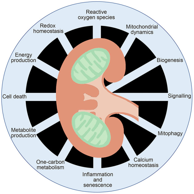
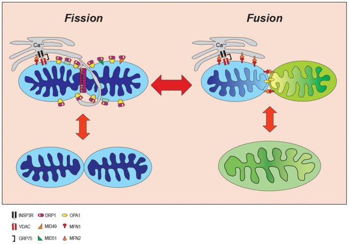
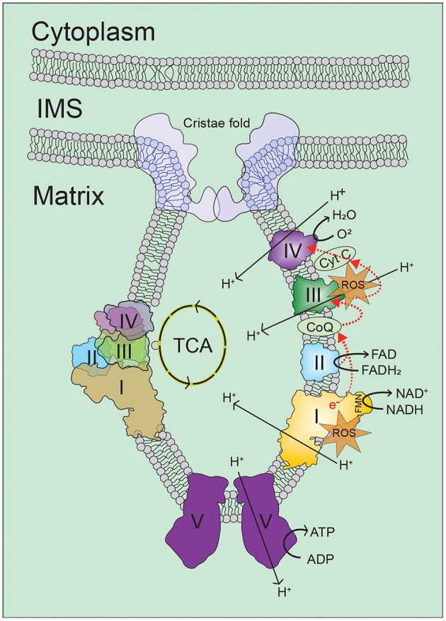
The role and nature of mitochondrial dysfunction in diabetic kidney disease (DKD) has been extensively studied. Yet, the molecular drivers of mitochondrial remodeling in DKD are poorly understood. Diabetic kidney cells exhibit a cascade of mitochondrial dysfunction ranging from changes in mitochondrial morphology to significant alterations in mitochondrial biogenesis, biosynthetic, bioenergetics and production of reactive oxygen species (ROS). How these changes individually or in aggregate contribute to progression of DKD remain to be fully elucidated. Nevertheless, because of the remarkable progress in our basic understanding of the role of mitochondrial biology and its dysfunction in DKD, there is great excitement on future targeted therapies based on improving mitochondrial function in DKD. This review will highlight the latest advances in understanding the nature of mitochondria dysfunction and its role in progression of DKD, and the development of mitochondrial targets that could be potentially used to prevent its progression.

Keywords: bioenergetics; diabetic kidney disease; mitochondria; mitochondrial dynamics; mitochondrial respiratory complexes; oxidative phosphorylation.


Conflict of interest statement

The authors declare that the research was conducted in the absence of any commercial or financial relationships that could be construed as a potential conflict of interest.

Figures

Figure 1
Figure 1
Multifaceted functions of mitochondrial function. Mitochondria are known to generate ATP and metabolites critical for signal transduction, as well as playing key roles in inflammation, calcium homeostasis, redox homeostasis and cell death.
Figure 2
Figure 2
Mitochondrial dynamic. Mitochondria continuously change their size and shape by two opposing processes: mitochondrial fission and fusion. During the mitochondrial fission, mitochondria become fragmented in response to cell stress whereas they form an elongated shape increasing ATP production to adjust to cellular stresses. INSP3R, Inositol trisphosphate receptor; DRP1, dynamin-related protein 1; OPA1, optic atrophy 1; VDAC, voltage-dependent anion channel; MID49/51, mitochondrial dynamics proteins of 49 and 51kD; MFN1/2, mitofusin proteins 1 and 2; GRP75, glucose-regulated protein 75.
Figure 3
Figure 3
Oxidative phosphorylation. Cellular energy in the form of ATP is mainly generated in mitochondria by the oxidative phosphorylation (OXPHOS) process, in which electrons on the inner-membrane of the mitochondria are passed through a series of mitochondrial complexes (Complexes I-V) in redox reactions. Energy released in these reactions is then coupled to ATP generation. Increase in intracellular levels of NADH and FADH2 drives oxidative phosphorylation, which leads to increase of oxygen consumption and ATP production by ATP synthesis. For more information, refer to the main text. ROS, reactive oxygen species; TCA, tricarboxylic acid; I/II/III/IV/V, mitochondrial respiratory complex I/II/III/IV/V; NADH, reduced nicotinamide adenine dinucleotide; NAD, nicotinamide adenine dinucleotide; FADH2, reduced flavin adenine dinucleotide; FAD, flavin adenine dinucleotide; FMN, flavin mononucleotide; CoQ, coenzyme Q; Cyt C, cytochrome C.

References

    1. Gilbert RE. Proximal tubulopathy: prime mover and key therapeutic target in diabetic kidney disease. Diabetes. (2017) 66:791–800. 10.2337/db16-0796 - DOI - PubMed
    1. Pfaller W, Rittinger M. Quantitative morphology of the rat kidney. Int J Biochem. (1980) 12:17–22. 10.1016/0020-711X(80)90035-X - DOI - PubMed
    1. Brinkkoetter PT, Bork T, Salou S, Liang W, Mizi A, Ozel C, et al. . Anaerobic glycolysis maintains the glomerular filtration barrier independent of mitochondrial metabolism and dynamics. Cell Rep. (2019) 27:1551–66.e5. 10.1016/j.celrep.2019.04.012 - DOI - PMC - PubMed
    1. Abe Y, Sakairi T, Kajiyama H, Shrivastav S, Beeson C, Kopp JB. Bioenergetic characterization of mouse podocytes. Am J Physiol Cell Physiol. (2010) 299:C464–76. 10.1152/ajpcell.00563.2009 - DOI - PMC - PubMed
    1. Muller-Deile J, Schiffer M. The podocyte power-plant disaster and its contribution to glomerulopathy. Front Endocrinol. (2014) 5:209. 10.3389/fendo.2014.00209 - DOI - PMC - PubMed

LinkOut - more resources