. 2021 Sep 17:12:100142.
doi: 10.1016/j.mtbio.2021.100142. eCollection 2021 Sep.

The spatial form periosteal-bone complex promotes bone regeneration by coordinating macrophage polarization and osteogenic-angiogenic events

Affiliations

The spatial form periosteal-bone complex promotes bone regeneration by coordinating macrophage polarization and osteogenic-angiogenic events

C Zhao et al. Mater Today Bio. .

Abstract

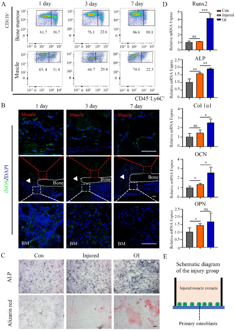
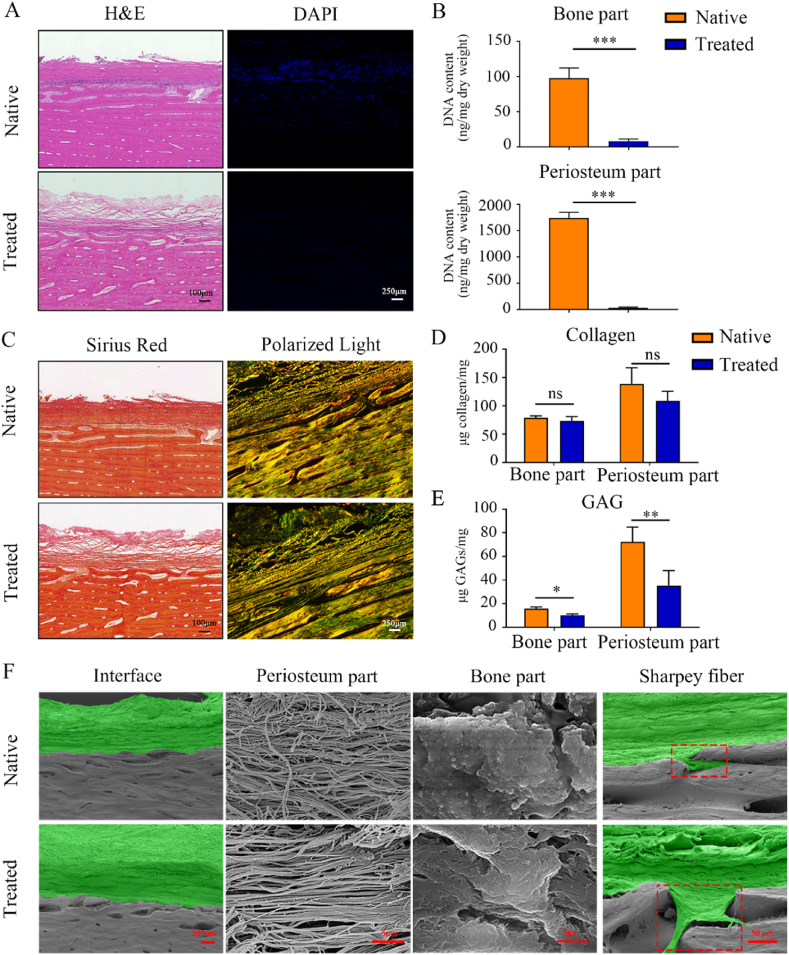
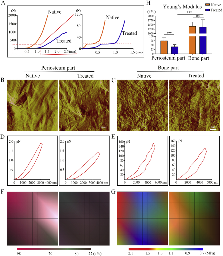
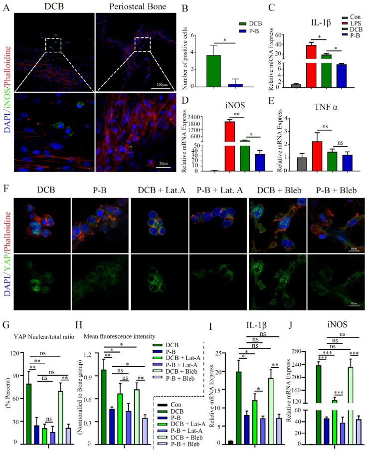
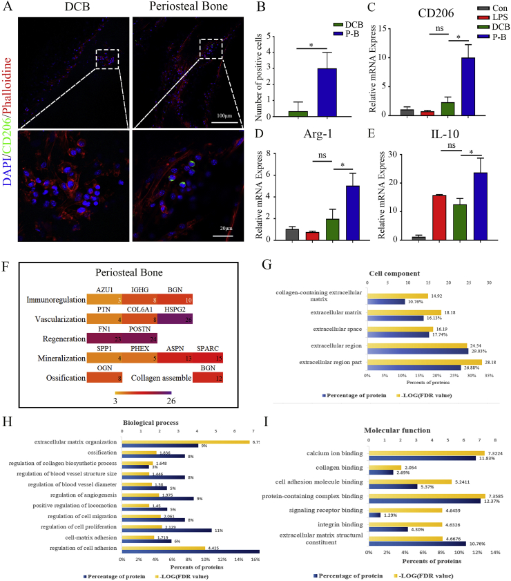
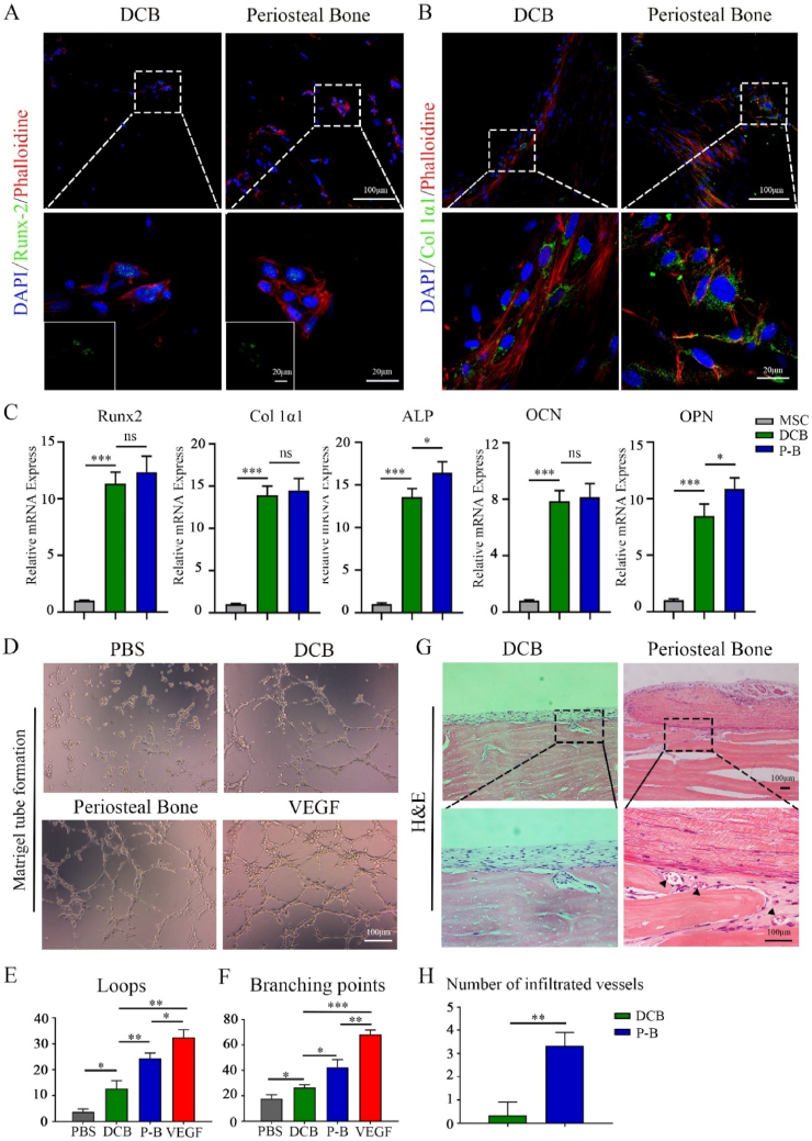
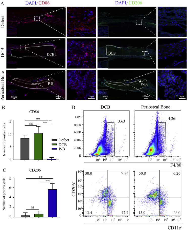
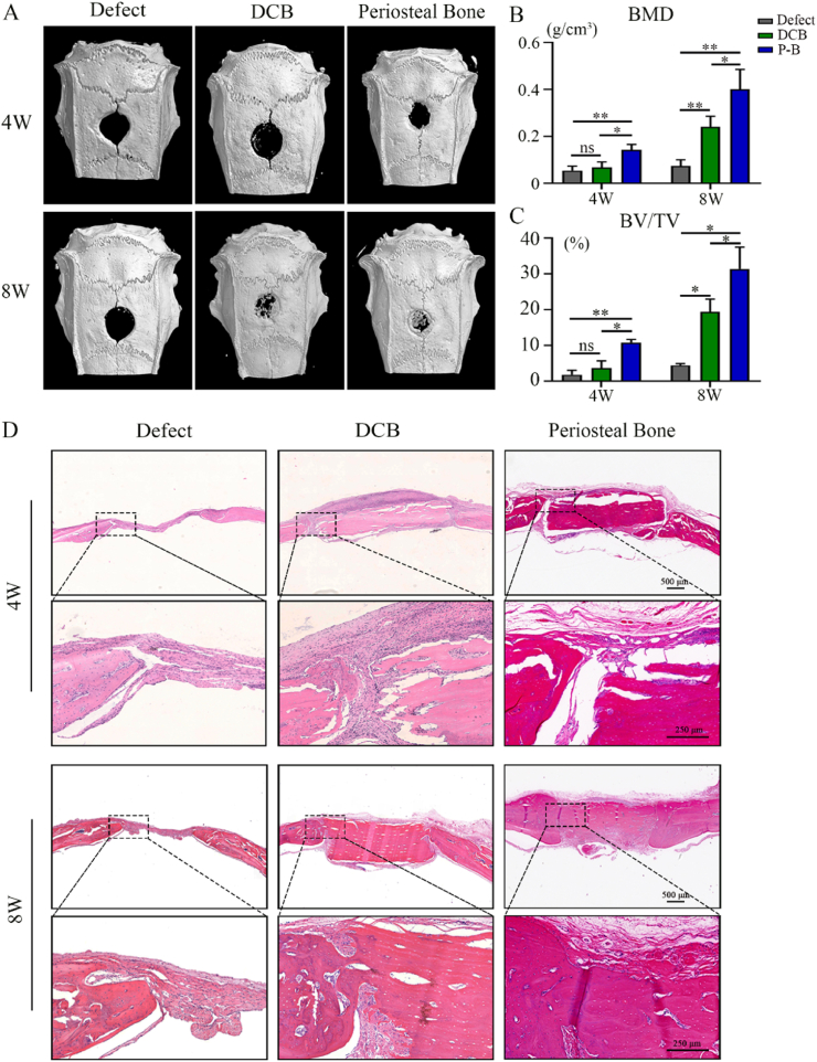
Bone defects associated with soft tissue injuries are an important cause of deformity that threatens people's health and quality of life. Although bone substitutes have been extensively explored, effective biomaterials that can coordinate early inflammation regulation and subsequent repair events are still lacking. We prepared a spatial form periosteal bone extracellular matrix (ECM) scaffold, which has advantages in terms of low immunogenicity, good retention of bioactive ingredients, and a natural spatial structure. The periosteal bone ECM scaffold with the relatively low-stiffness periosteum (41.6 ± 3.7 kPa) could inhibit iNOS and IL-1β expression, which might be related to actin-mediated YAP translocation. It also helped to promote CD206 expression with the potential influence of proteins related to immune regulation. Moreover, the scaffold combined the excellent properties of decalcified bone and periosteum, promoted the formation of blood vessels, and good osteogenic differentiation (RUNX2, Col 1α1, ALP, OPN, and OCN), and achieved good repair of a cranial defect in rats. This scaffold, with its natural structural and biological advantages, provides a new idea for bone healing treatment that is aligned with bone physiology.

Keywords: Angiogenesis; Bone healing; Macrophage polarization; Osteogenesis; Periosteal-bone complex; Stiffness.


Conflict of interest statement

The authors declare that they have no known competing financial interests or personal relationships that could have appeared to influence the work reported in this paper.

Figures

Image 1
Graphical abstract
Fig. 1
Fig. 1
Infiltration of inflammatory cells around the skeletal muscle–femur injury site and the influence of injured skeletal muscles on osteogenesis. (A) Representative flow cytometry analysis to examine the changes in the number of CD45, CD11b, and Ly6C positive cells in the muscles and bone marrow at the injury site from day 1 to day 7. (B) Representative immunofluorescence staining of iNOS positive cells around the injured site from day 1 to day 7 (green: iNOS; blue: cell nucleus; red box: surrounding muscle; white box: bone marrow; white triangle: defect area; BM: bone marrow). (C) Alkaline phosphatase and alizarin red staining of primary calvarial osteoblasts in the control, injured, and osteogenic induction (OI) groups. (D) Relative mRNA expression of RUNX2, ALP, Col 1α1, OCN, and OPN from primary calvarial osteoblasts in the control, injured, and OI groups after culturing for 7 days. (E) Schematic diagram of osteogenic induction in the muscle injury group. Data are presented as mean ± SD (n = 4). (Scale bar = 100 μm; ∗: P < 0.05, ∗∗: P < 0.01, and ∗∗∗: P < 0.001).
Fig. 2
Fig. 2
Validation of the spatial form periosteal bone ECM scaffold and three-dimensional structure evaluation. (A) H&E and DAPI staining of the native and treated periosteal bone ECM scaffolds. (B) DNA contents of the periosteum part and cortical bone part before and after treatment. (C) Sirius red staining and polarized light microscopy observations of the types and orientations of collagen in the native and treated periosteal bone ECM scaffold. (D, E) Collagen (D) and GAGs (E) contents in the periosteum part and cortical bone part before and after treatment. (F) Scanning electron microscopy (SEM) images of the periosteum part, cortical bone part, interfaces, and Sharpey fibers of the native and treated periosteal bone ECM scaffolds (green: periosteum; gray: cortical bone; red dotted frame: Sharpey fiber). Native: before treatment; Treated: after treatment. Data are presented as mean ± SD (n = 4). (Scale bars are listed above; ∗: P < 0.05, ∗∗: P < 0.01, and ∗∗∗: P < 0.001).
Fig. 3
Fig. 3
Mechanical properties of the spatial form periosteal bone ECM scaffold before and after treatment. (A) Representative stress–strain curves of the periosteal bone ECM scaffold before and after treatment. (B, C) Representative atomic force microscopy images of the periosteum (B) and cortical bone part (C) before and after treatment. (D, E) Representative nanoindentation curves of the periosteum (D) and cortical bone part (E) before and after treatment. (F, G) Distribution maps of Young’s modulus of the periosteum (F) and cortical bone part (G) before and after treatment. (H) Values of Young’s modulus in the periosteum and cortical bone part before and after treatment. Native: before treatment; Treated: after treatment. Data are presented as mean ± SD (n = 4). (Scale bar = 1 μm; ∗: P < 0.05, ∗∗: P < 0.01, and ∗∗∗: P < 0.001).
Fig. 4
Fig. 4
Periosteal bone scaffold inhibits M1 polarization might through actin-mediated YAP translocation. (A) Representative immunofluorescence images of iNOS in BMMs cultured on DCB and the periosteal bone scaffolds for 24 h (green: iNOS; red: phalloidin, blue: nucleus; 20× and 80×). (B) Quantitative analysis of iNOS positive cells cultured in the same conditions as those in (A). (C, D, E) Relative mRNA expression of IL-1β (C), iNOS (D), and TNF-α (E) in BMMs cultured in the same conditions as those in (A). (F) Representative immunofluorescence images of YAP in BMMs cultured on DCB and the periosteal bone scaffold for 24 h and treated with LAT. A or blebbistatin for 6 h (green: YAP; red: phalloidin; blue: nucleus). (G) Quantitative analysis of YAP nuclear/total ratio in BMMs in (F). (H) Mean fluorescence intensity of YAP in BMMs in (F); the values were normalized to the DCB group. (I, J) Relative mRNA expression of IL-1β (I) and iNOS (J) in BMMs cultured on DCB and the periosteal bone scaffold for 24 h and treated with LAT. A or blebbistatin for 6 h. LPS: lipopolysaccharide; DCB: Decellularized decalcified bone scaffold; P–B: Periosteal bone scaffold. Data are presented as mean ± SD (n = 4). (Scale bars are listed above; ∗: P < 0.05, ∗∗: P < 0.01, and ∗∗∗: P < 0.001).
Fig. 5
Fig. 5
The periosteal bone scaffold promoted M2 polarization and presented a benign repair microenvironment. (A) Representative immunofluorescence images of CD206 in BMMs cultured on DCB and the periosteal bone scaffolds for 24 h (green: CD206; red: phalloidin; blue: nucleus; 20 × and 80 × ). (B) Quantitative analysis of CD206 positive cells cultured in the same conditions as those in (A). (C, D, E) Relative mRNA expression of CD206 (C), Arg-1 (D), and IL-10 (E) in BMMs cultured in the same conditions as those in (A). (F) Proteins that might be involved in immune regulation, vascularization, and regeneration in the periosteum, and proteins that might participate in mineralization, ossification, and collagen assembly in cortical bone. (G, H, I) Mass spectrometry results of the periosteal bone scaffold in aspects of cell components (G), biological process (H), and molecular functions (I). LPS: lipopolysaccharide; DCB: Decellularized decalcified bone scaffold; P–B: Periosteal bone scaffold. Data are presented as mean ± SD (n = 4). (Scale bars are listed above; ∗: P < 0.05, ∗∗: P < 0.01, and ∗∗∗: P < 0.001).
Fig. 6
Fig. 6
The spatial form periosteal bone scaffold promoted osteogenesis and angiogenesis. (A, B) Representative immunofluorescence images showing the expression of RUNX2 (A) and Col 1α1 (B) in the MSCs cultured on the decellularized decalcified cortical bone (DCB), and periosteal bone scaffold for 7 and 14 days respectively (green: RUNX2 or Col 1α1; red: phalloidin; blue: nucleus). (C) Relative mRNA expression of RUNX2, ALP, Col1α1, OCN, and OPN in MSCs cultured in polystyrene plate, the DCB and periosteal bone scaffolds for 14 days. (D) Tube formation images in the phosphate buffer solution (PBS), DCB, periosteal bone scaffold, and vascular endothelial growth factor (VEGF) groups. (E, F) Quantitative analysis of loops (E) and intersection nodes (F) in D), three representative pictures were selected in each group for statistics. (G) H&E images revealed vascular infiltration 4 weeks after subcutaneous implantation in the DCB and periosteal bone scaffold groups. (H) Quantitative analysis of infiltrated vessels in the DCB and periosteal bone scaffold groups 4 weeks after subcutaneous implantation. DCB: Decellularized decalcified bone scaffold; P–B: Periosteal bone scaffold. Black triangle: blood vessel. Data are presented as mean ± SD (n = 4). (Scale bars are listed above; ∗: P < 0.05, ∗∗: P < 0.01, and ∗∗∗: P < 0.001).
Fig. 7
Fig. 7
The spatial form periosteal bone scaffold regulated macrophage polarization at an early stage of implantation of cranial defects. (A) Representative immunofluorescence images of CD86 (red) and CD206 (green) positive macrophages in empty defect, DCB, and periosteal bone scaffold groups at day 7 (green: CD206; red: CD86; blue: nucleus; the white dotted box represented the implanted scaffolds). (B, C) Quantitative analysis of CD86 (B) and CD206 (C) positive cells in the empty defect, DCB, and periosteal bone scaffold groups. (D) Representative flow cytometry analysis of F4/80, CD11c, and CD206 positive macrophages in the DCB and periosteal bone scaffold groups on day 7 of the subcutaneous embedding experiments. DCB: Decellularized decalcified bone. P–B: Periosteal bone scaffold. White triangle: periosteum. Data are presented as mean ± SD (n = 4). (Scale bars are listed above; ∗: P < 0.05, ∗∗: P < 0.01, and ∗∗∗: P < 0.001).
Fig. 8
Fig. 8
The spatial form periosteal bone scaffold promoted to achieve good bone healing. (A) Mineralization of the calvarial defects evaluated by micro-CT 4 and 8 weeks after implantation. (B, C) Quantitative analysis of the BMD (B) and BV/TV (C) of the regenerated bone 4 and 8 weeks after implantation. (D) H&E staining of the calvarial defects 4 and 8 weeks after implantation. High magnification images of the regions highlighted by the black box are shown below. DCB: Decellularized decalcified bone; P–B: Periosteal bone scaffold. Data are presented as mean ± SD (n = 4). (Scale bars are listed above; ∗: P < 0.05, ∗∗: P < 0.01, and ∗∗∗: P < 0.001).

Similar articles

Cited by

References

    1. Hertz A., Bruce I.J. Inorganic materials for bone repair or replacement applications. Nanomedicine. 2007;2(6):899–918. doi: 10.2217/17435889.2.6.899. (Lond) - DOI - PubMed
    1. Engler A.J., Sen S., Sweeney H.L., Discher D.E. Matrix elasticity directs stem cell lineage specification. Cell. 2006;126(4):677–689. doi: 10.1016/j.cell.2006.06.044. - DOI - PubMed
    1. Sadowska J.M., Ginebra M.P. Inflammation and biomaterials: role of the immune response in bone regeneration by inorganic scaffolds. J. Mater. Chem. B. 2020;8(41):9404–9427. doi: 10.1039/d0tb01379j. - DOI - PubMed
    1. Kim H.D., Amirthalingam S., Kim S.L., Lee S.S., Rangasamy J., Hwang N.S. Biomimetic materials and fabrication approaches for bone tissue engineering. Adv. Healthc Mater. 2017;6(23) doi: 10.1002/adhm.201700612. - DOI - PubMed
    1. Bai L., Liu Y., Du Z., Weng Z., Yao W., Zhang X., Huang X., Yao X., Crawford R., Hang R., Huang D., Tang B., Xiao Y. Differential effect of hydroxyapatite nano-particle versus nano-rod decorated titanium micro-surface on osseointegration. Acta Biomater. 2018;76:344–358. doi: 10.1016/j.actbio.2018.06.023. - DOI - PubMed