. 2021 Oct 13;116(1):57.
doi: 10.1007/s00395-021-00899-z.

Prevention of vascular calcification by the endogenous chromogranin A-derived mediator that inhibits osteogenic transdifferentiation

Affiliations

Prevention of vascular calcification by the endogenous chromogranin A-derived mediator that inhibits osteogenic transdifferentiation

Setareh Orth-Alampour et al. Basic Res Cardiol. .

Abstract

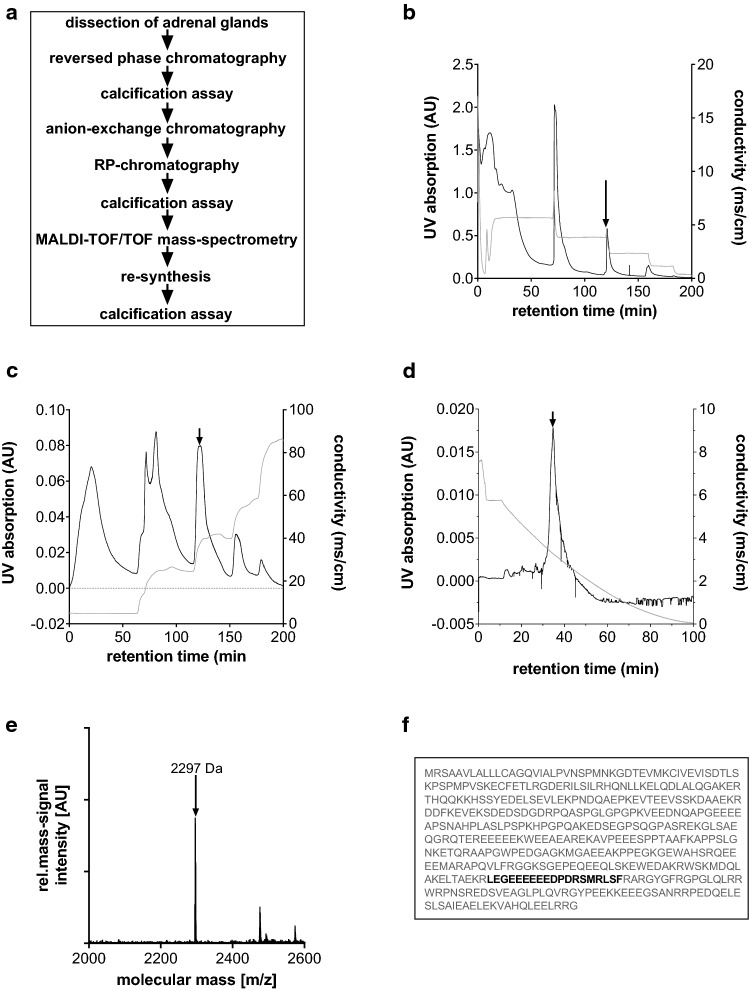
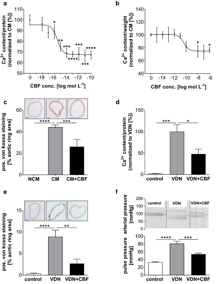
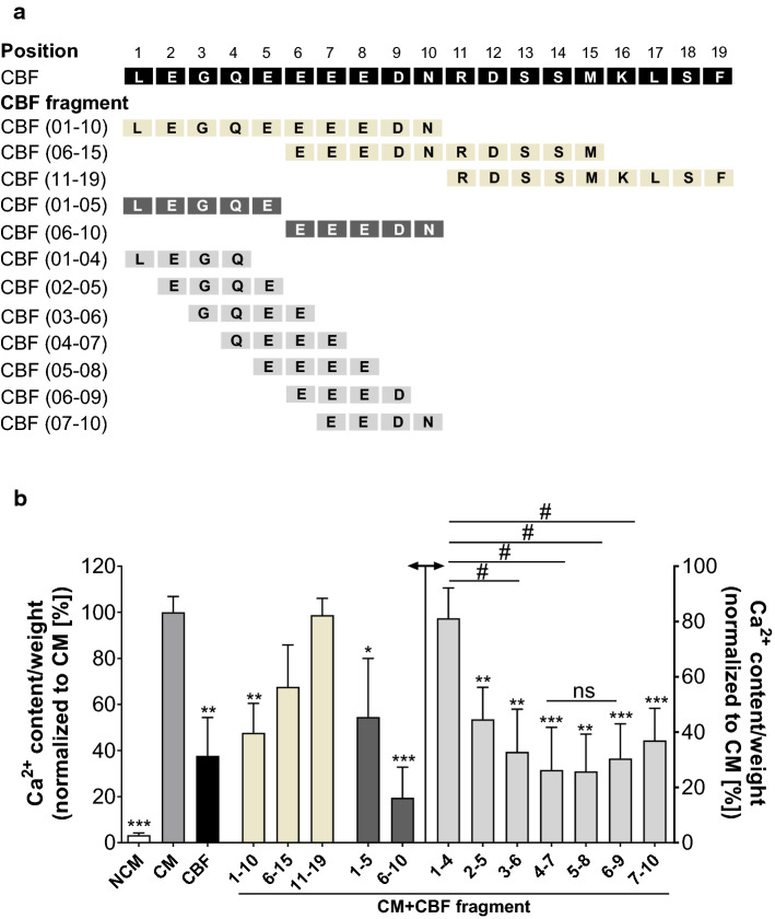
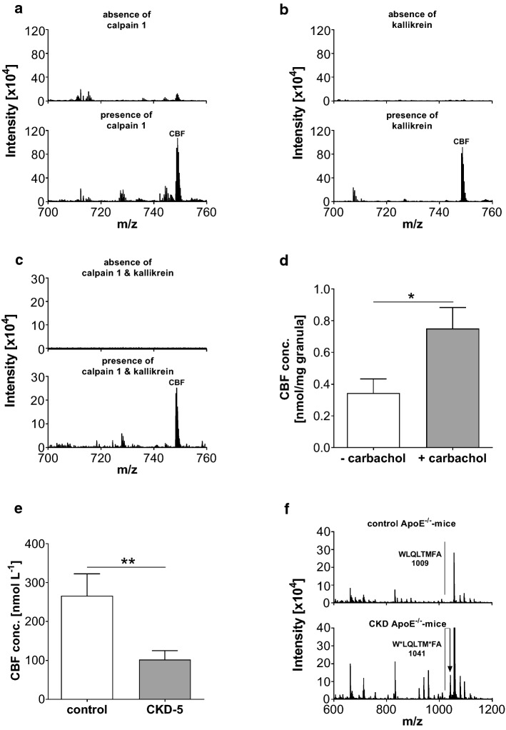
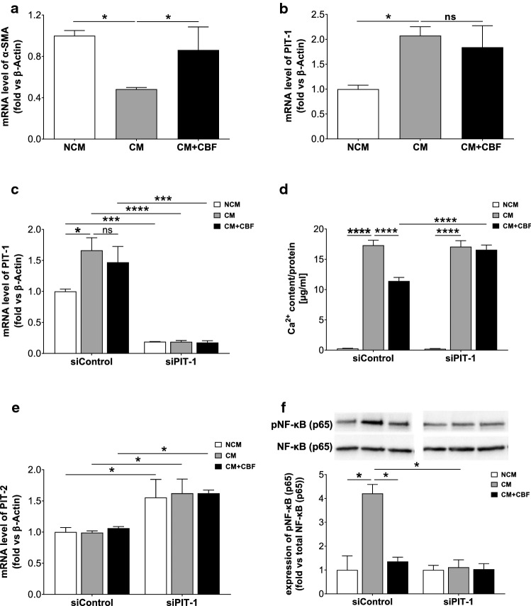
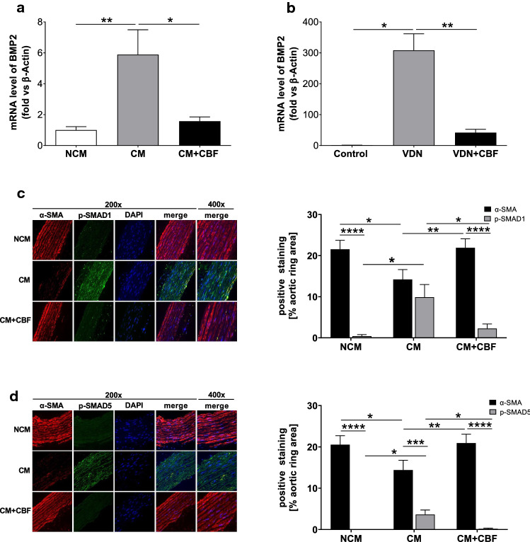
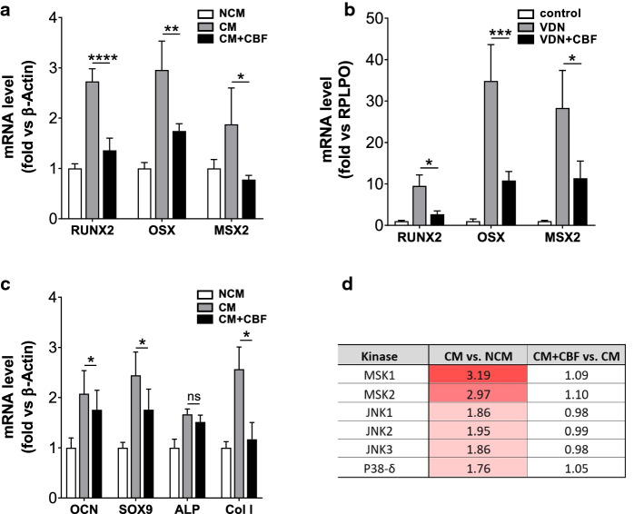
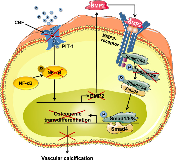
The adrenal glands participate in cardiovascular (CV) physiology and the pathophysiology of CV diseases through their effects on sodium and water metabolism, vascular tone and cardiac function. In the present study, we identified a new adrenal compound controlling mesenchymal cell differentiation that regulates osteoblastic differentiation in the context of vascular calcification. This peptide was named the "calcification blocking factor" (CBF) due to its protective effect against vascular calcification and is released from chromogranin A via enzymatic cleavage by calpain 1 and kallikrein. CBF reduced the calcium content of cells and thoracic aortic rings under calcifying culture conditions, as well as in aortas from animals treated with vitamin D and nicotine (VDN animals). Furthermore, CBF prevented vascular smooth muscle cell (VSMC) transdifferentiation into osteoblast-like cells within the vascular wall via the sodium-dependent phosphate transporter PIT-1 and by inhibition of NF-κB activation and the subsequent BMP2/p-SMAD pathway. Pulse pressure, a marker of arterial stiffness, was significantly decreased in VDN animals treated with CBF. In line with our preclinical data, CBF concentration is significantly reduced in diseases characterized by increased calcification, as shown in patients with chronic kidney disease. In preparation for clinical translation, the active site of the native 19-AS long native CBF was identified as EGQEEEED. In conclusion, we have identified the new peptide CBF, which is secreted from the adrenal glands and might prevent vascular calcification by inhibition of osteogenic transdifferentiation. The anti-calcific effects of CBF and short active site may therefore promote the development of new tools for the prevention and/or treatment of vascular calcification.

Keywords: Adrenal glands; Cardiorenal syndrome; Vascular calcification; Vascular smooth muscle cell transdifferentiation.


Conflict of interest statement

All the authors declare that they have no conflict of interest.

Figures

Fig. 1
Fig. 1
Identification and characterization of “calcification blocking factor” (CBF) from bovine adrenal glands. a Outline of experimental workflow for the identification and characterization of CBF, isolated from bovine adrenal glands. b Stepwise preparative reversed-phase chromatography of the bovine adrenal extracts (chromatographic conditions: column: Lichroprep RP C 18 (Merck); eluent A: 0.1% trifluoroacetic acid in water; eluent B: 80% acetonitrile in water; gradient: 20, 40, 60, 80, and 100% eluent B; UV absorption 280 nm). The fraction labelled by an arrow caused an inhibitory effect on vascular calcification processes of thoracic aortic rings. c Stepwise preparative anion-exchange chromatography of the fraction labelled by an arrow in suppl. Figure 1a (chromatographic conditions: column: SuperformanceTM 16 (Merck); eluent A: 20 mmol L−1 K2HPO4 in water; eluent B: 1 mol L−1 NaCl in 20 mmol L−1 K2HPO4 in water; gradient: 20, 40, 60, 80, and 100% eluent B; UV absorption 280 nm). The fraction labelled by an arrow caused an inhibitory effect on vascular calcification processes of thoracic aortic rings. d Linear gradient reversed-phase chromatography of the desalted fraction labelled by an arrow in suppl. Figure 1b (chromatographic conditions: column: LiChroprep RP C 18e (Merck); eluent A: 0.1% trifluoroacetic acid in water; eluent B: 80% acetonitrile in water; gradient: 1–100% eluent B over 90 min; UV absorption 280 nm). The fraction labelled by an arrow caused an inhibitory effect on vascular calcification processes of thoracic aortic rings. e The fraction shown in (d) was analysed by MALDI-TOF/TOF mass spectrometry to identify the sequence of the inhibitory factor of calcification. The mass signal (m/z) at 2297 Da [M + H]+ corresponds to the peptide sequence LEGEEEEEEDPDRSMRLSF, which was named “calcification blocking factor” (CBF). f Sequence matching: the peptide sequence of CBF represented in black characters matches the 350–369 amino acid position of bovine chromogranin A protein represented in grey characters
Fig. 2
Fig. 2
CBF inhibits calcification in thoracic aortic rings and vascular smooth muscle cells and influences pulse pressure in vivo. a Dose–response effect of CBF on Ca2+ content of cultivated human aortic smooth muscle cells. Data are shown as mean ± SEM. ***P ≤ 0.001, ****P ≤ 0.0001 compared to the calcifying condition in the absence of CBF based on one-way ANOVA. Bonferroni’s multiple comparisons were used as a post-test (N = 10 per group). b Dose–response effect of CBF on Ca2+ content of isolated thoracic aortic rings. Data are shown as mean ± SEM. *P < 0.05 compared to the calcifying condition in the absence of CBF based on one-way ANOVA. Bonferroni’s multiple comparisons were used as a post-test (N = 3–9 per group). c Quantification of von Kossa-stained thoracic aortic rings (original magnification × 40, scale bar 1000 μm), incubated in non-calcifying conditions (NCM; white bar) and under calcifying conditions in the absence (CM; grey bar) or presence of CBF (100 nmol L−1) (CM + CBF; black bar), respectively. Bars represent the mean ± SEM of the percentage of the stained area. ***P ≤ 0.001, ****P ≤ 0.0001 compared to the calcifying condition in the absence of CBF based on one-way ANOVA. Bonferroni’s multiple comparisons were used as a post-test (N = 9 per group). d Calcium content was measured in the aorta of untreated control Wistar rats (control: white bar), and VDN rats receiving a vehicle infused (VDN; grey bar) or CBF infused (31 µg kg−1 per day for 4 weeks) (VDN + CBF; black bars) via an osmotic pump. Data are shown as mean ± SEM. *P < 0.05, ***P ≤ 0.0001 compared with VDN group based on one-way ANOVA. Bonferroni’s multiple comparisons were used as a post-test (N = 6 per group). e Quantification of von Kossa-stained thoracic aortic rings (original magnification × 40, scale bar 1000 μm) of rats from each experimental group are shown. Bars represent the mean ± SEM of the percentage of the stained area. **P ≤ 0.001, ****P ≤ 0.00001 compared with the VDN group based on one-way ANOVA. Bonferroni’s multiple comparisons were used as a post-test (N = 10 per group). f CBF prominently prevented the VDN-induced rise in pulse pressure. Representative graphs of carotid arterial pressure recorded in a rat from each experimental group are shown. The graph represents the mean ± SEM of pulse pressure values. Pulse pressure was increased twofold in VDN rats. CBF counteracted the changes in both systolic and diastolic pressure associated with VDN and reduced the pulse pressure by 63%. ***P ≤ 0.0001; ****P ≤ 0.00001 compared with VDN group based on one-way ANOVA. Bonferroni’s multiple comparisons were used as a post-test (N = 10 per group)
Fig. 3
Fig. 3
Identification of relevant amino acid sequence of CBF causing the calcification inhibitory effect. a Overview of the amino acid sequence of CBF fragments used for identification of the active site of CBF causing the calcification inhibitory effect. Black: amino acid sequence of CBF. Beige: CBF fragments with an amino acid sequence of 9–10 amino acids. Dark grey: CBF fragments with an amino acid sequence of five amino acids. Light grey: CBF fragments with an amino acid sequence of four amino acids. b Quantification of Ca2+ content of cultivated rat thoracic aortic rings incubated in non-calcifying conditions (NCM; white bars) and under calcifying conditions in the absence (CM; grey bar) or presence of CBF (CM + CBF; black bar) or CBF fragments (CM + CBF; beige, dark and light grey bars) as indicated in (a) (100 nmol L−1). Data are shown as mean ± SEM. **P ≤ 0.001 compared with the CM group based on one-way ANOVA. #P < 0.05 demonstrates significant differences in Ca2+ contents caused by CBF fragments with an amino acid sequence of four amino acids based on one-way ANOVA. Bonferroni’s multiple comparisons were used as a post-test (N = 6–9 per group). ns no significant difference
Fig. 4
Fig. 4
Identification of enzymes cleaving CBF from chromogranin A. a LC-qTOF mass spectrometric analyses of LAKELTAEKR-LEGQEEEEDNRDSSMKLSF incubated in the absence (upper panel) and presence (lower panel) of kallikrein. The molecular mass (m/z) of 748.66 [M + 3H]3+ of the average spectrum at retention time range 18.8–19.7 corresponds to CBF. b LC-qTOF mass-spectrometric analyses of LEGQEEEEDNRDSSMKLSF-RARAYG FRGP incubated in the absence (upper panel) and presence (lower panel) of calpain 1. The molecular mass (m/z) of 748.66 [M + 3H]3+ of the average spectrum at retention time range 18.8–19.7 corresponds to CBF. c LC-qTOF mass-spectrometric analyses of chromogranin A incubated in the absence (upper panel) and presence (lower panel) with both kallikrein and calpain 1. The molecular mass (m/z) of 748.66 [M + 3H]3+ of the average spectrum of retention time range 18.8–19.7 corresponds to CBF. d Quantification of CBF in the supernatant of granules from bovine adrenal glands unstimulated and stimulated with carbachol (0.1 mmol L−1). The release of CBF from granula of adrenal glands into the culture media was doubled after stimulation. Data are shown as mean ± SEM. *P < 0.05 compared with the group without carbachol treatment based on unpaired t tests (N = 5 in each group). e The concentration of CBF in plasma of healthy controls (N = 13) and CKD-5 patients (N = 17) is reported in nmol L−1. CBF concentrations were decreased in patients suffering from CKD-5. ** indicates P ≤ 0.001compared to healthy based on unpaired t tests. f Characteristic MALDI mass spectra of the chromogranin A cleaving enzyme calpain 1 isolated from control C57BL/6J ApoE−/− mice (upper panel) and CKD adenine C57BL/6J ApoE−/− mice (lower panel) demonstrating CKD-associated enzyme modification by oxidation. The induction of CKD was performed by a 1.5-week diet of 0.3% adenine. A stable CKD condition was subsequently achieved with 0.15% adenine for 4.5 weeks. In addition, the animal received a high-fat diet during the whole time of experiments starting 4 weeks before the induction phase (N = 5 per group)
Fig. 5
Fig. 5
The sodium-dependent phosphate cotransporter 1 (PIT-1) mediates the inhibitory effect of CBF. a Relative quantification of α-smooth muscle actin protein (α-SMA) gene expression by RT-qPCR analyses. The mRNA level of α-SMA significantly decreased in the case of cultivating the cells under calcifying conditions (calcifying medium (CM)) compared to non-calcifying conditions (non-calcifying medium (NCM)), but not in the presence of CBF. Data are shown as mean ± SEM. *P < 0.05 compared with CM based on one-way ANOVA. Bonferroni’s multiple comparisons were used as a post-test (N = 3 per group). b Relative quantification of PIT-1 gene expression by RT-qPCR analyses. The increased gene expression in human aortic smooth muscle cells cultured for 7 days in calcifying medium (CM; grey bar) vs. non-calcifying medium (NCM; white bar) was not affected by CBF (100 nmol L−1) (black bar). Data are shown as mean ± SEM. *P < 0.05 compared with CM based on one-way ANOVA. Bonferroni’s multiple comparisons were used as a post-test (N = 3 per group). c Quantification of PIT-1 gene expression in VSMCs transfected with a control and PIT-1 siRNA, respectively, after incubation in non-calcifying medium (NCM; white bar) or calcifying medium in the absence (CM; grey bar) or presence (black bar) of CBF. Data are shown as mean ± SEM. *P < 0.05; ***P ≤ 0.001 and ****P ≤ 0.0001. The data based on one-way ANOVA. Bonferroni’s multiple comparisons were used as a post-test (N = 6 per group). d Quantification of Ca2+ content in transfected human aortic smooth muscle cells with 120 µM control siRNA or 120 µM PIT-1 siRNA after incubation with non-calcifying conditions (non-calcifying medium (NCM; white bar) and under calcifying conditions in the absence (CM; grey bar) or presence (black bar) of CBF (100 nmol L−1). Data are shown as mean ± SEM. ****P ≤ 0.0001 based on two-way ANOVA. Bonferroni’s multiple comparisons were used as a post-test (N = 3 per group). e Quantification of phosphate transporter 2 (PIT-2) gene expression in VSMCs transfected with a control and PIT-1 siRNA, respectively, after incubation in non-calcifying medium (NCM; white bar) or calcifying medium in the absence (CM; grey bar) or presence of CBF (black bar). Data are shown as mean ± SEM. *P < 0.05. The data based on one-way ANOVA. Bonferroni’s multiple comparisons were used as a post-test (N = 5 per group). f Western blot and quantitative analysis of NF-κB (p65) activation in VSMCs transfected with a control and PIT-1 siRNA, respectively, after incubation in non-calcifying medium (NCM; white bar) or calcifying medium in the absence (CM; grey bar) or presence of CBF (black bar). Data are shown as mean ± SEM. *P < 0.05. The data based on one-way ANOVA. Bonferroni’s multiple comparisons were used as a post-test (N = 3 per group)
Fig. 6
Fig. 6
CBF inhibits the BMP2/p-SMAD pathway. a Relative quantification of bone morphogenetic protein 2 (BMP2) gene expression in vitro by RT-qPCR analyses. The increased gene expression in human aortic smooth muscle cells cultured for 7 days in calcifying medium ((CM); grey bar) vs. non-calcifying medium ((NCM); white bar) was diminished in the presence of CBF (100 nmol L−1) (black bar). Data are shown as mean ± SEM. *P < 0.05; **P ≤ 0.01 compared with CM based on one-way ANOVA. Bonferroni’s multiple comparisons were used as a post-test (N = 6 per group). b Relative quantification of bone morphogenetic protein 2 (BMP2) gene expression in vivo by RT-qPCR analyses. The increased gene expression of the aorta of VDN rats (grey bar) vs. control rats (white bar) was limited in rats treated with CBF (31 µg kg−1 per day for 4 weeks) (black bar). Data are shown as mean ± SEM. *P < 0.05; **P ≤ 0.01 compared with VDN group based on one-way ANOVA Bonferroni’s multiple comparisons were used as a post-test (N = 5 per group). c, d Relative quantification of (c) α-smooth muscle actin (α-SMA) and phospho-SMAD1 (p-SMAD1) and d α-SMA and phospho-SMAD5 (p-SMAD5) of rat thoracic aortic rings cultivated in non-calcifying medium (NCM; white bar) and calcifying medium in the absence (CM; grey bar) or presence of CBF (100 nmol L−1) (black bar). Representative images (original magnification × 200, Scale bar 200 μm) of c α-SMA (red) and p-SMAD1 or d α-SMA (red) and p-SMAD5 (green), DAPI staining of nuclei (blue), and the corresponding merged images are shown. Data are shown as mean ± SEM. *P < 0.05; **P ≤ 0.01; ***P ≤ 0.0001, ****P ≤ 0.00 = 01. The data based on one-way ANOVA. Bonferroni’s multiple comparisons were used as a post-test (N = 6–9 in each group)
Fig. 7
Fig. 7
CBF reduces the expression of genes involved in osteoblast differentiation. a, b Relative quantification of runt-related transcription factor 2 (RUNX2), osterix (OSX) and msh homeobox 2 (MSX2) gene expression in (a) in vitro using hAoSMCs and (b) in vivo, respectively, by RT-qPCR analyses. Data are shown as mean ± SEM. *P < 0.05, **P ≤ 0.01, ***P ≤ 0.001, ****P ≤ 0.0001 compared with CM based on one-way ANOVA. Bonferroni’s multiple comparisons were used as a post-test (N = 4–6 in each group). c Relative quantification of osteocalcin (OCN), SOX9, alkaline phosphatase (ALP) and type 1 collagen (Col l) gene expression in vitro using hAoSMCs by RT-qPCR analyses. Data are shown as mean ± SEM. *P < 0.05 compared with CM based on one-way ANOVA. Bonferroni’s multiple comparisons were used as a post-test (N = 5 in each group). d Based on kinase activity profiling, kinase ranking scores (based on their significance and specificity in terms of the set of peptides used for the corresponding kinase) are shown for BMP-2-related kinases (N = 3 per condition)
Fig. 8
Fig. 8
Effect of CBF on vascular calcification processes is mediated by the PIT-1/NF-κB/BMP2/p-SMAD pathway. CBF affects the transport of phosphates by the type III NaPi cotransporter PIT-1. CBF inhibits the phosphorylation and subsequent activation of NF-κB (p65), decreased the BMP2 expression and inhibits the BMP2 pathway via inhibition of SMAD1/5/8 phosphorylation, the associated expression of osteogenic transdifferentiation genes and ultimately the vascular calcification of VSMCs

References

    1. Alexopoulos N, Raggi P. Calcification in atherosclerosis. Nat Rev Cardiol. 2009;6:681–688. doi: 10.1038/nrcardio.2009.165. - DOI - PubMed
    1. Balderman JA, Lee HY, Mahoney CE, Handy DE, White K, Annis S, Lebeche D, Hajjar RJ, Loscalzo J, Leopold JA. Bone morphogenetic protein-2 decreases microRNA-30b and microRNA-30c to promote vascular smooth muscle cell calcification. J Am Heart Assoc. 2012;1:e003905. doi: 10.1161/JAHA.112.003905. - DOI - PMC - PubMed
    1. Baulmann J, Schillings U, Rickert S, Uen S, Dusing R, Illyes M, Cziraki A, Nickering G, Mengden T. A new oscillometric method for assessment of arterial stiffness: comparison with tonometric and piezo-electronic methods. J Hypertens. 2008;26:523–528. doi: 10.1097/HJH.0b013e3282f314f7. - DOI - PubMed
    1. Blacher J, Guerin AP, Pannier B, Marchais SJ, London GM. Arterial calcifications, arterial stiffness, and cardiovascular risk in end-stage renal disease. Hypertension. 2001;38:938–942. doi: 10.1161/hy1001.096358. - DOI - PubMed
    1. Bokui N, Otani T, Igarashi K, Kaku J, Oda M, Nagaoka T, Seno M, Tatematsu K, Okajima T, Matsuzaki T, Ting K, Tanizawa K, Kuroda S. Involvement of MAPK signaling molecules and Runx2 in the NELL1-induced osteoblastic differentiation. FEBS Lett. 2008;582:365–371. doi: 10.1016/j.febslet.2007.12.006. - DOI - PMC - PubMed

Publication types