. 2022 Jan 5;23(1):e202100449.
doi: 10.1002/cbic.202100449. Epub 2021 Nov 5.

Xanthopsin-Like Systems via Site-Specific Click-Functionalization of a Retinoic Acid Binding Protein

Affiliations

Xanthopsin-Like Systems via Site-Specific Click-Functionalization of a Retinoic Acid Binding Protein

Giusy Tassone et al. Chembiochem. .

Abstract

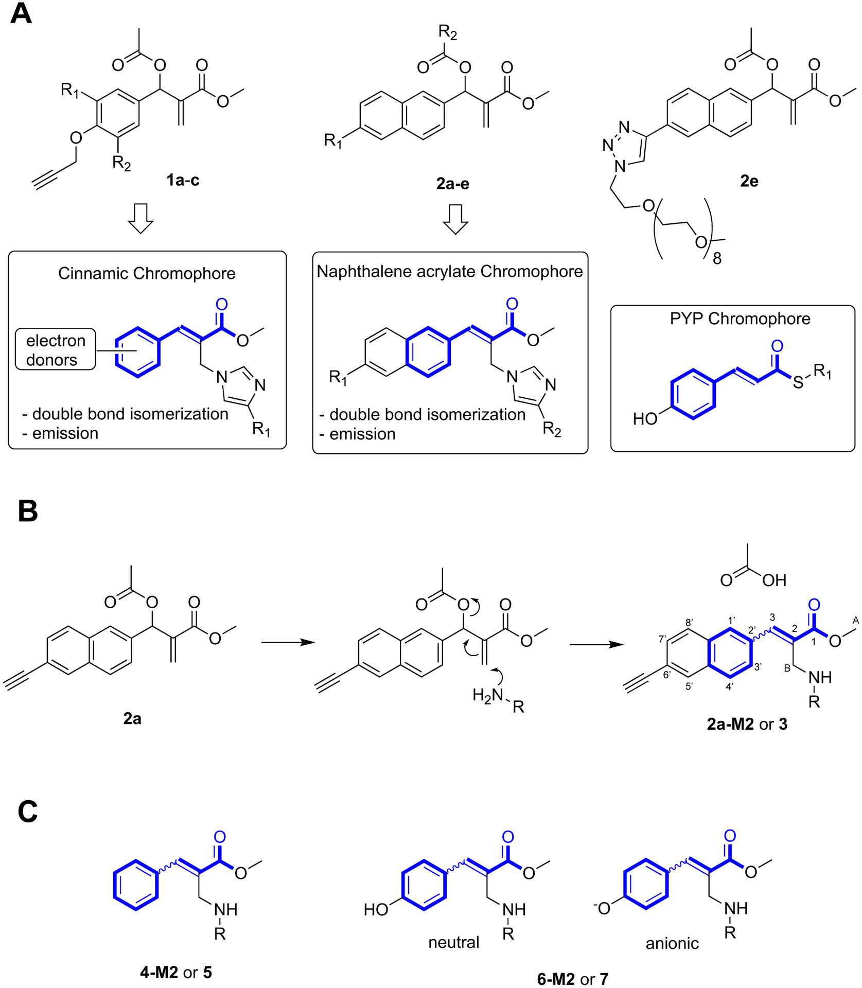
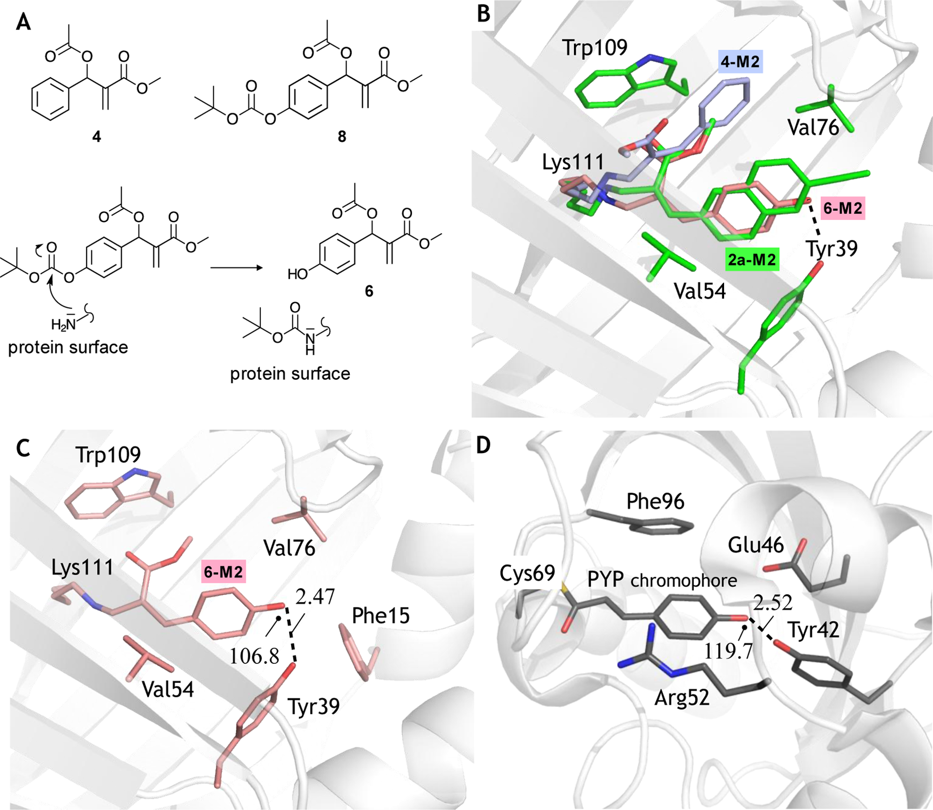
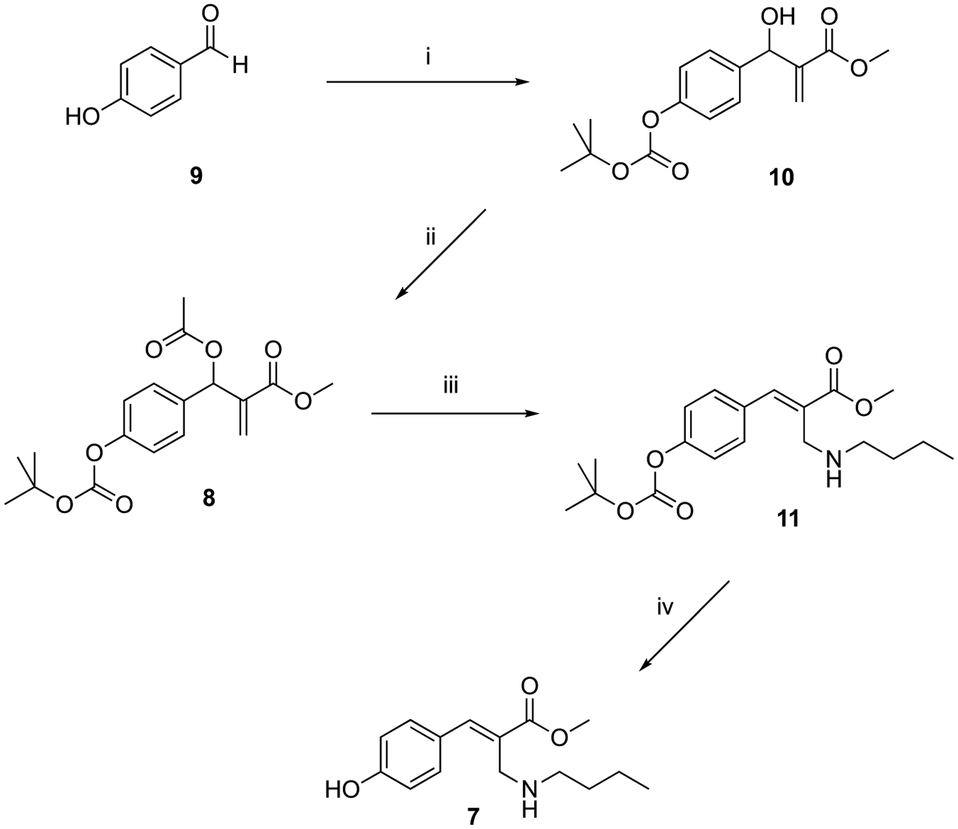
The use of light-responsive proteins to control both living or synthetic cells, is at the core of the expanding fields of optogenetics and synthetic biology. It is thus apparent that a richer reaction toolbox for the preparation of such systems is of fundamental importance. Here, we provide a proof-of-principle demonstration that Morita-Baylis-Hillman adducts can be employed to perform a facile site-specific, irreversible and diastereoselective click-functionalization of a lysine residue buried into a lipophilic binding pocket and yielding an unnatural chromophore with an extended π-system. In doing so we effectively open the path to the in vitro preparation of a library of synthetic proteins structurally reminiscent of xanthopsin eubacterial photoreceptors. We argue that such a library, made of variable unnatural chromophores inserted in an easy-to-mutate and crystallize retinoic acid transporter, significantly expand the scope of the recently introduced rhodopsin mimics as both optogenetic and "lab-on-a-molecule" tools.

Keywords: Morita-Baylis-Hillman adducts; PYP-like chromophores; light-sensitive proteins; site-specific reactions; synthetic xanthopsin-like proteins.


Figures

Figure 1.
Figure 1.
MBHAs reactivity. A. Structures of protein-reactive MBHAs 1a-c (1a: R1 = H, R2 = H; 1b: R1 = H R2 = OMe, 1c: R1 = OMe, R2 = OMe) and 2a-e (2a: R1 = C≡CH, R2 = OAc; 2b: R1 = OCH2C≡CH, R2 = OAc, 2c: R1 = OCH2C≡CH, R2 = COCH2CH2(OCH2CH2)11OMe, 2d: R1 = C≡CH, R2 = COCH2CH2(OCH2CH2)11OMe) and of their products of conjugation with proteins compared (blue-bold) with the PYP chromophore. B. Reaction of MBHA 2a with a primary amino group leading to 2a-M2 (R = side-chain of Lys111) and 3 (R = n-butyl, reagent n-butylamine, CHCl3). C. Phenyl-acrylate and p-hydroxyphenyl-acrylate protein constructs and corresponding models. 4-M2 (R = side-chain of Lys111) and 5 (R = n-butyl). 6-M2 (R = side-chain of Lys111) and 7 (R = n-butyl).
Figure 2.
Figure 2.
X-ray crystallographic structure of M2 (PDB: 6Z2U). Cartoon representation of the M2 tertiary structure. α-helices are colored liliac-violet, β strands in magenta and loops in black. Histidine (orange carbon atoms), arginine (green carbon atoms) and lysine (yellow carbon atoms) are displayed in sticks. A label marks the reactive Lys111 site.
Figure 3.
Figure 3.
Binding pocket structure. A. M2 pocket with its hydrophobic residues displayed in sticks (carbon atoms in green). B. 2a-M2 pocket (PDB: 6Z2Z). The chromophore and surrounding amino acid residues are shown as sticks (orange and green colors indicate carbon atoms. Oxygen, nitrogen, and sulphur atoms are colored in red, blue, and yellow, respectively). C. Superposition of the binding pockets of 2a-M2 and retinal-M2 rhodopsin mimics (PDB: 4YFQ) featuring the all-trans retinal protonated Schiff base chromophore in cyan. Residues are color coded as the respective chromophores. Oxygen and nitrogen atoms are colored red and blue, respectively. D. The same comparison with the 15-cis retinal protonated Schiff base (PDB: 4YFP).
Figure 4.
Figure 4.
Structural characterization of Xanthopsin-like systems. A. Top. MBHAs used to prepare the conjugates 4-M2 and 6-M2 (see Fig. 1). Bottom. Possible mechanism for the in situ production of the MBHA 6. B. Chromophore-binding cavity view of the superimposition between 2a-M2 construct (PDB id: 6Z2Z) (white cartoon and green carbons) with the phenolic chromophore of 6-M2 (PDB id: 6ZSW) (in stick pink carbons) and phenolic chromophore of 4-M2 (PDB id: 6ZSX) (in stick light blue carbons). Residues are color coded as 2a carbons. In the figure, oxygen and nitrogen atoms are colored red and blue, respectively. C. Chromophore-binding cavity view of 6-M2 construct. White color was used in the cartoon for the M2 portion, while the generated chromophore and the amino acid surroundings are shown as stick pink carbons. D. Chromophore-binding cavity view of the PYP X-ray crystallographic structure (PDB id: 2ZOI). White color was used in the cartoon for the protein portion, while its p-hydroxythiocinnamate chromophore and the surroundings residues are shown as stick grey carbons. Oxygen atoms are colored in red, nitrogen atoms in blue, and the sulfur atom in yellow. In panels C and D the marked (see dashed line) hydrogen bond lengths is given Å and the relative angles in degrees.
Figure 5.
Figure 5.
UV-Vis absorption spectra. A. Comparison of the normalized absorption spectra of M2, 7 (pure E diastereoisomer), and 8 in tris buffer solution and anionic form of 7 obtained by addition of KOH. B. Absorption spectra of M2, of the M2+8 mixture registered after 24 hours in tris buffer solution at concentration of 2.6 × 10⁻⁵ M and differential spectrum obtained by subtracting the spectrum of the first one from that of the second.
Figure 6.
Figure 6.
Photoisomerization of the neutral form of 7. A. Excitation energies (kcal/mol) of the S0 and S1 equilibrium (eq.) structures of the neutral form of Z-7. The relaxation from the allowed S2 state (exp. 318 nm in solution vs. comp. 257 nm (#303 nm after correcting the geometry for electron correlation effects) in vacuo may involve a S2/S1 conical intersection. The relevant bond lengths (Å) and torsion (degrees) of the same structures are also given. Red (negative) and blue (positive) balloons represent charge magnitudes (positive electron units). B. 1H NMR spectra of E-7 in methanol-d4. C. 1H NMR spectra of the E-7 and Z-7 mixture obtained after irradiating E-7 for two hours with a UV-B light lamp. D. The absorption spectra of neutral E-7 in methanol (black line) and of its E/Z mixture (red line) obtained after continuous irradiation at 313 nm for 18 minutes is consistent with a blue-shifted absorption for Z-7 with respect to E-7.
Figure 7.
Figure 7.
Kinetic isomerization of 6-M2 monitored by UV-vis spectra registered under continuous irradiation at 280 nm in tris buffer solution pH 8 (A) and in tris buffer solution added with NaOH until pH 12.7 (B). In panel B, the arrows indicate the progressive increase or decrease of the spectrum as a consequence of the irradiation.
Scheme 1.
Scheme 1.
Synthesis of MBHA derivative 8 and its reaction with n-butylamine. Reagents: (i) BOC anhydride, methyl acrylate, DABCO, CH3OH, THF; (ii) CH3COCl, TEA, CH2Cl2; (iii) n-butylamine, THF-H2O (5:1); (iv) CF3COOH, CH2Cl2.

References

    1. Rodriguez EA, Campbell RE, Lin JY, Lin MZ, Miyawaki A, Palmer AE, Shu X, Zhang J, Tsien RY, Trends Biochem. Sci 2017, 42, 111–129. - PMC - PubMed
    1. Schmidt D, Cho YK, Trends Biotechnol. 2015, 33, 80–91. - PubMed
    1. Stano P, Mavelli F, Life 2015, 5, 1700–1702.
    1. Zhang F, Vierock J, Yizhar O, Fenno LE, Tsunoda S, Kianianmomeni A, Prigge M, Berndt A, Cushman J, Polle J, Magnuson J, Hegemann P, Deisseroth K, Cell 2011, 147, 1446–1457. - PMC - PubMed
    1. Kandori H, Bull. Chem. Soc. Jpn 2019, 93, 76–85.

Publication types