. 2021 Nov 19;16(11):2137-2143.
doi: 10.1021/acschembio.1c00542. Epub 2021 Oct 14.

Rapid Analysis of ADP-Ribosylation Dynamics and Site-Specificity Using TLC-MALDI

Affiliations

Rapid Analysis of ADP-Ribosylation Dynamics and Site-Specificity Using TLC-MALDI

Sean R Wallace et al. ACS Chem Biol. .

Abstract

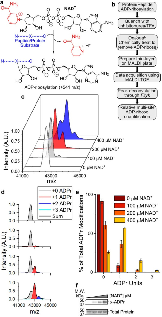
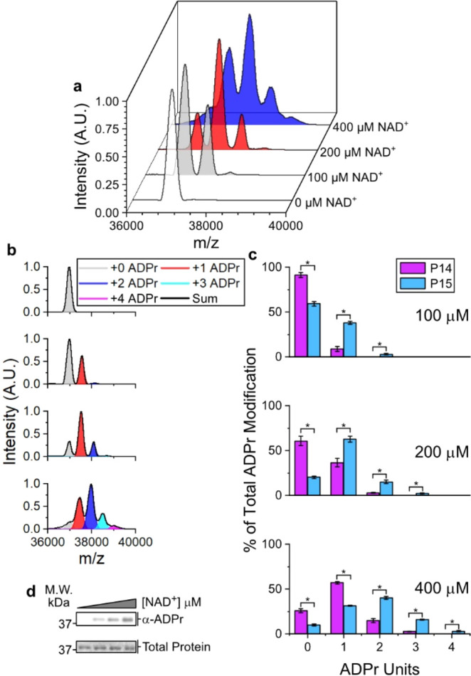
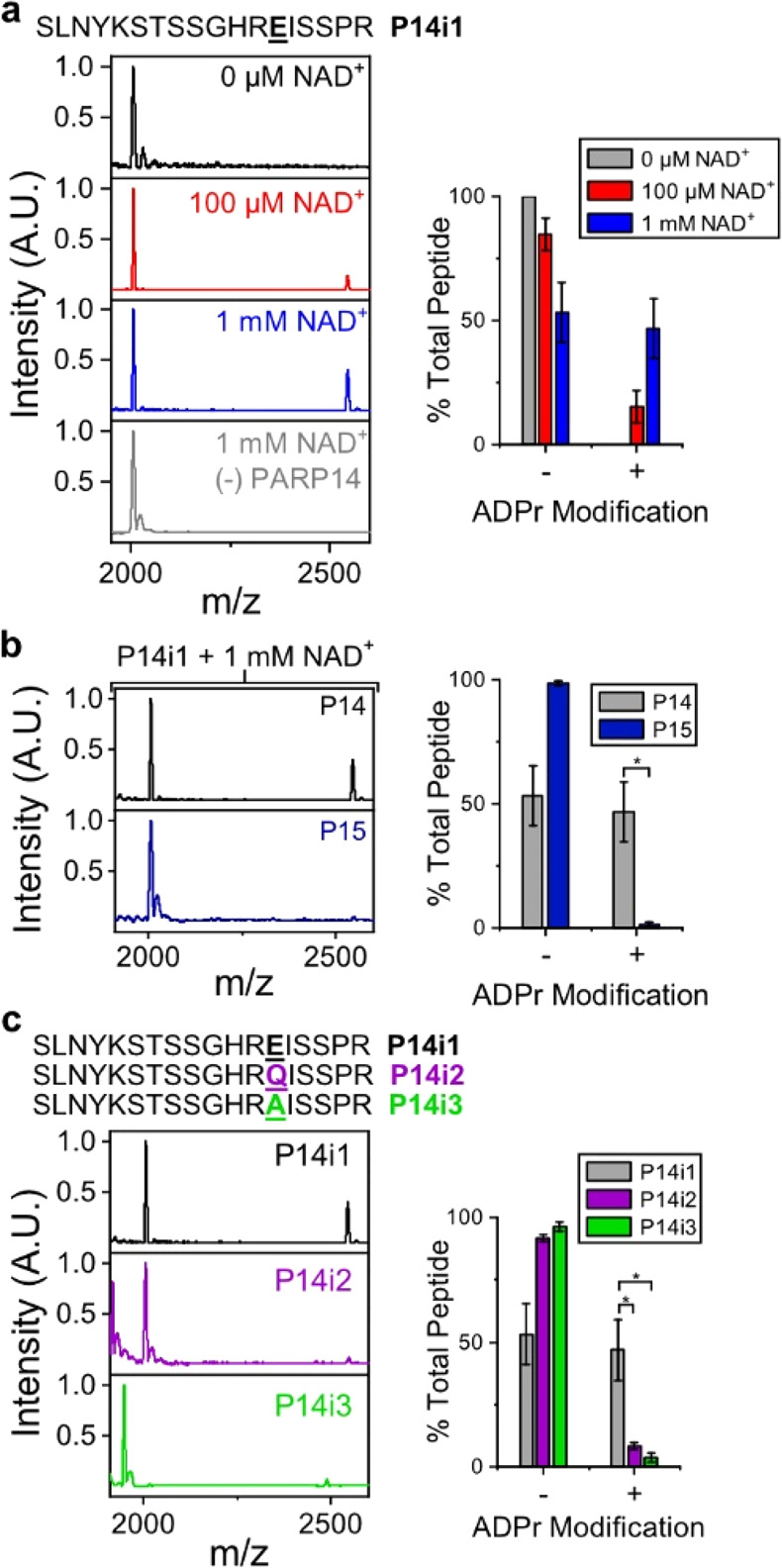
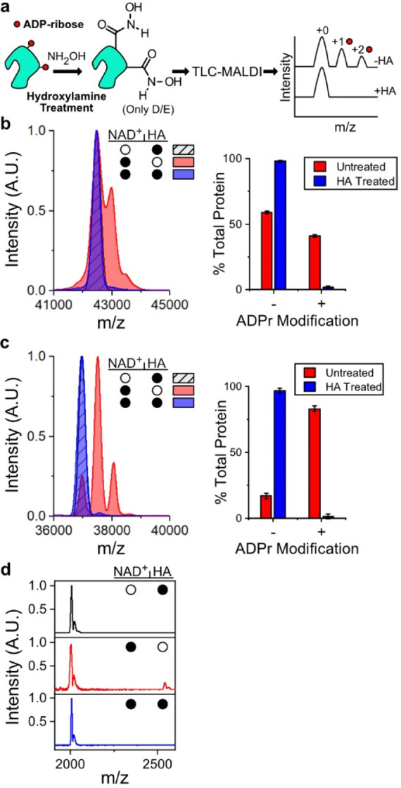
Poly(ADP-ribose) polymerases, PARPs, transfer ADP-ribose onto target proteins from nicotinamide adenine dinucleotide (NAD+). Current mass spectrometric analytical methods require proteolysis of target proteins, limiting the study of dynamic ADP-ribosylation on contiguous proteins. Herein, we present a matrix-assisted laser desorption/ionization time-of-flight (MALDI-TOF) method that facilitates multisite analysis of ADP-ribosylation. We observe divergent ADP-ribosylation dynamics for the catalytic domains of PARPs 14 and 15, with PARP15 modifying more sites on itself (+3-4 ADP-ribose) than the closely related PARP14 protein (+1-2 ADP-ribose)─despite similar numbers of potential modification sites. We identify, for the first time, a minimal peptide fragment (18 amino-acids) that is preferentially modified by PARP14. Finally, we demonstrate through mutagenesis and chemical treatment with hydroxylamine that PARPs 14/15 prefer acidic residues. Our results highlight the utility of MALDI-TOF in the analysis of PARP target modifications and in elucidating the biochemical mechanism governing PARP target selection.


Conflict of interest statement

The authors declare no competing financial interest.

Figures

Figure 1
Figure 1
P14 auto-ADP-ribosylates at a single site under physiological conditions. (a) ADP-ribosylation reaction catalyzed by PARP enzymes. (b) TLC-MALDI workflow. (c) P14 was incubated in the presence of increasing concentrations of NAD+ and was subjected to TLC-MALDI to visualize the resulting increase in m/z due to ADPr (+541). (d) MS spectra were integrated to determine the relative levels of auto-ADP-ribosylation. (e) Quantification of the results in (b). The bar graphs depict the fraction of the total P14 protein that has been modified at 0, 1, or 2 distinct sites (mean ± S.E.M., n = 3). (f) In vitro P14 ADP-ribosylation assay.
Figure 2
Figure 2
P15 auto-ADP-ribosylates at multiple sites under physiological conditions. (a) P15 was incubated in the presence of increasing concentrations of NAD+ and was subjected to TLC-MALDI to visualize the resulting increase in m/z due to ADP-ribosylation (+541). (b) MS spectra were integrated to determine the relative levels of auto-ADP-ribosylation. (c) Quantification of ADPr levels for both P14 and P15. The bar graphs depict the fraction of the total protein that has been modified at 0, 1, 2, 3, or 4 distinct sites (mean ± S.E.M., n = 3). *represents p-value <0.05, two-tailed Student’s t test. The concentration of NAD+ is given. (d) In vitro P15 ADP-ribosylation assay.
Figure 3
Figure 3
TLC-MALDI performed with minimal peptide fragments reveals proximal PARP selectivity. (a) P14 was incubated with P14i1 in the presence of increasing concentrations of NAD+, and the resulting MS spectra were normalized and plotted against each other to visualize the increase in m/z due to ADPr (+541). The bar graphs depict the fraction of the total peptide that has been modified (mean ± S.E.M., n = 3). The sequence of the P14i1 peptide fragment is provided. (b) P15 is unable to transfer ADPr to the P14i1 peptide. Experiments were performed as in (a) in the presence of 1 mM NAD+. (c) Loss of the acidic modification site prevents ADP-ribosylation. Experiments were performed as in (a) in the presence of 1 mM NAD+. *represents p-value <0.05, two-tailed Student’s t test.
Figure 4
Figure 4
TLC-MALDI performed following HA treatment confirms acidic residues as the targets of P14 and P15 ADP-ribosylation. (a) HA treatment workflow. (b) P14 was incubated with 400 μM NAD+ prior to treatment with HA and was subjected to TLC-MALDI to visualize the resulting increase in m/z due to ADP-ribosylation (+541). The bar graphs depict the fraction of the total protein that has been modified (mean ± S.E.M., n = 3). (c) The same experiment from (a) was performed with P15. (d) P14 was incubated with P14i1 and treated as in (a). The filled circles represent treatment with NAD+, HA, or both.

References

    1. Citarelli M.; Teotia S.; Lamb R. S. Evolutionary History of the Poly(ADP-Ribose) Polymerase Gene Family in Eukaryotes. BMC Evol. Biol. 2010, 10 (1), 308.10.1186/1471-2148-10-308. - DOI - PMC - PubMed
    1. Krueger K. M.; Barbieri J. T. The Family of Bacterial ADP-Ribosylating Exotoxins. Clin. Microbiol. Rev. 1995, 8 (1), 34–47. 10.1128/CMR.8.1.34. - DOI - PMC - PubMed
    1. Hottiger M. O.; Hassa P. O.; Lüscher B.; Schüler H.; Koch-Nolte F. Toward a Unified Nomenclature for Mammalian ADP-Ribosyltransferases. Trends Biochem. Sci. 2010, 35 (4), 208–219. 10.1016/j.tibs.2009.12.003. - DOI - PubMed
    1. Vyas S.; Matic I.; Uchima L.; Rood J.; Zaja R.; Hay R. T.; Ahel I.; Chang P. Family-Wide Analysis of Poly(ADP-Ribose) Polymerase Activity. Nat. Commun. 2014, 5, 4426.10.1038/ncomms5426. - DOI - PMC - PubMed
    1. Leidecker O.; Bonfiglio J. J.; Colby T.; Zhang Q.; Atanassov I.; Zaja R.; Palazzo L.; Stockum A.; Ahel I.; Matic I. Serine Is a New Target Residue for Endogenous ADP-Ribosylation on Histones. Nat. Chem. Biol. 2016, 12 (12), 998–1000. 10.1038/nchembio.2180. - DOI - PMC - PubMed

Publication types

MeSH terms

Substances