. 2021 Nov 9;144(19):1567-1583.
doi: 10.1161/CIRCULATIONAHA.120.052023. Epub 2021 Oct 14.

Long Noncoding RNA MIAT Controls Advanced Atherosclerotic Lesion Formation and Plaque Destabilization

Affiliations

Long Noncoding RNA MIAT Controls Advanced Atherosclerotic Lesion Formation and Plaque Destabilization

Francesca Fasolo et al. Circulation. .

Abstract

Background: Long noncoding RNAs (lncRNAs) are important regulators of biological processes involved in vascular tissue homeostasis and disease development. The present study assessed the functional contribution of the lncRNA myocardial infarction-associated transcript (MIAT) to atherosclerosis and carotid artery disease.

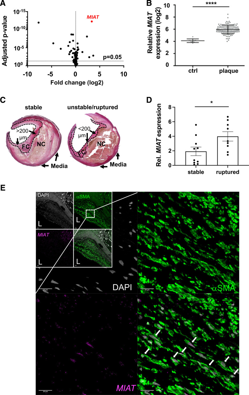
Methods: We profiled differences in RNA transcript expression in patients with advanced carotid artery atherosclerotic lesions from the Biobank of Karolinska Endarterectomies. The lncRNA MIAT was identified as the most upregulated noncoding RNA transcript in carotid plaques compared with nonatherosclerotic control arteries, which was confirmed by quantitative real-time polymerase chain reaction and in situ hybridization.

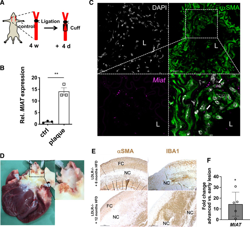
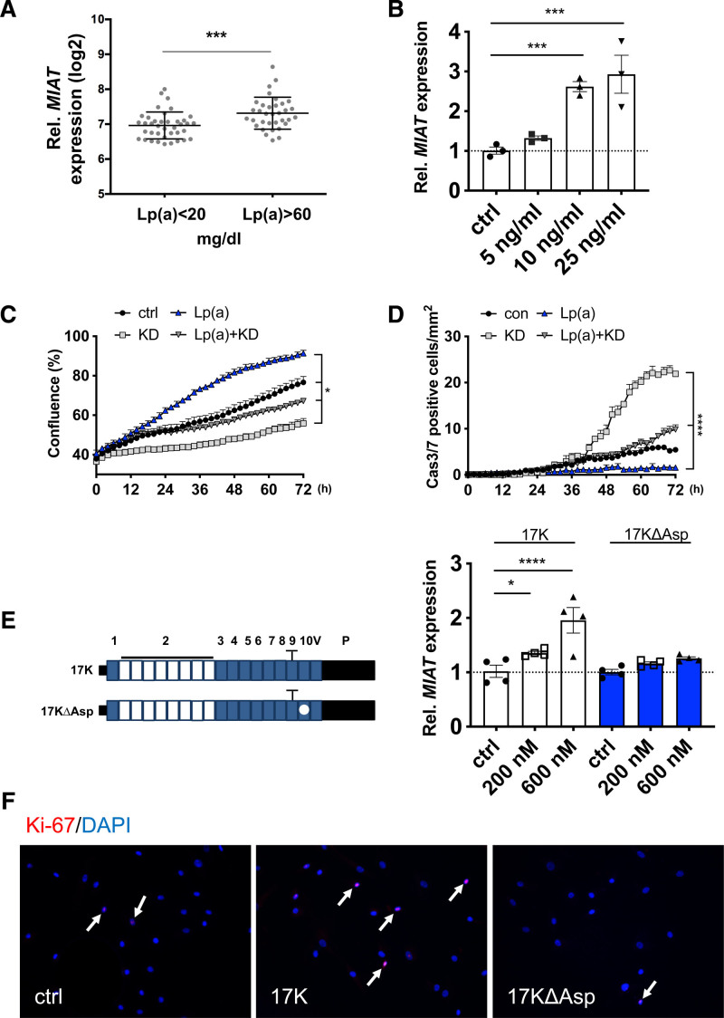
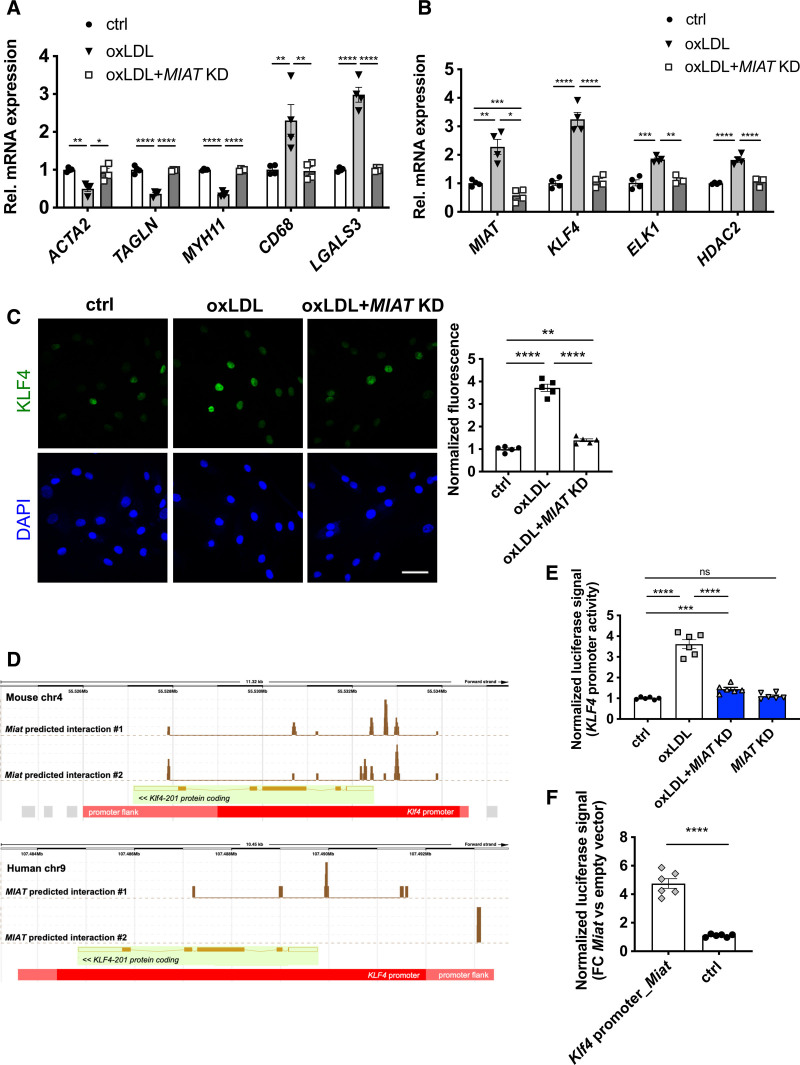
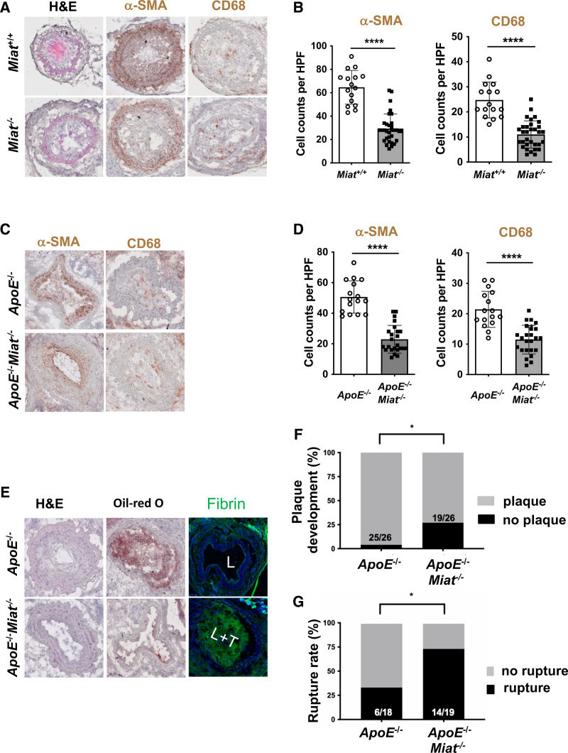
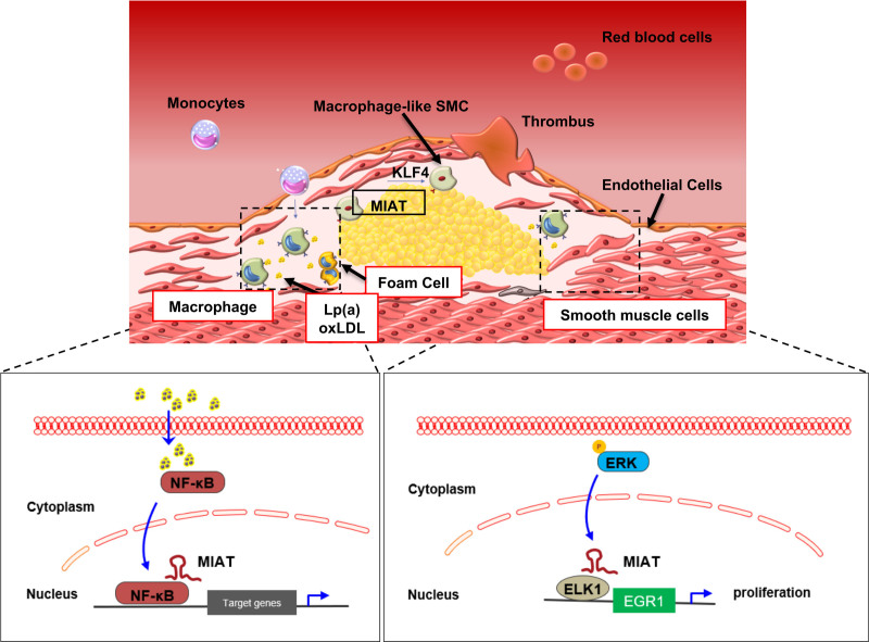
Results: Experimental knockdown of MIAT, using site-specific antisense oligonucleotides (LNA-GapmeRs) not only markedly decreased proliferation and migration rates of cultured human carotid artery smooth muscle cells (SMCs) but also increased their apoptosis. MIAT mechanistically regulated SMC proliferation through the EGR1 (Early Growth Response 1)-ELK1 (ETS Transcription Factor ELK1)-ERK (Extracellular Signal-Regulated Kinase) pathway. MIAT is further involved in SMC phenotypic transition to proinflammatory macrophage-like cells through binding to the promoter region of KLF4 and enhancing its transcription. Studies using Miat-/- and Miat-/-ApoE-/- mice, and Yucatan LDLR-/- mini-pigs, as well, confirmed the regulatory role of this lncRNA in SMC de- and transdifferentiation and advanced atherosclerotic lesion formation.

Conclusions: The lncRNA MIAT is a novel regulator of cellular processes in advanced atherosclerosis that controls proliferation, apoptosis, and phenotypic transition of SMCs, and the proinflammatory properties of macrophages, as well.

Keywords: RNA, long noncoding; atherosclerosis; carotid artery diseases; lipoprotein(a); myocytes, smooth muscle; stroke.


Figures

Figure 1.
Figure 1.
MIAT expression is increased in advanced stages of human carotid artery disease. A, Scatter plot of deregulated noncoding transcripts in human carotid plaques (n=127) compared with nonatherosclerotic controls (n=10) from the Biobank of Karolinska Endarterectomies. B, Expression plot for MIAT based on the results presented in Figure 1A. C, Hematoxylin and eosin–stained stable and unstable/ruptured atherosclerotic lesions from the Munich Vascular Biobank. Dotted line indicates the laser-catapulted fibrous cap (FC). NC represents necrotic core. FC >200 µm was considered a stable lesion; FC <200 µm was unstable/ruptured. D, MIAT expression in laser captured microdissected stable versus ruptured fibrous caps (mean±SEM; unpaired nonparametric Student t test). E, Colocalization analysis of MIAT and αSMA protein was performed by RNAscope protocol. MIAT and αSMA protein were detected through Opal 690 and Opal 520, visualized in purple and green color, respectively. Nuclei are stained with DAPI (gray). Upper Left, the boxes represent overview images of the carotid plaques, and the zoomed-in images are magnifications acquired within the plaque shoulder region. White arrows highlight the nuclear detection of MIAT in cells expressing the smooth muscle cell marker (αSMA). Bar=100 µm. *P<0.05; ****P<0.0005. Data are presented as mean±SD (SEM in D) with Student t test and corrected for multiple comparison (A) using Bonferroni correction. Ctrl indicates control; DAPI, 4′,6-diamidino-2-phenylindole; Rel., relative; and αSMA, smooth muscle cell α-actin.
Figure 2.
Figure 2.
MIAT is significantly increased in murine carotid plaques and regulates proliferation in vascular smooth muscle cells. A, Illustration of the inducible plaque rupture model using incomplete ligation and cuff placement in ApoE–/– mice. B, Expression of Miat determined by quantitative real-time polymerase chain reaction from advanced plaques and contralateral controls from the inducible plaque rupture model presented in Figure 2B. C, Colocalization analysis of Miat and αSMA protein was performed by RNAscope in murine carotid plaques. Miat and αSMA protein were detected through Opal 690 (purple) and Opal 520 (green). Nuclei are stained with DAPI (gray). D, Ruptured plaque in the coronary ostia from an advanced lesion from LDLR–/– Yucatan mini-pigs being fed 12 months of high-fat diet (HFD). E, Immunostaining of smooth muscle cell α-actin (αSMA) and macrophage marker-ionized calcium-binding adapter molecule 1 (IBA1) in plaques from early (6 months HFD) and advanced (12 months HFD) carotid artery lesions (FC indicates fibrous cap; NC, necrotic core). F, Relative MIAT expression in advanced (12 months HFD) versus early (6 months HFD) carotid plaques. Data were analyzed by Student t test. *P<0.05; ***P<0.001. Ctrl indicates control; DAPI, 4′,6-diamidino-2-phenylindole; L, lumen; and Rel., relative.
Figure 3.
Figure 3.
MIAT regulates human carotid artery smooth muscle cell proliferation through the ERK/ELK1/EGR1 pathway. A and B, Proliferation (A) or apoptosis (B) of human carotid smooth muscle cells (hCASMCs) on knockdown of MIAT (KD) monitored through live-cell imaging over time (0–72 hours). Data were analyzed by 2-way ANOVA. C and D, Proliferation (C) or apoptosis (D) of hCASMCs on KD of MIAT determined by Ki-67 or Caspase 3 immunofluorescent staining (white arrows indicate Ki-67/Caspase 3–positive cells). Bar=50 µm. E, In silico analysis of transcription factors associated with MIAT (predicted binding free energy of <–30 Kcal/mol) using the regulatory RNA elements/RegRNA tool. F, MIAT fragment containing 2 predicted ELK1 binding sites (MIAT-ELK1_BS1_BS2) was transiently transfected and coimmunoprecipitated with endogenous ELK1 in hCASMCs. IgGs were used as control of immunoprecipitation (IP) specificity. MIAT or GAPDH (as unrelated target) enrichment in ELK1 IP fraction was quantified with quantitative real-time polymerase chain reaction and expressed as (2ΔCt)×100 ELK1 IP÷(2ΔCt)×100 IgG. ΔCt was calculated based on input. RNA content in IP or IgG was normalized on RPLPO mRNA. ELK1 IP efficiency was monitored by Western blot using an anti-ELK1 antibody. G, Transfected cells were lysed after 24 hours, and total protein was extracted. P-ERK normalized to total ERK was monitored using Western Blot. Quantification of the Western Blot was done with Fiji Image J software. H, MIAT-ELK1_BS1_BS2 was transiently transfected, and proliferation was monitored using the IncuCyte Live Cell Imaging System. Bar=50 µm. Data were analyzed by 2-way repeated-measures ANOVA (A), area under the curve (B), and Student t test (C, D, F, G). *P<0.05; **P<0.01; ***P<0.001; ****P<0.0001. Casp indicates caspase; Ctrl, control; DAPI, 4′,6-diamidino-2-phenylindole; EV, empty vector (control); and IgG, immunoglobulin G.
Figure 4.
Figure 4.
MIAT is increased in patients with elevated circulating lipoprotein(a) levels. A, Expression of MIAT in patients in Biobank of Karolinska Endarterectomies with low (<20 mg/dL) and high (>60 mg/dL) lipoprotein(a) (Lp[a]) serum levels. B. Expression of MIAT in hCASMCs on Lp(a) stimulation. C and D, Proliferation (C) or apoptosis (D) of hCASMCs on MIAT knockdown (KD), Lp(a) or combined treatment monitored live-cell imaging over time (data were analyzed by 2-way ANOVA). E, Left, Schematic of APO(a) peptide constructs with (17K) or without (17KDelta) an attached oxidized phospholipid. E, Right, Expression of MIAT in hCASMCs on 17K or 17KDelta stimulation. F, Immunostaining of Ki-67 in hCASMCs with 17K or 17KDelta stimulation (white arrows indicate Ki-67–positive cells). Data were analyzed by Student t test (A), 2-way repeated-measures ANOVA (C), area under the curve (D), or 1-way ANOVA (B, E). *P<0.05; ***P<0.001; ****P<0.0001. Cas indicates caspase; ctrl, control; DAPI, 4′,6-diamidino-2-phenylindole; and Rel., relative.
Figure 5.
Figure 5.
MIAT triggers inflammation and macrophage activity in advanced atherosclerotic lesions. A, Expression of MIAT in human monocyte-derived macrophages on MIAT knockdown (KD). B, Uptake of florescence-labeled oxLDL in human monocyte-derived macrophages on MIAT KD monitored through live-cell imaging and analyzed by 1-way ANOVA. C, Expression of MIAT in human monocyte-derived macrophages on stimulation with oxLDL. D and E, Expression of MIAT in THP1-derived macrophages stimulated with LPS (D) or IL4 (E). F, Immunostaining of NF-κB in human monocyte-derived macrophages on oxLDL stimulation with or without MIAT KD. Quantification is shown on the right side. G, Binding of endogenous MIAT to NFKB2/p52/p100 in THP1 cells. MIAT expression in THP1 cells was triggered by oxLDL stimulation and immunoprecipitation with NFKB2/p52/p100 or control IgG performed. MIAT or GAPDH (as unrelated target) enrichment in NFKB2 IP fraction were quantified with real-time quantitative polymerase chain reaction. Data were analyzed by Student t test (A, C–E, G), AUC (B), or Mann-Whitney U test (F). Bar=50 µm. **P<0.01; ***P<0.001; ****P<0.0001. ctrl indicates control; DAPI, 4′,6-diamidino-2-phenylindole; FITC, fluorescein isothiocyanate; IgG, immunoglobulin G; IL4, interleukin 4; IP, immunoprecipitation; LPS, lipopolysaccharide; NF-κB, Nuclear Factor κ-light-chain enhancer of activated B cells; oxLDL, oxidized low-density lipoprotein; and Rel., relative.
Figure 6.
Figure 6.
MIAT participates in smooth muscle cell transdifferentiation into inflammatory macrophage-like cells through KLF4 activation. A, Expression of SMCs, phagocytosis and/or macrophage markers in hCASMCs stimulated with oxLDL and with or without MIAT knockdown (KD). B, Expression of MIAT, transcription factor KLF4, coeffectors ELK1 and HDAC2 in hCASMCs stimulated with oxLDL and with or without MIAT KD. C, Left, Immunostaining of KLF4 in hCASMCs stimulated with oxLDL and with or without MIAT KD; Right, fluorescence quantification. D, Interaction of Miat/MIAT with Klf4/KLF4 promoter as predicted by LongTarget v2.1. Peaks indicate predicted binding sites of Miat/MIAT within Klf4/KLF4 promoter region. E, Luciferase reporter assay with human KLF4 promoter on oxLDL stimulation/MIAT KD in hCASMCs. F, Luciferase reporter assay with murine Klf4 promoter (containing Miat predicted binding sites; Klf4 promoter_Miat) or promoter flanking regions (harboring no predicted Miat binding sites; Ctrl) in mouse aortic SMCs on Miat overexpression (pCAG-Miat, murine). Bar=50 µm. Data were analyzed by 1-way ANOVA.*P<0.05; **P<0.01; ***P<0.001; ****P<0.0001; P value >0.05 indicates no significance. ctrl indicates control; DAPI, 4′,6-diamidino-2-phenylindole; FC, fibrous cap; hCASMC, human carotid smooth muscle cell; KLF4, Krüppel-like factor 4; oxLDL, oxidized low-density lipoprotein; Rel., relative; and SMC, smooth muscle cell.
Figure 7.
Figure 7.
MIAT deletion affects smooth muscle cell proliferation and plaque vulnerability in vivo. A, Morphology (H&E), immunostaining for CD68 and smooth muscle cell α-actin (αSMA) in Miat–/– mice versus littermate Miat wildtype controls on carotid ligation injury. B, Analysis of cell counts (8 times/animal) per high-power field (HPF) for αSMA and CD68 from A. C, Morphology (H&E), immunostaining for CD68 and αSMA in ApoE–/–Miat–/– and ApoE–/– (Miat wildtype) controls on exposure to the inducible plaque rupture model (incomplete ligation and cuff placement). D, Cell counts (8 times/animal) per HPF for αSMA and CD68 from C. E, H&E, Oil-Red O (indicating lipid deposition), and cross-linked immunofluorescent fibrin staining (indicating an atherothrombotic event) in ApoE–/–Miat–/– versus Apo–/– Miat+/+ littermate controls (L indicates lumen; L+T, lumen with thrombus). F, Plaque development (in %) when using the inducible plaque rupture model in ApoE–/–-Miat–/– versus ApoE–/–Miat+/+ mice. G, Rupture ratio (in %) in the inducible plaque rupture model comparing ApoE–/–Miat–/– versus ApoE–/– (Miat wildtype) controls. Data were analyzed by Student t test (B, D). Fisher exact test was used to determine plaque development and rupture ratio. *P<0.05; **P<0.01. H&E indicates hematoxylin and eosin.
Figure 8.
Figure 8.
Proposed mechanism of action for MIAT in advanced atherosclerosis and plaque destabilization. KLF4 indicates Krüppel-like factor 4; Lp(a), lipoprotein(a); NF-κB, Nuclear Factor κ-light-chain enhancer of activated B cells; oxLDL, oxidized low-density lipoprotein; and SMC, smooth muscle cell.

References

    1. Sadat U, Jaffer FA, van Zandvoort MA, Nicholls SJ, Ribatti D, Gillard JH. Inflammation and neovascularization intertwined in atherosclerosis: imaging of structural and molecular imaging targets. Circulation. 2014; 130:786–794. doi: 10.1161/CIRCULATIONAHA.114.010369 - PMC - PubMed
    1. van Lammeren GW, den Ruijter HM, Vrijenhoek JE, van der Laan SW, Velema E, de Vries JP, de Kleijn DP, Vink A, de Borst GJ, Moll FL, et al. . Time-dependent changes in atherosclerotic plaque composition in patients undergoing carotid surgery. Circulation. 2014; 129:2269–2276. doi: 10.1161/CIRCULATIONAHA.113.007603 - PubMed
    1. Fernández-Friera L, Peñalvo JL, Fernández-Ortiz A, Ibañez B, López-Melgar B, Laclaustra M, Oliva B, Mocoroa A, Mendiguren J, Martínez de Vega V, et al. . Prevalence, vascular distribution, and multiterritorial extent of subclinical atherosclerosis in a middle-aged cohort: the PESA (Progression of Early Subclinical Atherosclerosis) study. Circulation. 2015; 131:2104–2113. doi: 10.1161/CIRCULATIONAHA.114.014310 - PubMed
    1. Donnan GA, Fisher M, Macleod M, Davis SM. Stroke. Lancet. 2008; 371:1612–1623. doi: 10.1016/S0140-6736(08)60694-7 - PubMed
    1. Libby P, Ridker PM, Hansson GK. Progress and challenges in translating the biology of atherosclerosis. Nature. 2011; 473:317–325. doi: 10.1038/nature10146 - PubMed

Publication types

Substances