. 2021 Nov 17;13(620):eabj7790.
doi: 10.1126/scitranslmed.abj7790. Epub 2021 Nov 17.

Lung epithelial and endothelial damage, loss of tissue repair, inhibition of fibrinolysis, and cellular senescence in fatal COVID-19

Affiliations

Lung epithelial and endothelial damage, loss of tissue repair, inhibition of fibrinolysis, and cellular senescence in fatal COVID-19

Felice D'Agnillo et al. Sci Transl Med. .

Abstract

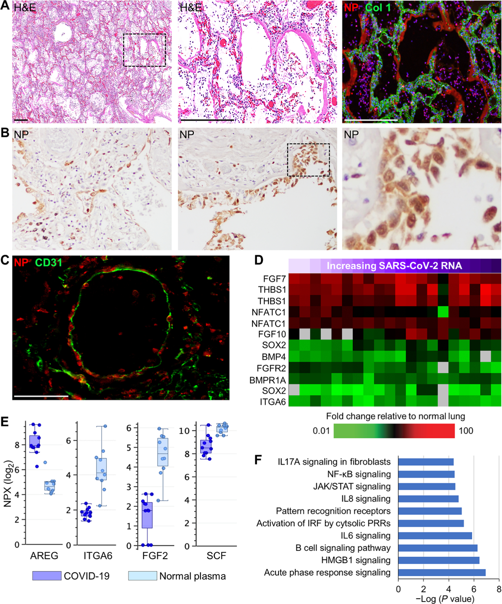
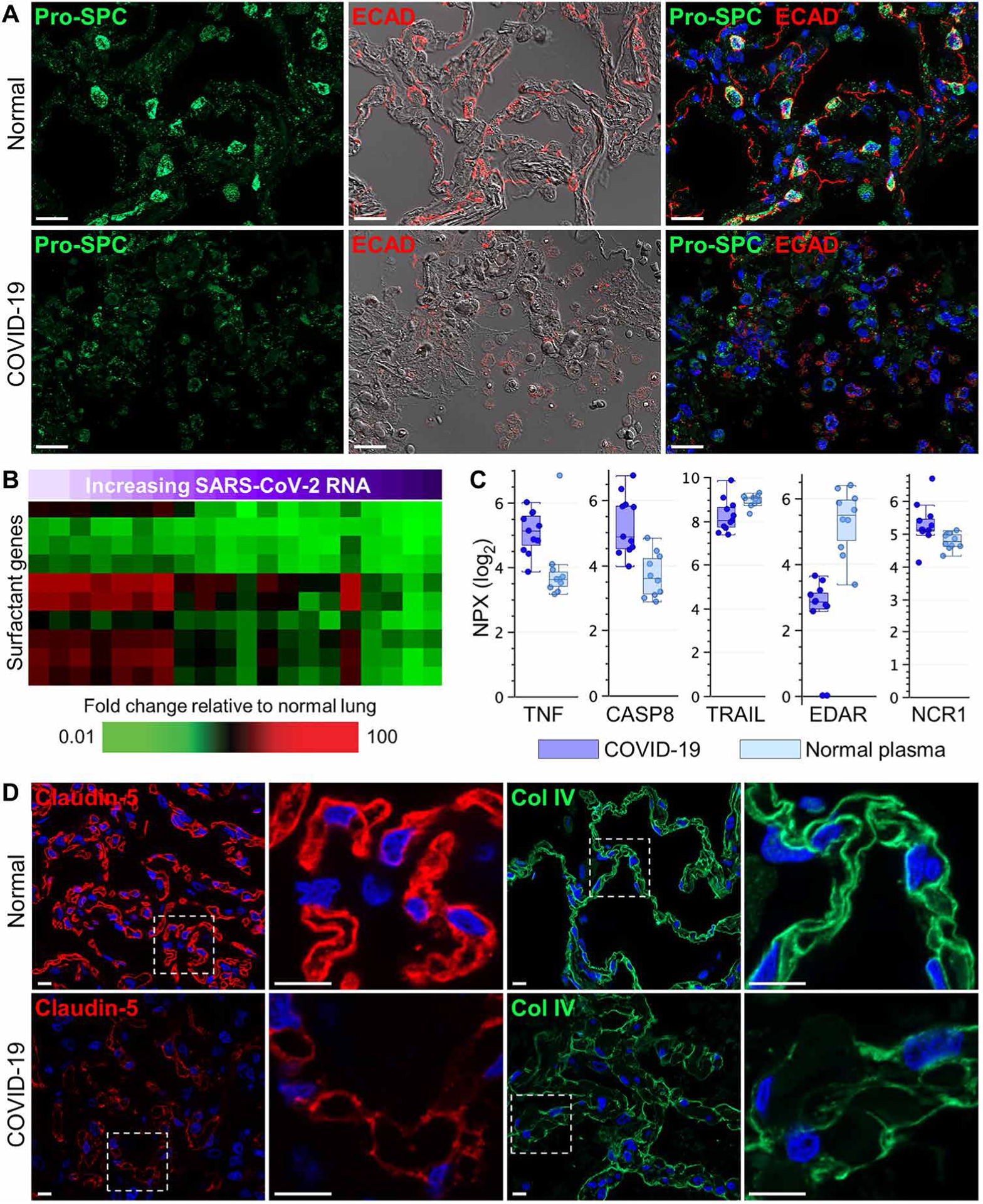
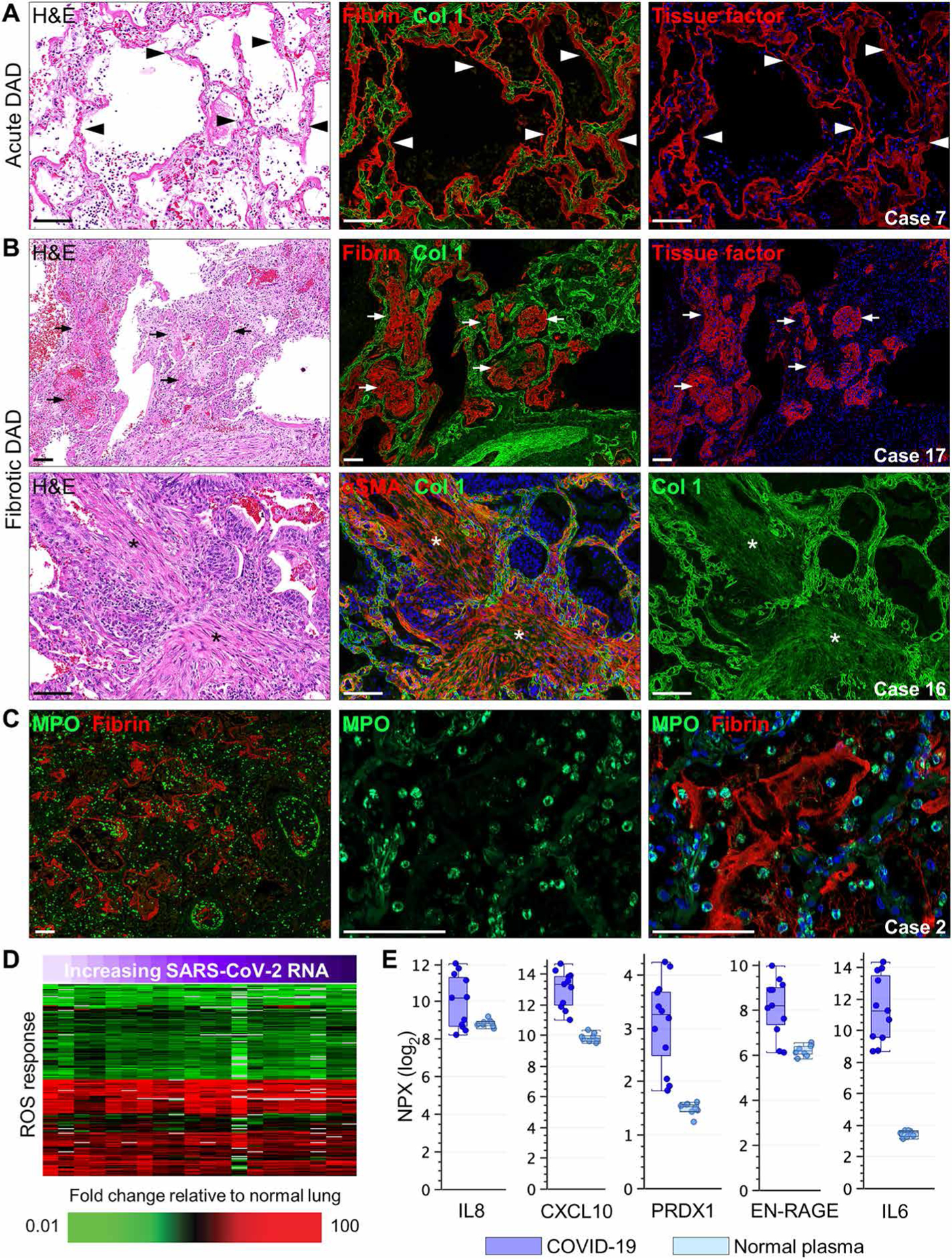
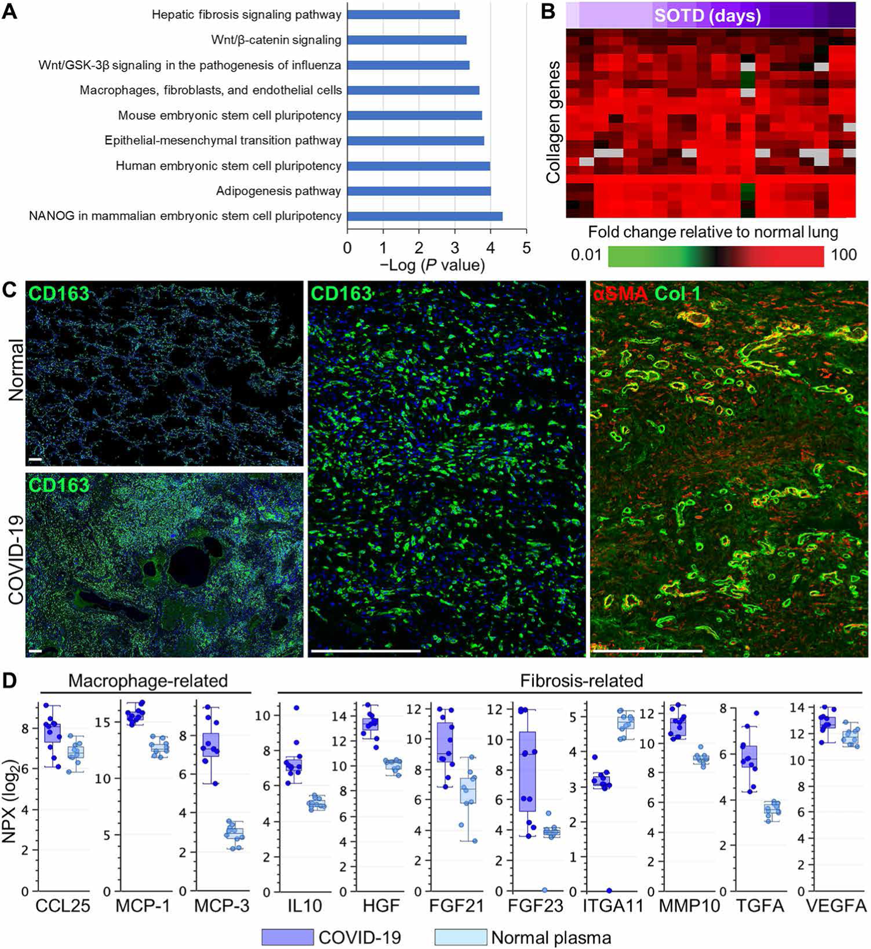
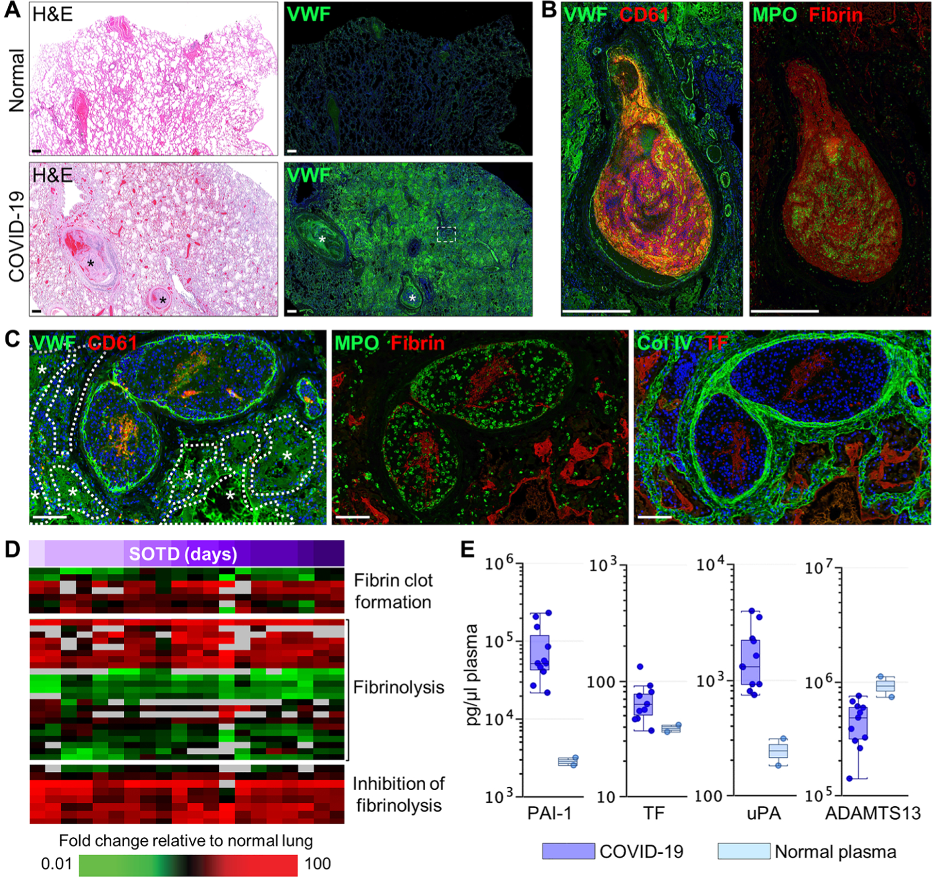
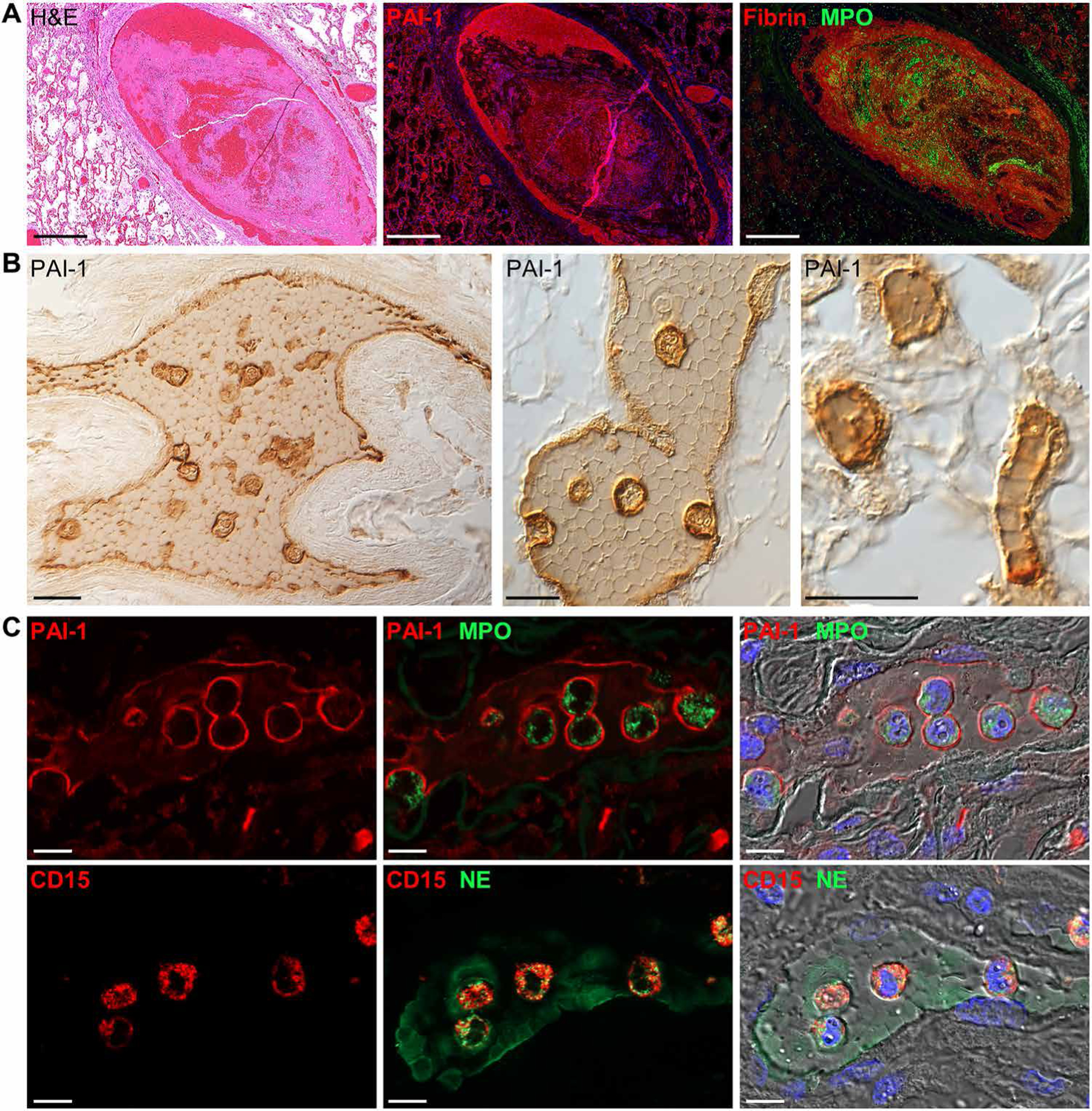
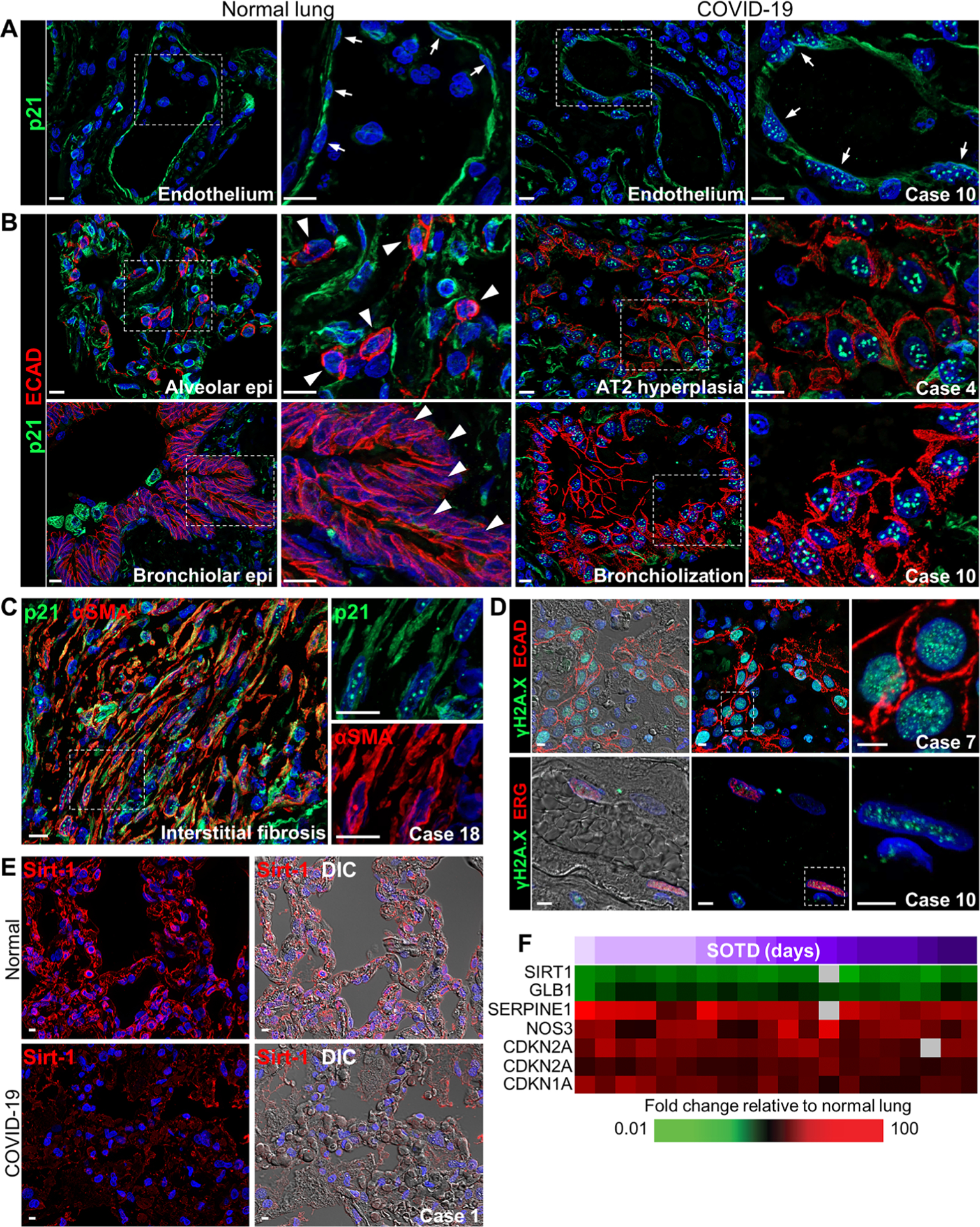
Coronavirus disease 2019 (COVID-19), caused by the severe acute respiratory syndrome coronavirus 2 (SARS-CoV-2), is characterized by respiratory distress, multiorgan dysfunction, and, in some cases, death. The pathological mechanisms underlying COVID-19 respiratory distress and the interplay with aggravating risk factors have not been fully defined. Lung autopsy samples from 18 patients with fatal COVID-19, with symptom onset-to-death times ranging from 3 to 47 days, and antemortem plasma samples from 6 of these cases were evaluated using deep sequencing of SARS-CoV-2 RNA, multiplex plasma protein measurements, and pulmonary gene expression and imaging analyses. Prominent histopathological features in this case series included progressive diffuse alveolar damage with excessive thrombosis and late-onset pulmonary tissue and vascular remodeling. Acute damage at the alveolar-capillary barrier was characterized by the loss of surfactant protein expression with injury to alveolar epithelial cells, endothelial cells, respiratory epithelial basal cells, and defective tissue repair processes. Other key findings included impaired clot fibrinolysis with increased concentrations of plasma and lung plasminogen activator inhibitor-1 and modulation of cellular senescence markers, including p21 and sirtuin-1, in both lung epithelial and endothelial cells. Together, these findings further define the molecular pathological features underlying the pulmonary response to SARS-CoV-2 infection and provide important insights into signaling pathways that may be amenable to therapeutic intervention.


Conflict of interest statement

S.J.O. has received funding from Agenus, Amgen, Biothera, Bristol Myers Squibb, Exicure, Genocea, Incyte, Merck, Ultimovacs, and Viralytics. S.J.O. has consulted for Biothera, Bristol Myers Squibb, BioNTech, Exicure, Immunsys, and Merck. K. Sadtler is an inventor on a provisional U.S. patent application no. 63/092,350 entitled “Antibody specific for SARS-CoV-2 receptor binding domain and therapeutic methods.” The other authors declare that they have no competing interests.

Figures

Fig. 1.
Fig. 1.. SARS-CoV-2 detection and host gene expression in COVID-19 lung autopsy samples.
(A) Shown are representative images from serial lung autopsy sections from a patient with COVID-19 with acute DAD (case 1). Sections were stained with H&E or immunostained for the SARS-CoV-2 nucleocapsid antigen (NP) and collagen type 1 (Col 1). The digitally magnified boxed area shows marked NP immunoreactivity in hyaline membranes lining alveoli and intra-alveolar epithelial debris and cellular infiltrates. Nuclei were counterstained with Hoechst 33342 dye (blue). (B) Representative images of NP immunohistochemistry show positive staining in bronchiolar epithelium, epithelial basal cells, and alveolar epithelium (case 2). Black dashed box shows area of digital enlargement. Original magnification, ×200. (C) Immunofluorescence image of a medium-sized blood vessel stained for NP and endothelial CD31 (case 7). (D) Heatmap shows expression of genes encoding lung repair–related proteins in lung tissue from 13 COVID-19 cases (table S5). Each column represents gene expression data from a microarray experiment comparing RNA from COVID-19 lung tissue to pooled RNA isolated from normal lung tissue (n = 3). Genes shown in red were significantly increased (twofold, P < 0.05, using standard t test with Benjamini-Hochberg correction), genes shown in green were decreased, and genes in black indicate no change in expression in COVID-19 lung tissue relative to normal lung tissue. An increase in SARS-CoV-2 RNA in lung tissue is indicated by a purple gradient bar with values ranging from 0 to 44.9. Viral CT values were normalized to the calibrator gene GAPDH, and final CT values were inverted (40 − Δt) such that a higher value represents a higher viral load. (E) Basal cell and lung repair–related proteins in plasma from patients with COVID-19 (n = 6) and healthy volunteers (n = 10) were measured by the Olink platform. Values were statistically significant (P < 0.05) between the groups using standard t test with Benjamini-Hochberg correction. Scale of normalized protein expression (NPX) is log2. Each circle represents an individual plasma sample, and 25th, median, and 75th quartiles are indicated with box-and-whisker plots. SCF, stem cell factor. (F) Shown is a gene ontology analysis of transcripts with expression correlating (R ≥ 0.6) with pulmonary SARS-CoV-2 RNA levels. Scale bars, 250 μm (A) and 100 μm (C). NF-κB, nuclear factor κB; JAK, Janus kinase; STAT, signal transducer and activator of transcription; IRF, IFN regulatory factor.
Fig. 2.
Fig. 2.. Alveolar epithelial and endothelial damage in COVID-19 lung autopsy samples.
(A) Representative immunofluorescence and differential interference contrast (DIC) images showing prosurfactant protein C (Pro-SPC) and E-cadherin (ECAD) expression in a COVID-19 lung autopsy sample (case 1) and in normal lung tissue. Normal lung tissue alveolar septa show prominent Pro-SPC and E-cadherin expression in lung AT2 cells and epithelial junctions, respectively, compared to COVID-19 lung tissue which shows marked loss of alveolar septal Pro-SPC and E-cadherin staining and intra-alveolar accumulation of positive-stained epithelial debris. (B) Heatmap shows expression of genes encoding lung surfactant proteins that show differential expression in COVID-19 lung tissue samples (n = 13 cases; table S5) compared to normal lung tissue. SARS-CoV-2 RNA in lung tissue is indicated by a purple gradient bar with values ranging from 0 to 44.9. Viral CT values were normalized to the calibrator gene GAPDH, and final CT values were inverted (40 − Δt) such that a higher value represents a higher viral load. (C) Cell death–related proteins in plasma from patients with COVID-19 (n = 6) and healthy volunteers (n = 10) were measured by Olink platform. Values were statistically significant (P < 0.05) between the groups using standard t test with Benjamini-Hochberg correction. Scale of NPX is log2. Each circle represents an individual plasma sample, and 25th, median, and 75th quartiles are indicated with box-and-whisker plots. (D) Shown are representative immunofluorescence images of alveolar capillary claudin-5 expression and alveolar basement membrane collagen type 4 (Col IV) expression in normal lung tissue and COVID-19 lung tissue (case 12). Digitally magnified boxed areas highlight the reduced and discontinuous staining of claudin-5 and Col IV in the COVID-19 lung tissue compared to normal lung tissue. Scale bars, 20 μm (A) and 10 μm (D).
Fig. 3.
Fig. 3.. Progressive DAD histopathology and neutrophil responses in COVID-19 lung autopsy samples.
(A and B) Representative lung sections show immunostaining of (A) acute and (B) fibrotic DAD in COVID-19 lung tissue (cases 7, 16, and 17). For (A) and (B), serial lung sections were stained with H&E or were immunostained for fibrin, collagen type 1 (Col 1), tissue factor, or αSMA. Black/white arrowheads in (A) showing acute DAD lung tissue indicate hyaline membrane formation with marked colocalization of fibrin and tissue factor lining alveolar spaces. Black/white arrows in (B) showing fibrotic DAD lung tissue depict late lesions consisting of colocalized intra-alveolar fibrin and tissue factor in “fibrin balls” within alveolar spaces. Black/white asterisks show areas of interstitial fibrosis with marked colocalized expression of αSMA and Col 1. (C) Representative immunofluorescence staining of neutrophil myeloperoxidase (MPO) and fibrin in a COVID-19 autopsy lung tissue sample with histological evidence of acute DAD (case 2). (D) Heatmap presents expression of genes associated with the ROS response that show differential expression in COVID-19 lung tissue samples (n = 13 cases; table S5) compared to normal lung tissue. SARS-CoV-2 RNA in lung tissue is indicated by a purple gradient bar with values ranging from 0 to 44.9. Viral CT values were normalized to the calibrator gene GAPDH, and final CT values were inverted (40 − Δt) such that a higher value represents a higher viral load. (E) Measurement of proteins associated with neutrophil infiltration and oxidative stress in plasma from patients with COVID-19 (n = 6) and healthy volunteers (n = 10) was performed using the Olink platform. Values were statistically significant (P < 0.05) between the groups using standard t test with Benjamini-Hochberg correction. Each circle represents an individual plasma sample, and 25th, median, and 75th quartiles are indicated with box-and-whisker plots. Scale of NPX is log2. Scale bars, 100 μm.
Fig. 4.
Fig. 4.. Macrophage activation and fibrogenesis in COVID-19 lung autopsy samples.
(A) Gene ontology analysis of transcripts with expression correlating (R ≥ 0.6) with time of symptom onset to death (SOTD). (B) Heatmap shows expression of genes encoding collagen proteins that show differential expression in COVID-19 lung tissue samples (n = 13 cases; table S5) compared to normal lung tissue. SOTD is indicated by a purple gradient bar. (C) Representative immunofluorescence staining of macrophage CD163 or αSMA and collagen type 1 (Col 1) in COVID-19 lung tissue with histological evidence of fibrotic DAD (case 18). Low-power images highlight the increased patchy distribution of CD163-positive macrophages in regions undergoing fibrosis in the COVID-19 lung section compared to lower CD163 expression in normal lung tissue. High-power images of serial sections depict interstitial fibrotic areas filled with CD163-positive macrophages that colocalize with diffuse deposition of αSMA and Col 1 surrounded by brightly stained αSMA- and Col 1–positive blood vessels. (D) Measurement of proteins associated with macrophage infiltration and fibrogenesis in plasma from patients with COVID-19 (n = 6) and healthy volunteers (n = 10) using the Olink platform. Values were statistically significant (P < 0.05) between the groups using standard t test with Benjamini-Hochberg correction. Scale of NPX is log2. Each circle represents an individual plasma sample, and 25th, median, and 75th quartiles are indicated with box-and-whisker plots. Scale bars, 250 μm.
Fig. 5.
Fig. 5.. Vascular injury and mediators of coagulopathies in COVID-19 lung autopsy samples.
(A) Serial sections of normal and COVID-19 (case 7) lung tissue were stained with H&E or immunostained for von Willebrand Factor (VWF). Widespread intra- and extravascular VWF staining is observed in the COVID-19 lung section including large blood clots with high VWF expression (asterisks). (B) Large blood clots [region indicated by white asterisks in (A)] show marked expression of VWF, platelet CD61, fibrin, and neutrophil MPO. (C) Medium-sized blood clots [white boxed region in (A)] show endothelial- and clot-associated VWF and CD61, MPO and fibrin, and tissue factor (TF) and Col IV. Asterisks and dotted white outlines depict intra-alveolar spaces flooded with extravascular VWF. (D) Heatmap shows expression of genes associated with coagulopathy with differential expression in COVID-19 lung tissue samples (n = 13 cases; table S5) compared to normal lung tissue. Time (days) of SOTD is indicated by a purple gradient bar. (E) Measurement of coagulation-related proteins in plasma from patients with COVID-19 (n = 6) and healthy volunteers (n = 10) was performed by multiplex enzyme-linked immunosorbent assay. Each circle represents an individual plasma sample, and 25th, median, and 75th quartiles are indicated with box-and-whisker plots. Scale bars, 500 μm (A and B) and 100 μm (C).
Fig. 6.
Fig. 6.. Vascular- and clot-associated PAI-1 expression in COVID-19 lung autopsy samples.
(A) Representative images of a pulmonary clot in lung tissue from a patient with COVID-19 (case 7). Serial lung sections were stained with H&E or were immunostained for PAI-1 or fibrin and MPO. Marked PAI-1 expression was observed along the inner endothelial lining and colocalized with fibrin-rich regions of the clot. (B) Representative immunohistochemical images of clot-associated PAI-1 expression in a medium-sized pulmonary artery (left), small-sized pulmonary vein (middle), and alveolar capillaries (right) (case 10). PAI-1 was detected on vessel endothelium and was lightly expressed between tightly packed polyhedrocyte-shaped red blood cells and markedly expressed on clot-embedded cells. (C) Shown are immunofluorescence and DIC images of clot-embedded cells expressing PAI-1 and the neutrophil markers, MPO, neutrophil elastase (NE), and CD15. Nuclei were counterstained with Hoechst 33342 dye (blue). Scale bars, 500 μm (A) and 10 μm (B and C).
Fig. 7.
Fig. 7.. Expression of cellular senescence markers in COVID-19 lung autopsy samples.
(A and B) Representative images show p21 staining in (A) vascular endothelium and (B) alveolar and airway epithelium in normal lung tissue and COVID-19 lung tissue (cases 4 and 10). (A) Distinctive punctate nuclear p21 staining is observed in endothelial cells in the COVID-19 lung section but not in the normal lung section (white arrows). (B) Punctate p21 nuclear staining is seen in E-cadherin–labeled epithelial cells in abnormal hyperplastic and bronchiolization lesions in COVID-19 lung tissue. White arrowheads depict the lack of nuclear p21 staining in normal lung AT2 cells and bronchiolar epithelial cells. (C) Representative immunofluorescence images show colocalized staining of p21 and α–smooth muscle actin (αSMA) in cells forming interstitial fibrotic lesions in lung tissue from a patient with COVID-19 (case 18) with a longer SOTD. White dotted box shows area of digital enlargement. (D) Immunofluorescence and DIC images show nuclear γH2A.X foci in epithelial (top) and endothelial (bottom) cells costained for E-cadherin or the erythroblast transformation specific (ETS)–related gene (ERG) in COVID-19 lung tissue (cases 7 and 10). (E) Representative immunofluorescence and DIC images show sirtuin-1 (Sirt-1) expression in the alveolar septa of a normal lung section and a COVID-19 lung section (case 1). (F) Heatmap shows expression of genes encoding senescence markers with differential expression in COVID-19 lung tissue samples (n = 13 cases; table S5) compared to normal lung tissue. Time (days) of SOTD is indicated by a purple gradient bar. Scale bars, 10 μm (A to C) and 5 μm (D and E). Epi, epithelium.

References

    1. Batah SS, Fabro AT, Pulmonary pathology of ARDS in COVID-19: A pathological review for clinicians. Respir. Med 176, 106239 (2021). - PMC - PubMed
    1. Bösmüller H, Matter M, Fend F, Tzankov A, The pulmonary pathology of COVID-19. Virchows Arch. 478, 137–150 (2021). - PMC - PubMed
    1. Polak SB, Van Gool IC, Cohen D, von der Thüsen JH, van Paassen J, A systematic review of pathological findings in COVID-19: A pathophysiological timeline and possible mechanisms of disease progression. Mod. Pathol 33, 2128–2138 (2020). - PMC - PubMed
    1. Wiersinga WJ, Rhodes A, Cheng AC, Peacock SJ, Prescott HC, Pathophysiology, transmission, diagnosis, and treatment of coronavirus disease 2019 (COVID-19): A review. JAMA 324, 782–793 (2020). - PubMed
    1. Sauter JL, Baine MK, Butnor KJ, Buonocore DJ, Chang JC, Jungbluth AA, Szabolcs MJ, Morjaria S, Mount SL, Rekhtman N, Selbs E, Sheng ZM, Xiao Y, Kleiner DE, Pittaluga S, Taubenberger JK, Rapkiewicz AV, Travis WD, Insights into pathogenesis of fatal COVID-19 pneumonia from histopathology with immunohistochemical and viral RNA studies. Histopathology 77, 915–925 (2020). - PMC - PubMed

Publication types