. 2021 Nov 26;374(6571):1099-1106.
doi: 10.1126/science.abj8430. Epub 2021 Oct 14.

Replication and single-cycle delivery of SARS-CoV-2 replicons

Affiliations

Replication and single-cycle delivery of SARS-CoV-2 replicons

Inna Ricardo-Lax et al. Science. .

Abstract

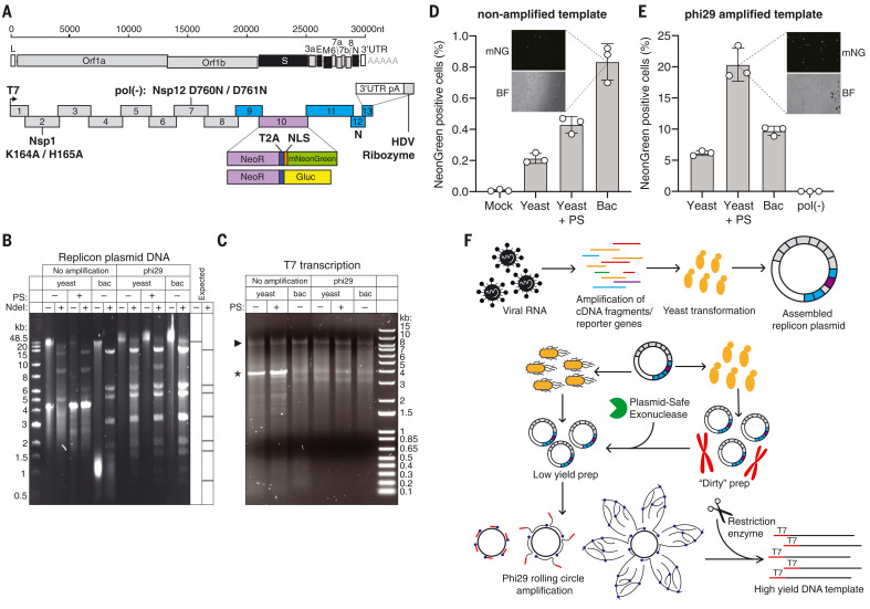
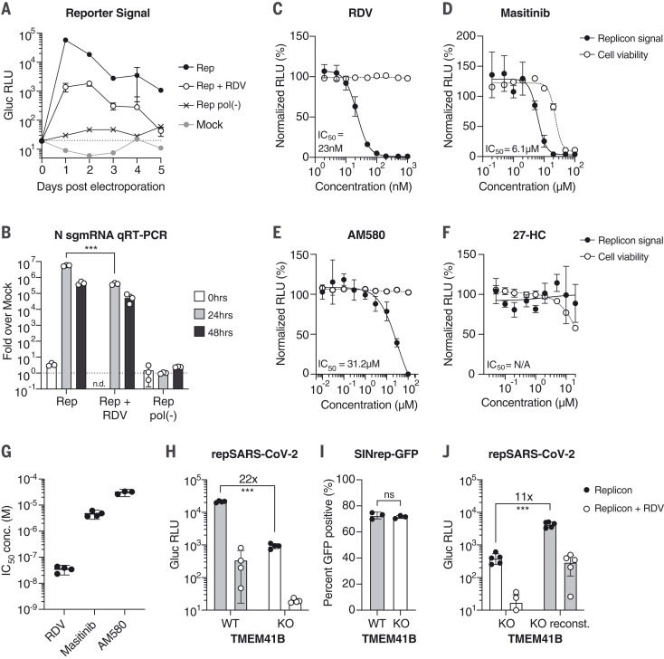
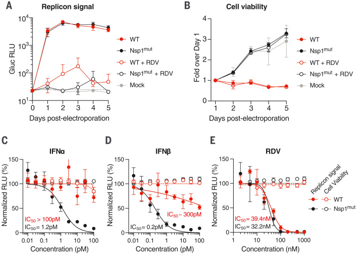
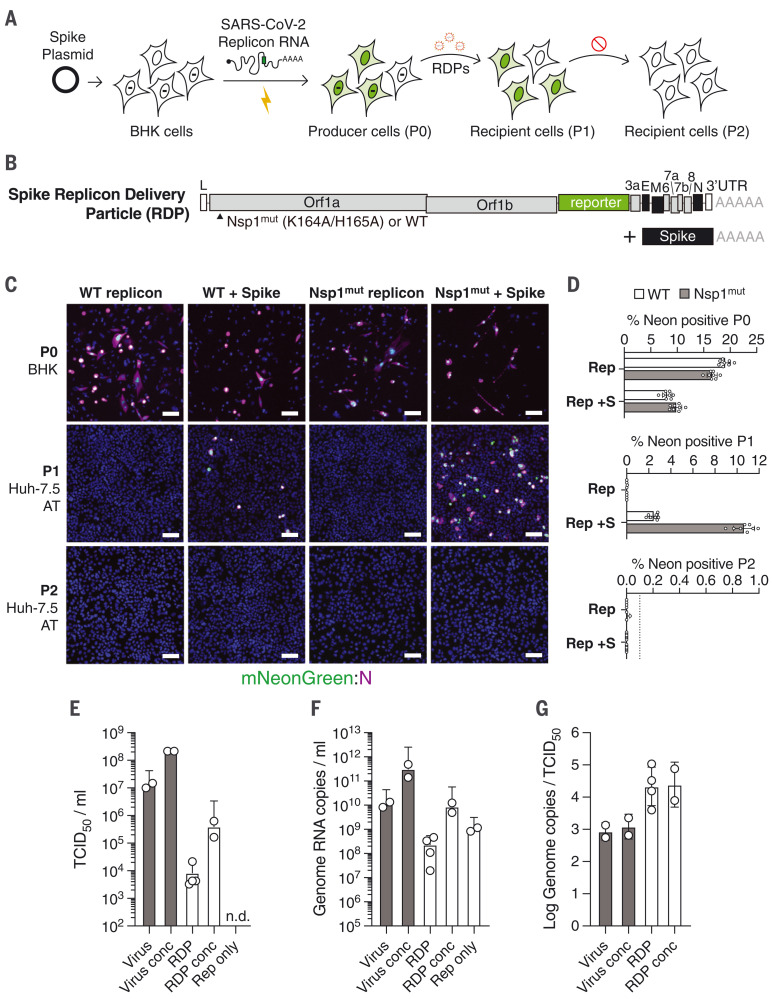
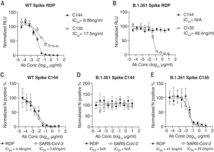
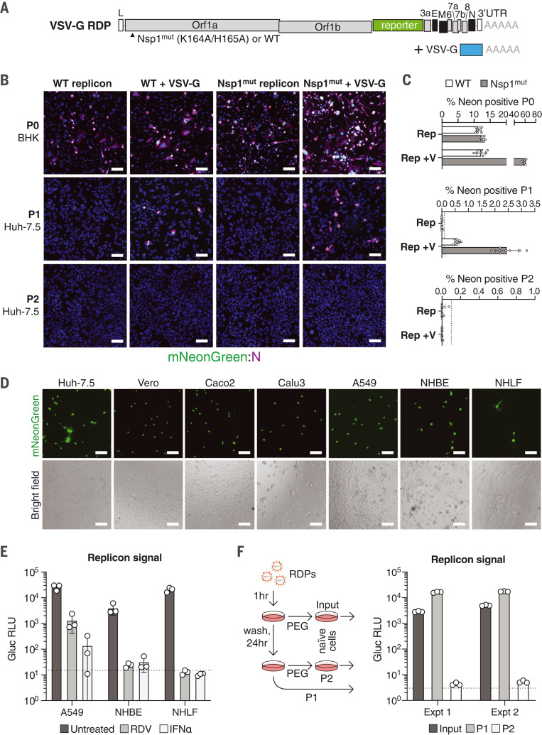
Molecular virology tools are critical for basic studies of the severe acute respiratory syndrome coronavirus 2 (SARS-CoV-2) and for developing new therapeutics. Experimental systems that do not rely on viruses capable of spread are needed for potential use in lower-containment settings. In this work, we use a yeast-based reverse genetics system to develop spike-deleted SARS-CoV-2 self-replicating RNAs. These noninfectious self-replicating RNAs, or replicons, can be trans-complemented with viral glycoproteins to generate replicon delivery particles for single-cycle delivery into a range of cell types. This SARS-CoV-2 replicon system represents a convenient and versatile platform for antiviral drug screening, neutralization assays, host factor validation, and viral variant characterization.


Figures

Fig. 1.
Fig. 1.. SARS-CoV-2 replicon design and launch optimization.
(A) (Upper schematic) SARS-CoV-2 genome, with structural proteins in black. (Lower schematic) Replicon amplicon fragments for yeast assembly. Fragments from (10) are shown in gray; fragments harboring mutations in Nsp1 or Nsp12 [pol(-)] are marked as such. The reporter gene cassette in place of spike is shown in purple; reengineered flanking regions are in blue. nt, nucleotides; L, leader; UTR, untranslated region; pA: polyA, HDV, hepatitis delta virus; NLS, nuclear localization sequence. (B) Agarose gel of replicon DNA recovered from yeast or bacteria (bac). Phi29 amplification or plasmid-safe (PS) DNAse treatment is indicated. Expected NdeI digest is depicted at right. (C) Agarose gel of T7 RNA transcription reactions from the DNA plasmids in (B). The arrowhead indicates the expected size of full-length RNA; the asterisk denotes truncated product. (D and E) Percent of mNeonGreen replicon–positive BHK-21 cells from nonamplified (D) or phi29-amplified (E) DNA templates measured by flow cytometry. Insets show representative mNeonGreen (mNG) and bright-field (BF) images. N = 3 biological replicates. Error bars indicate SEM; “Mock” indicates no RNA electroporation. (F) Optimized RNA production for SARS-CoV-2 replicons. Overlapping PCR fragments are assembled in yeast and propagated in bacteria or yeast, in which case they are treated with PS DNAse. Subsequent phi29 amplification ensures full-length DNA template availability for transcription.
Fig. 2.
Fig. 2.. SARS-CoV-2 replicons are sensitive to antiviral compounds, host factor loss, and viral mutant phenotypes.
(A) Gaussia luciferase (Gluc) in Huh-7.5 supernatant from cells electroporated with Gluc replicon RNA (Rep), seeded with 100 nM remdesivir (RDV) or vehicle. Mock electroporation and pol(-) replicons were used as controls. The dashed line indicates the limit of detection. N = 4. Error bars indicate SD. RLU, relative light units. (B) qRT-PCR measurements for subgenomic N RNA for cells in (A). Signal from mock-infected cells was used for normalization (dashed line). N = 3. Error bars indicate SD. ***P < 0.001; n.d., not determined. sgmRNA, subgenomic mRNA. (C to F) Representative experiments in Huh-7.5 cells were electroporated with the Gluc replicon RNA and seeded with (C) remdesivir (N = 4), (D) masitinib (N = 4), (E) AM580 (N = 3), or (F) 27-hydroxycholesterol (27-HC) (N = 3). After 24 hours, Gluc signal in the supernatant (filled circles) and cell viability (empty circles) were measured and normalized to vehicle-treated cells. Error bars indicate SEM. (G) IC50 values from independent experiments using the compounds presented in (C) to (E). (H) Parental Huh-7.5 (WT) and clonal TMEM41B KO cells were electroporated as indicated in (A). Gluc was measured 24 hours after electroporation. N = 4. (I) Cells as in (H) were electroporated with SINrep-GFP alphavirus replicon RNA. After 24 hours, GFP-positive cells were quantified by flow cytometry. N = 3. (J) As in (H), using cells reconstituted with TMEM41B. N = 5. Error bars indicate SD. ***P < 0.001 (two-sided Student’s t test); ns, not significant.
Fig. 3.
Fig. 3.. Nsp1-deficient replicons are hypersensitive to interferons.
(A and B) Time course measurements of Gluc in the supernatant (A) or cell viability (B) of Huh-7.5 cells electroporated with WT or Nsp1 K164A/H165A double mutant (Nsp1mut) Gluc replicon RNA. Cells were seeded with 100 nM remdesivir or vehicle and were washed in phosphate-buffered saline 24 hours before each respective time-point collection. Mock-electroporated cells were used as controls for post-electroporation cell viability. The dashed line indicates the limit of detection. N = 4. Error bars indicate SD. (C to E) Huh-7.5 cells were electroporated with WT or Nsp1mut replicon RNA and seeded on 96-well plates containing the indicated concentrations of (C) IFNα, (D) IFNβ, or (E) remdesivir. Gluc activity (filled circles) and cell viability (empty circles) were measured 24 hours after electroporation. N = 4. Error bars indicate SD.
Fig. 4.
Fig. 4.. Trans-complementation of replicons with spike yields single-cycle SARS-CoV-2.
(A) A scheme to trans-complement replicons with ectopically expressed spike for single-cycle virion production. BHK-21 cells are transfected with a spike-encoding plasmid; 24 hours later, they are electroporated with ΔS mNeonGreen SARS-CoV-2 replicon RNA. Supernatant from these producer cells (P0) is collected and passed onto naïve recipient cells (P1), yielding reporter activity. A second round of passaging onto naïve recipient cells (P2) fails to propagate the replicon. (B) A spike trans-complemented replicon consists of spike-deleted replicon RNA alongside plasmid-driven spike expression. Nsp1 mutations relative to the WT sequence are indicated. (C) BHK-21 producer cells (P0) alone or transfected with a spike-encoding plasmid were electroporated with WT or Nsp1mut replicon RNAs. The RDPs in resulting supernatants were concentrated after 24 hours and passaged onto Huh-7.5 cells that overexpress ACE2 and TMPRSS2 (Huh-7.5 AT cells; P1 and P2), as in (A). Immunofluorescence images at 4× magnification of the mNeonGreen signal (green) and N antibody staining (magenta) are shown. Scale bars, 100 μm. (D) Quantification of the percentage of NeonGreen-positive cells in each passage for the results in (C). The dashed line denotes the lower limit of quantification. N = 8. Error bars indicate SD. (E) TCID50 per milliliter of independently prepared SARS-CoV2 and RDP stocks were calculated by end-point dilution assay on Huh-7.5 AT cells. Conc, stocks concentrated by PEG precipitation; n.d., not detected. (F) Genome RNA copies per milliliter from the virus and RDP stocks indicated in (E) were calculated by qRT-PCR. (G) The ratio between RNA copies per milliliter indicated in (F) and TCID50 per milliliter indicated in (E) was calculated to reflect specific infectivity. In (E) to (G), error bars indicate SD.
Fig. 5.
Fig. 5.. Neutralization assays with RDPs recapitulate authentic SARS-CoV-2 antibody phenotypes.
(A and B) Antibody neutralization assays in Huh-7.5 cells of Gluc RDPs trans-complemented with WA1/2020 spike (A) or the B.1.351 South African variant (B) in the presence of increasing concentrations of C144 and C135 neutralizing antibodies. Data are representative of two independent experiments and are normalized to infected cells without antibody. N = 3. Error bars indicate SEM. Ab Conc, antibody concentration; IC50, concentration resulting in 50% reduction in relative RLU values; N/A, not applicable. (C to E) Neutralization assays for SARS-CoV-2 RDPs or virus in the presence of increasing antibody concentrations. WT spike with C144 antibody (C), B.1.351 spike with C144 antibody (D), and B.1.351 with C135 antibody (E) are shown, representative of two independent experiments. Data are normalized to infected cells without antibody. N = 3. Error bars indicate SEM.
Fig. 6.
Fig. 6.. VSV-G pseudotyping for efficient SARS-CoV-2 replicon delivery.
(A) Schematic of the elements required for production of VSV-G RDPs. (B) BHK-21 producer cells (P0) alone or transfected with VSV-G were electroporated with WT or Nsp1mut replicons. After 48 hours, RDPs in supernatants were serially passaged onto Huh-7.5 cells (P1 and P2). mNeonGreen signal and N antibody staining is depicted at 4× magnification. Scale bars, 100 μm. (C) Percentage of mNeonGreen-positive cells in each passage from (B). N = 8. Error bars indicate SD; dashed lines indicate the lower limit of quantification. (D) Indicated cell lines were incubated with mNeonGreen VSV-G RDPs for 24 hours. Bright-field and fluorescent images were taken at 10× magnification. Scale bars, 100 μm. (E) NHBE, NHLF, and A549 cells were pretreated for 24 hours with 100 nM remdesivir or 100 pM IFNα and infected with Gluc VSV-G RDPs. Gluc activity was measured 24 hours after infection. N = 3. Error bars indicate SD; the dashed line represents the limit of detection. (F) Single-cycle infectivity of VSV-G RDPs. Supernatant from RDP-infected cells (P1) was read for Gluc activity and passaged onto naive cells (P2) after PEG concentration. Input supernatant serves as a positive control for concentrated input. Schema are shown at left and results at right for two experiments. The dashed line represents the limit of detection.

Similar articles

Cited by

References

    1. French R., Ahlquist P., Intercistronic as well as terminal sequences are required for efficient amplification of brome mosaic virus RNA3. J. Virol. 61, 1457–1465 (1987). 10.1128/jvi.61.5.1457-1465.1987 - DOI - PMC - PubMed
    1. Bredenbeek P. J., Frolov I., Rice C. M., Schlesinger S., Sindbis virus expression vectors: Packaging of RNA replicons by using defective helper RNAs. J. Virol. 67, 6439–6446 (1993). 10.1128/jvi.67.11.6439-6446.1993 - DOI - PMC - PubMed
    1. Lohmann V., Körner F., Koch J., Herian U., Theilmann L., Bartenschlager R., Replication of subgenomic hepatitis C virus RNAs in a hepatoma cell line. Science 285, 110–113 (1999). 10.1126/science.285.5424.110 - DOI - PubMed
    1. Blight K. J., Kolykhalov A. A., Rice C. M., Efficient initiation of HCV RNA replication in cell culture. Science 290, 1972–1974 (2000). 10.1126/science.290.5498.1972 - DOI - PubMed
    1. Thiel V., Herold J., Schelle B., Siddell S. G., Viral replicase gene products suffice for coronavirus discontinuous transcription. J. Virol. 75, 6676–6681 (2001). 10.1128/JVI.75.14.6676-6681.2001 - DOI - PMC - PubMed

Publication types

MeSH terms