. 2021 Oct 14;17(10):e1009967.
doi: 10.1371/journal.ppat.1009967. eCollection 2021 Oct.

Genetic targeting of Card19 is linked to disrupted NINJ1 expression, impaired cell lysis, and increased susceptibility to Yersinia infection

Affiliations

Genetic targeting of Card19 is linked to disrupted NINJ1 expression, impaired cell lysis, and increased susceptibility to Yersinia infection

Elisabet Bjanes et al. PLoS Pathog. .

Abstract

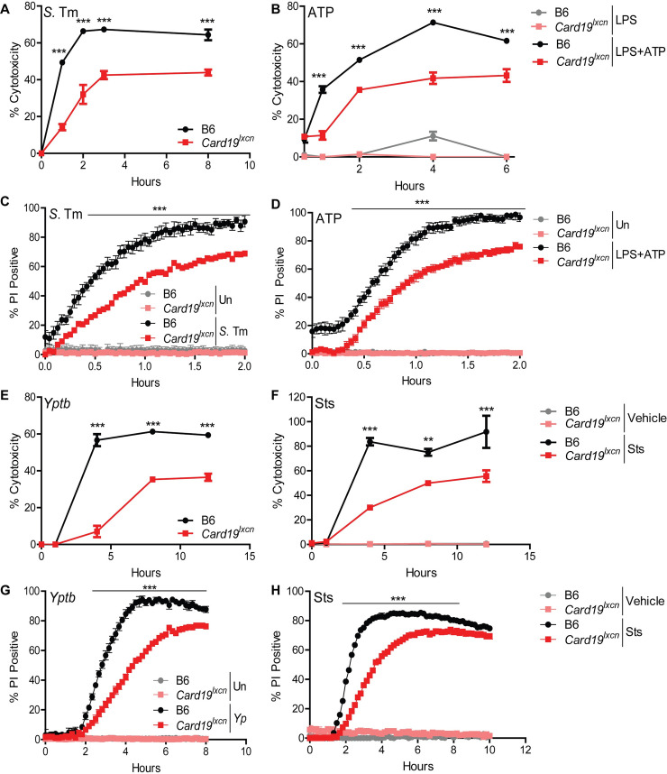
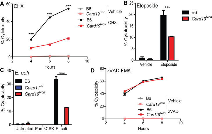
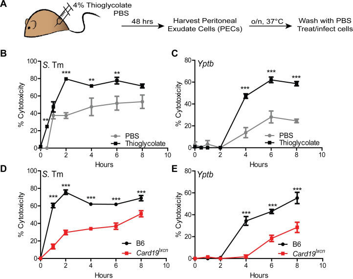
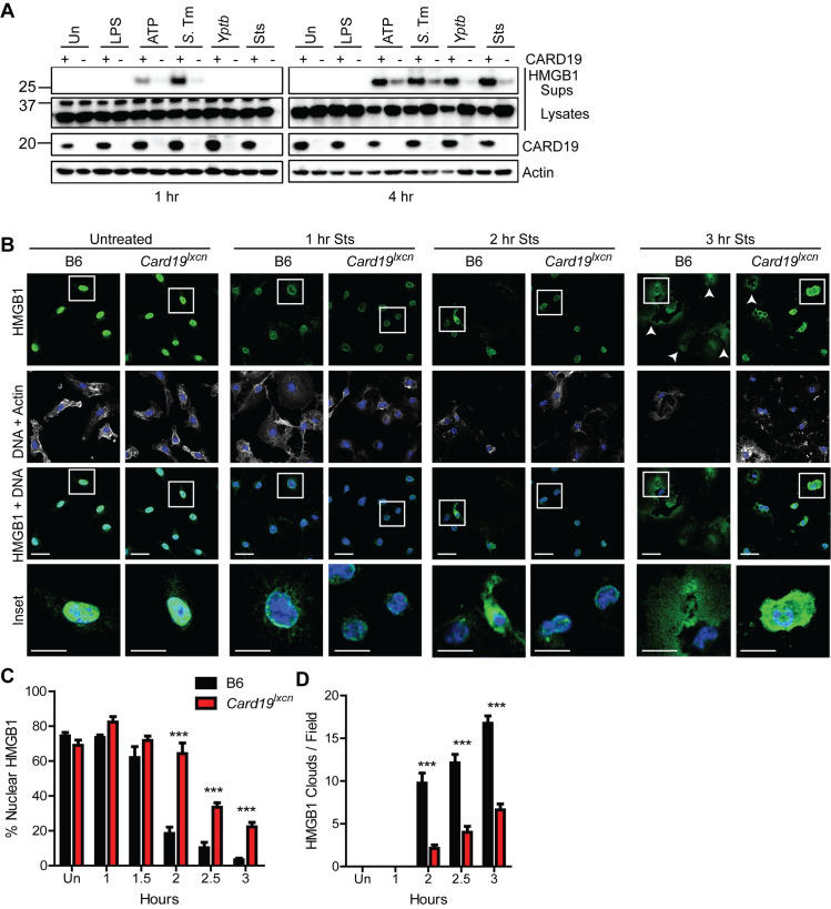
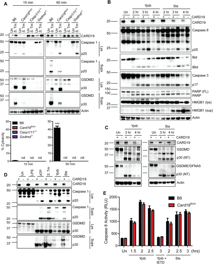
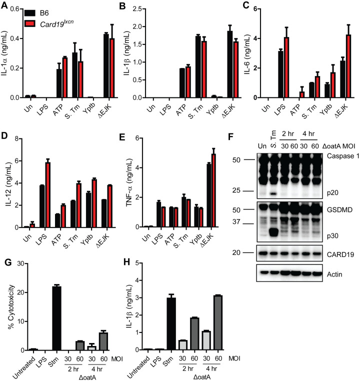
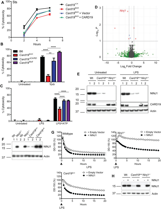
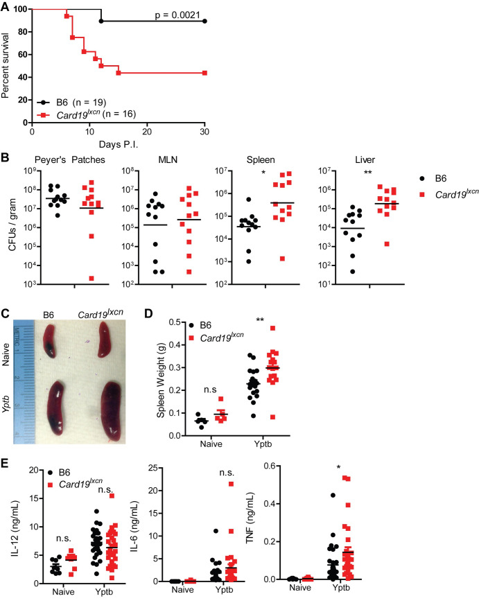
Cell death plays a critical role in inflammatory responses. During pyroptosis, inflammatory caspases cleave Gasdermin D (GSDMD) to release an N-terminal fragment that generates plasma membrane pores that mediate cell lysis and IL-1 cytokine release. Terminal cell lysis and IL-1β release following caspase activation can be uncoupled in certain cell types or in response to particular stimuli, a state termed hyperactivation. However, the factors and mechanisms that regulate terminal cell lysis downstream of GSDMD cleavage remain poorly understood. In the course of studies to define regulation of pyroptosis during Yersinia infection, we identified a line of Card19-deficient mice (Card19lxcn) whose macrophages were protected from cell lysis and showed reduced apoptosis and pyroptosis, yet had wild-type levels of caspase activation, IL-1 secretion, and GSDMD cleavage. Unexpectedly, CARD19, a mitochondrial CARD-containing protein, was not directly responsible for this, as an independently-generated CRISPR/Cas9 Card19 knockout mouse line (Card19Null) showed no defect in macrophage cell lysis. Notably, Card19 is located on chromosome 13, immediately adjacent to Ninj1, which was recently found to regulate cell lysis downstream of GSDMD activation. RNA-seq and western blotting revealed that Card19lxcn BMDMs have significantly reduced NINJ1 expression, and reconstitution of Ninj1 in Card19lxcn immortalized BMDMs restored their ability to undergo cell lysis in response to caspase-dependent cell death stimuli. Card19lxcn mice exhibited increased susceptibility to Yersinia infection, whereas independently-generated Card19Null mice did not, demonstrating that cell lysis itself plays a key role in protection against bacterial infection, and that the increased infection susceptibility of Card19lxcn mice is attributable to loss of NINJ1. Our findings identify genetic targeting of Card19 being responsible for off-target effects on the adjacent gene Ninj1, disrupting the ability of macrophages to undergo plasma membrane rupture downstream of gasdermin cleavage and impacting host survival and bacterial control during Yersinia infection.


Conflict of interest statement

I have read the journal’s policy and the authors of this manuscript have the following competing interests: Opher S. Kornfeld and Bettina L. Lee are employees of Genentech.

Figures

Fig 1
Fig 1. Card19lxcn BMDMs are deficient for caspase-dependent cell death.
Primary C57BL/6J (B6) or Card19lxcn BMDMs were infected with Salmonella enterica serovar Typhimurium (S. Tm) or Y. pseudotuberculosis (Yptb), or treated with LPS+ATP or staurosporine (Sts), as indicated, and the kinetics of cell death was assayed by release of lactate dehydrogenase (LDH) (A, B, E, F) or propidium iodide (PI) uptake (C, D, G, H). Each figure is representative of three or more independent experiments. LDH release was assayed at specific times post-infection. PI uptake was measured over a two or ten-hour timecourse with fluorometric measurements taken at 2.5 minute (C, D) or 10 minute (G, H) intervals. Mean ± SEM of triplicate wells is displayed. Each panel is representative of three or more independent experiments. *** p < 0.001, ** p < 0.01, * p < 0.05. n.s. not significant, analyzed by 2-way ANOVA with Bonferroni multiple comparisons post-test.
Fig 2
Fig 2. Card19lxcn BMDMs are deficient in apoptosis and non-canonical inflammasome-induced pyroptosis but not necroptosis.
Primary C57BL/6J (B6), Card19lxcn, Card19+/+, and Casp11-/- BMDMs were treated with (A) Cycloheximide (CHX) (B) etoposide, (C) Pam3CSK+E. coli or (D) z-VAD-FMK + LPS and cell death was assayed by LDH release. BMDMs were treated, supernatants were harvested from triplicate wells at (A) (D) indicated time points (B) 24 hours, or (C) 16 hours and measured for cytotoxicity. Mean ± SEM of triplicate wells is displayed. Each figure is representative of 2 or more independent experiments. *** p < 0.001, ** p < 0.01, * p < 0.05. n.s. not significant. 2-way ANOVA with Bonferroni multiple comparisons post-test.
Fig 3
Fig 3. Peritoneal macrophages from Card19lxcn mice have reduced levels of cell death.
(A) B6 and Card19lxcn mice were injected with 4% aged thioglycolate or PBS. 48 hrs later, PECS were harvested, RBC lysed, counted and plated in triplicate overnight at 37°C. PBS PECS were pooled prior to plating. Cells were washed with PBS to remove non-adherent cells and infected with Yptb or S. Tm (MOI 10) or treated with staurosporine (10 uM). Cytotoxicity was measured by LDH release at indicated time points. *** p < 0.001, ** p < 0.01, * p < 0.05. n.s. not significant. 2-way ANOVA with Bonferroni multiple comparisons post-test. Graphs are representative of two independent experiments with 3–6 mice per group, per genotype. (B, C) PBS: mean ± SEM for technical replicates of 3 pooled B6 mice. Thioglycolate: means of technical replicates for 3 B6 mice ± SEM. Cells were infected with (B) S. Tm or (C) Yptb. (D, E) PBS: mean ± SEM for technical replicates of 4–6 pooled B6 and Card19lxcn mice + SEM. Thioglycolate: means of technical replicates for 4 B6 and Card19lxcn mice ± SEM. Cells were infected with (B) S. Tm or (C) Yptb.
Fig 4
Fig 4. Card19lxcn BMDMs retain intracellular alarmin HMGB1 following activation of cell death.
(A) Primary BMDMs from B6 (+) or Card19lxcn (-) mice were treated with indicated treatments or infections, and TCA precipitated supernatants (Sups) or whole cell lysates (Lysates) were harvested at 1 or 4 hours post-infection and analyzed by western blotting for HMGB1, CARD19, and actin (loading control). ATP indicates cells that were primed with LPS for 3 hours and treated with ATP for 1 or 4 hours. S.Tm–Salmonella Typhimurium; Yptb Yersinia pseudotuberculosis. Sts–staurosporine. (B) Confocal microscopy images of untreated and staurosporine (Sts) treated B6 and Card19lxcn BMDMs fixed at indicated times post-Sts treatment and stained for HMGB1, Actin and DNA (Hoescht). White arrows indicate HMGB1 clouds. Scale bar 20 microns, inset 10 microns. (C) Quantification of nuclear HMGB1. n = 25–50 cells per field, 5–8 fields per condition, per timepoint. (D) Quantification of HMGB1 clouds in B6 and Card19lxcn BMDMs after staurosporine treatment. 5–8 fields quantified per condition, per timepoint. Mean ± SEM is displayed. *** p < 0.001, ** p < 0.01, * p < 0.05. n.s. not significant. 2-way ANOVA with Bonferroni multiple comparisons post-test. Representative of 3 (A-C) or 2 (D) independent experiments.
Fig 5
Fig 5. Card19lxcn macrophages are not deficient in activation of caspase-1 or -8.
(A) B6, Card19lxcn, Casp1/11-/- or Gsdmd-/- BMDMs were left untreated or infected with S. Tm, and cell lysates (Lys) and TCA-precipitated supernatants (Sups) were run on SDS-PAGE and analyzed by western blotting for cleaved Casp1, GSDMD, or Actin (cell lysate loading control). Cell death from these cells was assayed in parallel by LDH release (indicated below blots). Mean ± SEM is displayed. 2-way ANOVA with Bonferroni multiple comparisons post-test. *** p < 0.001, ** p < 0.01, * p < 0.05. n.d. not detectable. (B) B6 (+) or Card19lxcn (-) BMDMs were infected with Yptb or treated with Sts or 2, 3, or 4 hours, or left untreated (Un) as indicated. Cell lysates and TCA-supernatants (HMGB1 sup) were prepared at indicated times and analyzed for presence of CARD19, cleaved caspase-8, tBid, cleaved caspase-3, cleaved PARP, HMGB1 and Actin (loading control). (C) B6 and Card19lxcn BMDMs were left untreated, infected with Yptb or treated with staurosporine. Lysates were harvested at the indicated time points, run on SDS-PAGE and analyzed by western blotting for CARD19, GSDMD, GSDME/DFNA5, and actin (loading control). (D) B6 (+) and Card19lxcn (-) BMDMs were left untreated, treated with LPS, LPS+ATP, Sts, or infected with S. Tm or Yptb. Cell lysates (Lys) and supernatants (Sups) were harvested 1 hour post-ATP or S.Tm infection or 3 hours post Sts treatment and Yptb infection and assayed for CARD19, Caspase-1, and presence of cleaved GSDMD. (E) B6 and Card19lxcn BMDMs were left untreated, infected with Yptb, or treated with staurosporine. BMDMs were pretreated with the caspase-8 inhibitor IETD for one hour prior to infection. Caspase-8 activity was assessed by Caspase-8-Glo. Mean ± SEM is displayed. Blots, caspase-8 activity and LDH are representative of at least 3 independent experiments.
Fig 6
Fig 6. The cell death defect in Card19lxcn BMDMs is independent from cytokine release.
(A-E) B6 (black bars) and Card19lxcn (red bars) BMDMs were primed with LPS for 3 hours and treated with extracellular ATP, infected with S. Tm, Yptb, or the Yptb ΔEJK strain lacking effectors YopE, YopJ and YopK. Supernatants were harvested 2 hours post-infection or ATP treatment and analyzed by ELISA for release of (A) IL-1α, (B) IL-1B, (C) IL-6, (D) IL-12, and (E) TNF-α. Mean ± SEM is displayed. Each figure is representative of 3 or more independent experiments. (F-H) B6 BMDMs were primed with LPS for 4 hours and infected with ΔoatA S. aureus or S. Tm. (F) Lysates were harvested at indicated times, run on SDS-PAGE page and analyzed by western blotting for Caspase 1, GSDMD, CARD19, or Actin (loading control). Supernatants were harvested at indicated time points and analyzed by (G) LDH for cytotoxicity and (H) ELISA for release of IL-1β. Each figure is representative of two independent experiments.
Fig 7
Fig 7. Card19lxcn BMDMs are hypomorphic for Ninj1 and reconstitution with Ninj1 restores cell death.
(A) Card19lxcn immortalized murine progenitors were stably transduced with CARD19 or empty vector. Mature macrophages derived from untransduced immortalized progenitors Card19lxcn and Card19+/+ and transduced immortalized progenitors Card19lxcn + Vector and Card19lxcn + CARD19 were treated with sts. Cell death was assayed by LDH release at indicated time points. Representative of three independent experiments. (B,C) B6, Card19lxcn, Card19ΔCARD, and Card19Null BMDMs were treated with (B) Yptb or (C) LPS+ATP. Cell death was assayed by LDH release. Representative of three independent experiments. 2-way ANOVA with Bonferroni multiple comparisons post-test. **** p < 0.0001 *** p < 0.001 ** p < 0.01, * p < 0.05, n.s. not significant. (D) Volcano plot from RNA-seq analysis on untreated B6 and Card19lxcn BMDMs showing differentially regulated genes. Genes whose expression is significantly altered are in red. Ninj1 is marked. (E) BMDMs from B6, Card19lxcn, and Ninj1-/- mice were primed with LPS or left untreated. Cell lysates were harvested, run on SDS-PAGE and analyzed by western blotting for CARD19, NINJ1, and Actin (cell lysate loading control). (F) BMDMs from B6, Card19lxcn, Ninj1-/-, and Card19Null mice were left untreated. Cell lysates were harvested, run on SDS-PAGE and analyzed by western blotting for NINJ1, and Actin (cell lysate loading control). (G, H) Wildtype, Ninj1-/- and Card19lxcn iBMDMs were reconstituted with NINJ1 or empty vector. (G) Release of dextran dye-150 (DD-150) in live cell imaging analysis following LPS electroporation over a 20 hour time course. (H) Lysates of reconstituted cells were run on SDS-PAGE and analyzed by western blotting for NINJ1 and actin (cell lysate loading control). (E-H) Representative of two independent experiments.
Fig 8
Fig 8. NINJ1 mediates anti-Yersinia host defense.
(A) Survival of B6 and Card19lxcn mice following oral infection 108 CFUs of strain IP2777 Yptb. Data are pooled from two independent experiments that each gave similar results. Log-rank (Mantel-Cox) Survival test. (B) Bacterial CFUs per gram tissue in Peyer’s Patches, MLN, Spleen and Liver in B6 and Card19lxcn mice seven days post-oral infection with Yptb. Representative of two (PP) or four (MLN, Spleen, Liver) independent experiments. Unpaired Student’s t-test. ** p < 0.01, * p < 0.05. (C) Representative images of naive and Yptb-infected B6 and Card19lxcn spleens, seven days post-infection Representative of three independent experiments. (D) Quantification of naive and infected spleen weights. Data are pooled from three independent experiments. Naive (n = 5), Yptb (B6 n = 19, Card19lxcn n = 16) 2-way ANOVA with Bonferroni multiple comparisons post-test. ** p < 0.01. (E) Serum cytokines from naive and Yptb infected B6 and Card19lxcn mice. Data are pooled from four independent experiments. Naïve (n = 8), Yptb (B6 n = 31, Card19lxcn n = 28) 2-way ANOVA with Bonferroni multiple comparisons post-test. * p < 0.05, n.s. not significant.

References

    1. Mori C, Nakamura N, Kimura S, Irie H, Takigawa T, Shiota K. Programmed cell death in the interdigital tissue of the fetal mouse limb is apoptosis with DNA fragmentation. Anat Rec. 1995;242(1):103–10. doi: 10.1002/ar.1092420114 . - DOI - PubMed
    1. Rodriguez I, Ody C, Araki K, Garcia I, Vassalli P. An early and massive wave of germinal cell apoptosis is required for the development of functional spermatogenesis. EMBO J. 1997;16(9):2262–70. doi: 10.1093/emboj/16.9.2262 ; PubMed Central PMCID: PMC1169828. - DOI - PMC - PubMed
    1. Kajstura J, Cheng W, Reiss K, Clark WA, Sonnenblick EH, Krajewski S, et al.. Apoptotic and necrotic myocyte cell deaths are independent contributing variables of infarct size in rats. Lab Invest. 1996;74(1):86–107. . - PubMed
    1. Mariathasan S, Newton K, Monack DM, Vucic D, French DM, Lee WP, et al.. Differential activation of the inflammasome by caspase-1 adaptors ASC and Ipaf. Nature. 2004;430(6996):213–8. Epub 2004/06/11. doi: 10.1038/nature02664 nature02664 [pii]. . - DOI - PubMed
    1. Miao EA, Alpuche-Aranda CM, Dors M, Clark AE, Bader MW, Miller SI, et al.. Cytoplasmic flagellin activates caspase-1 and secretion of interleukin 1beta via Ipaf. Nat Immunol. 2006;7(6):569–75. Epub 2006/05/02. ni1344 [pii]. doi: 10.1038/ni1344 . - DOI - PubMed

Publication types

MeSH terms

Substances