. 2021 Oct 14;12(10):948.
doi: 10.1038/s41419-021-04253-y.

CircNPHP4 in monocyte-derived small extracellular vesicles controls heterogeneous adhesion in coronary heart atherosclerotic disease

Affiliations

CircNPHP4 in monocyte-derived small extracellular vesicles controls heterogeneous adhesion in coronary heart atherosclerotic disease

Feng Xiong et al. Cell Death Dis. .

Abstract

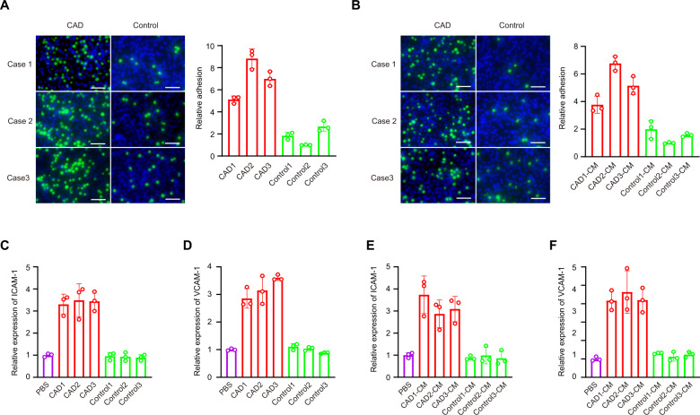
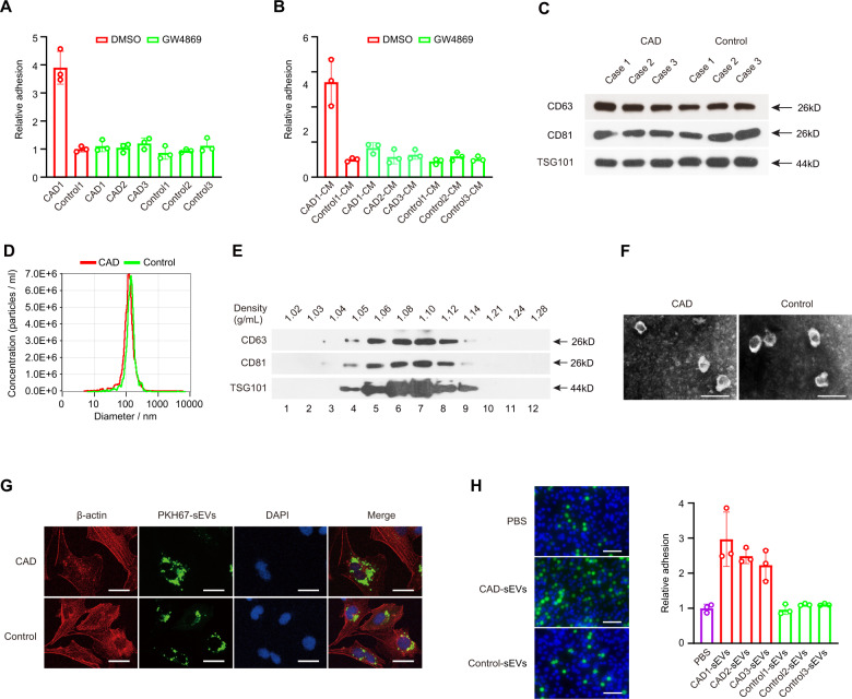
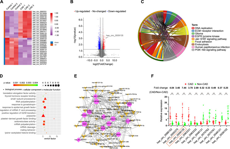
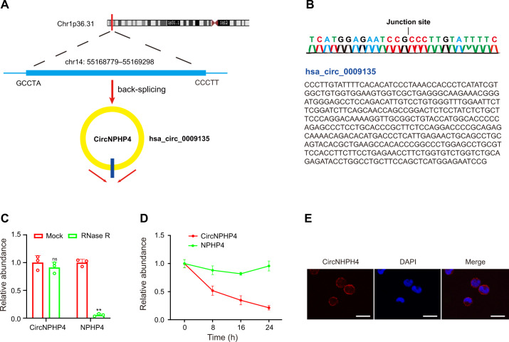
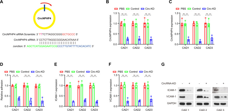
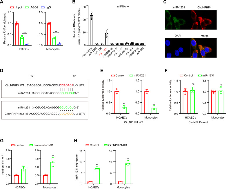
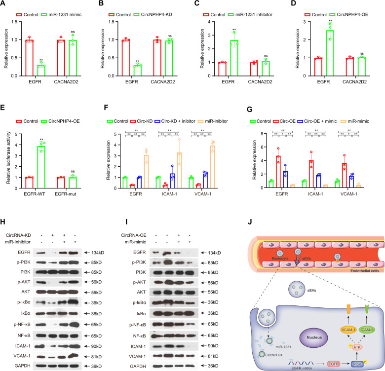
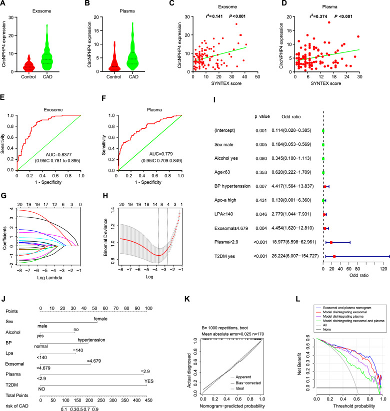
Small extracellular vesicles (sEVs)-derived circular RNAs (circRNAs) could regulate gene expression in recipient cells, and dysregulation of sEVs-derived circRNAs has been implicated in several diseases. However, the expression and function of sEVs-derived circRNAs in coronary heart atherosclerotic disease (CAD) remain unknown. In this study, we investigated global changes in the expression patterns of circRNAs in sEVs from CAD-related monocytes and identified circNPHP4 as a significantly upregulated circRNA. Knockdown of circNPHP4 inhibited heterogeneous adhesion between monocytes and coronary artery endothelial cells and reduced ICAM-1 and VCAM-1 expression. Investigations of the underlying mechanisms revealed that circNPHP4 contains a functional miR-1231-binding site. Mutation of the circNPHP4-binding sites in miR-1231 abolished the interaction, as indicated by a luciferase reporter assay. Furthermore, circNPHP4 affected the expression of miR-1231 and its target gene EGFR. Overexpression of miR-1231 blocked the inhibitory effect of circNPHP4 on heterogeneous adhesion. Moreover, downregulation of miR-1231 restored heterogeneous adhesion upon inhibition by circNPHP4 silencing. Additionally, circNPHP4 overexpression was correlated with aggressive clinicopathological characteristics in CAD patients. A multivariate logistic regression model and bootstrapping validation showed that circNPHP4 overexpression had a good risk prediction capability for CAD. The decision curve analysis revealed that using the CAD nomogram that included circNPHP4 overexpression to predict the risk of CAD was beneficial. Our results suggest that sEVs-derived circNPHP4 can serve as a potential target for CAD treatments or as a potential diagnostic marker for CAD patients.


Conflict of interest statement

The authors declare no competing interests.

Figures

Fig. 1
Fig. 1. Heterogeneous adhesion ability of the CAD-derived monocytes was increased compared with the adhesion ability of the control monocytes.
Heterogeneous adhesion ability of monocytes to HCAECs was analyzed using monocytes (A) or monocytes culture medium (CM) (B). ICAM-1 (C) and VCAM-1 (D) expression in HCAECs cocultured with monocytes were analyzed using qRT-PCR. ICAM-1 (E) and VCAM-1 (F) expression in HCAECs cocultured with monocytes culture medium were analyzed using qRT-PCR. Scale bar = 50 µm. Data are presented as means ± SD; a significant difference was identified with Student’s t test. *P < 0.05; **P < 0.01. ns (not significant).
Fig. 2
Fig. 2. sEVs from monocytes of CAD patients promoted heterogeneous adhesion.
Heterogeneous adhesion ability of monocytes to HCAECs was analyzed using monocytes (A) or monocytes culture medium (CM) (B) following GW4869 exposure. sEVs from monocytes were extracted to be verified using western blot assay (C) and the NTA method (D). CD63, CD81, and TSG101 expression were detected in fractions collected from OptiPrepTM density gradient centrifugation using western blot assay (E). sEVs obtained from fraction 7 (density 1.10 g/mL) was verified using transmission electron microscopy (F). Scale bar = 200 nm. G HCAECs were incubated directly with sEVs from CAD patients or control-related monocytes. Scale bar = 20 µm. H Heterogeneous adhesion ability of monocytes to HCAECs was analyzed using CAD-sEVs or Control-sEVs. Scale bar = 20 µm. Data are presented as means ± SD; a significant difference was identified with Student’s t test. *P < 0.05; **P < 0.01; ns (not significant).
Fig. 3
Fig. 3. Identification of differentially expressed circRNAs in sEVs from CAD patient monocytes.
A Clustered heatmap of the differentially expressed circRNAs in sEVs from monocytes. Upregulated circRNAs are shown in red and downregulated circRNAs are shown in green. B Volcano plots comparing circRNA expression between CAD patient and control. The red dots represent the significantly differentially expressed circRNAs (fold-change ≥1.5 and P < 0.01). C Top 15 classes of KEGG pathway enrichment terms. D Top 15 classes of disease enrichment terms (E) CircRNA-miRNA-mRNA network and pathway analysis. F The differential expression of 10 circRNAs in sEVs was validated in 10 monocytes from CAD patients and 10 monocytes from control using qRT-PCR. Data are presented as means ± SD; significant difference was identified with Student’s t test. *P < 0.05; **P < 0.01; ns (not significant).
Fig. 4
Fig. 4. Characterization of circNPHP4 in monocytes.
A The genomic location of the hNPHP4 gene and of circNPHP4. B Sanger sequencing showing the “head-to-tail” splicing of circNPHP4 in the monocytes. C qRT-PCR quantification of circNPHP4 and hNPHP4 mRNA expression in monocytes after treatment with RNase R. D qRT-PCR quantification of circNPHP4 and hNPHP4 mRNA expression in monocytes after treatment with Actinomycin D. Data are presented as means ± SD; significant difference was identified with Student’s t test. *P < 0.05; **P < 0.01; ns (not significant). E RNA FISH for circNPHP4. Nuclei were stained with DAPI. Scale bar = 20 µm.
Fig. 5
Fig. 5. CircNPHP4 from monocyte-derived sEVs promoted heterogeneous adhesion.
A Schematic representation of the siRNA sites specific to the back-splice junction of circNPHP4. Expression of circNPHP4 following siRNA treatment using qRT-PCR in monocytes (B) and in sEVs from monocytes (C). Heterogeneous adhesion was analyzed after transfection with circNPHP4 specific siRNA versus scramble controls (D). ICAM-1 (E) and VCAM-1 (F) were detected in HCAECs exposed to sEVs from circNPHP4-knockdown monocytes, as quantified by qRT-PCR and western blot analyses (G). Data are presented as means ± SD; a significant difference was identified with Student’s t test. *P < 0.05; **P < 0.01; ns (not significant).
Fig. 6
Fig. 6. CircNPHP4 acted as a miRNAs sponge for miR-1231.
A RIP was performed using an antibody against AGO2 on extracts from monocytes and HCAECs. B CircNPHP4 was performed using a circNPHP4-specific probe and control probe in HCAECs. The enrichment of circNPHP4 and microRNAs were detected by qRT-PCR and normalized to the control probe. C Co-localization between circNPHP4 and miR-1231 was observed by RNA in situ hybridization in HCAECs. Nuclei were stained with DAPI. Scale bar = 20 µm. D Schematic showing the predicted miR-1231 sites in circNPHP4. A Luciferase assay where monocytes were co-transfected with a scrambled control, miR-1231 mimic, and a luciferase reporter plasmid containing wild-type circNPHP4 (circNPHP4-WT) (E) or mutant circNPHP4 (circNPHP4-mut) (F). G qRT-PCR showed the level of circNPHP4 in the streptavidin-captured fractions from the monocytes and HCAECs lysates after transfection with biotinylated miR-1231 or control RNA. H Expression of miR-1231 was analyzed using qRT-PCR following circNPHP4 knockdown. Data are presented as means ± SD; a significant difference was identified with Student’s t test. *P < 0.05; **P < 0.01; ns (not significant).
Fig. 7
Fig. 7. CircNPHP4 promoted the EGFR/PI3K/AKT pathway through miR-1231.
CircNPHP4 promoted the EGFR/PI3K/AKT pathway through miR-1231 inhibition in HCAECs qRT-PCR quantification of EGFR and CACNA2D2 expression after miR-1231 overexpression (A) or circNPHP4 knockdown (B). EGFR and CACNA2D2 expression after transfection with miR-1231 inhibitor (C) or circNPHP4 expression vector (D) was quantified with qRT-PCR. Luciferase assay where HCAEC were co-transfected with a scrambled control, circNPHP4 expression plasmid, and a luciferase reporter plasmid containing either wild-type EGFR (EGFR-WT) or an EGFR construct with mutated miR-1231 binding sites (EGFR-mut) (E). F–I Reversion assays using vectors overexpressing or knocking down circNPHP4, as well as miR-1231 mimics or inhibitors. Data are presented as means ± SD; a significant difference was identified with Student’s t test. *P < 0.05; **P < 0.01; ns (not significant). J A proposed model illustrating the role of monocyte-derived sEVs circNPHP4 in regulating heterogeneous adhesion in vein endothelial cells.
Fig. 8
Fig. 8. CircNPHP4 upregulation in serum predicts aggressive clinicopathological characteristics.
A CircNPHP4 expression in serum from 109 CAD patients and 70 control. B CircNPHP4 expression in serum from 109 CAD patients and 70 control. C Pearson correlation between circNPHP4 expression and SYNTEX score in serum of CAD patients. D Pearson correlation between circNPHP4 expression and SYNTEX score in serum of CAD patients. E ROC curve for serum circNPHP4 that indicates a diagnostic value in CAD patients. F ROC curve for serum circNPHP4 that indicates a diagnostic value in CAD patients. G Lambda in the CAD and control cohort. H LASSO coefficient profiles of the 20 features. A coefficient profile plot was produced against the log (lambda) sequence. A vertical line was drawn at the value selected using fivefold cross-validation, where optimal lambda resulted in 9 features with nonzero coefficients. I Logistic regression analysis in the CAD and control cohort. J Nomogram using the circNPHP4 upregulation to estimate the diagnosis rate of CAD. K Calibration curves of the diagnosis prediction nomogram in the primary cohorts. L Decision curve analysis for the CAD nomogram in the cohort. The y-axis measures the net benefit. The black line represents the CAD risk nomogram. The thin solid line represents the assumption that all patients have CAD. The thick solid line represents the assumption that all patients are in Non-CAD. The decision curve shows that if the threshold probability of a patient and a doctor is >2% and <96%, respectively, using this CAD nomogram in the current study to predict CAD risk adds more benefit than the scheme.

References

    1. Hansson GK. Inflammation, atherosclerosis, and coronary artery disease. N Engl J Med. 2005;352:1685–95. doi: 10.1056/NEJMra043430. - DOI - PubMed
    1. Libby P, Ridker PM, Maseri A. Inflammation and atherosclerosis. Circulation. 2002;105:1135–43. doi: 10.1161/hc0902.104353. - DOI - PubMed
    1. Libby P, Buring JE, Badimon L, Hansson GK, Deanfield J, Bittencourt MS, et al. Atherosclerosis. Nat Rev Dis Prim. 2019;5:56. doi: 10.1038/s41572-019-0106-z. - DOI - PubMed
    1. Mirtschink P, Jang C, Arany Z, Krek W. Fructose metabolism, cardiometabolic risk, and the epidemic of coronary artery disease. Eur Heart J. 2018;39:2497–505. doi: 10.1093/eurheartj/ehx518. - DOI - PMC - PubMed
    1. Kristensen LS, Andersen MS, Stagsted L, Ebbesen KK, Hansen TB, Kjems J. The biogenesis, biology and characterization of circular RNAs. Nat Rev Genet. 2019;20:675–91. doi: 10.1038/s41576-019-0158-7. - DOI - PubMed

Publication types

MeSH terms