. 2021 Oct 14;6(1):63.
doi: 10.1038/s41536-021-00175-7.

Pain sensing neurons promote tissue regeneration in adult mice

Affiliations

Pain sensing neurons promote tissue regeneration in adult mice

Lise Rabiller et al. NPJ Regen Med. .

Abstract

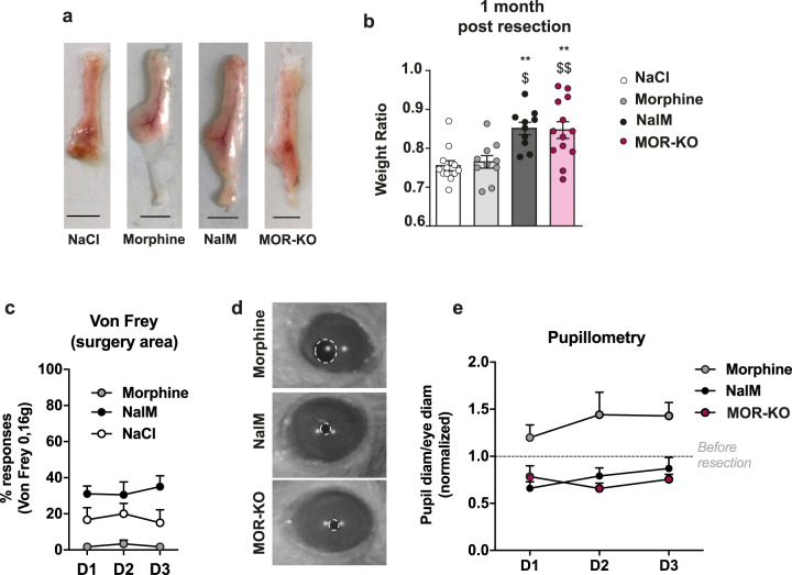
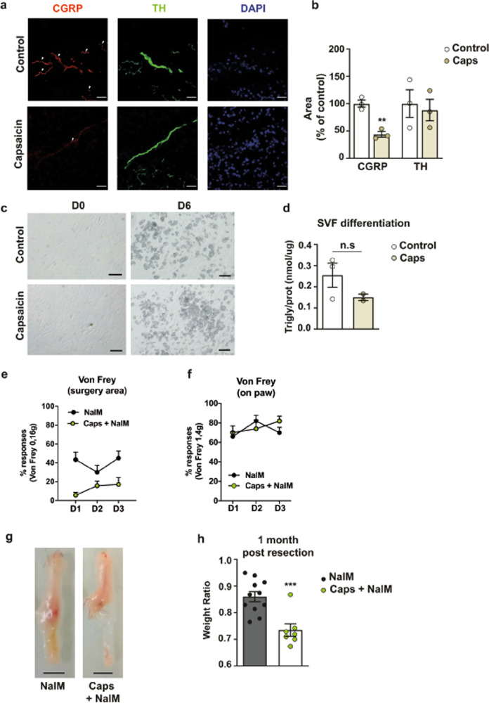
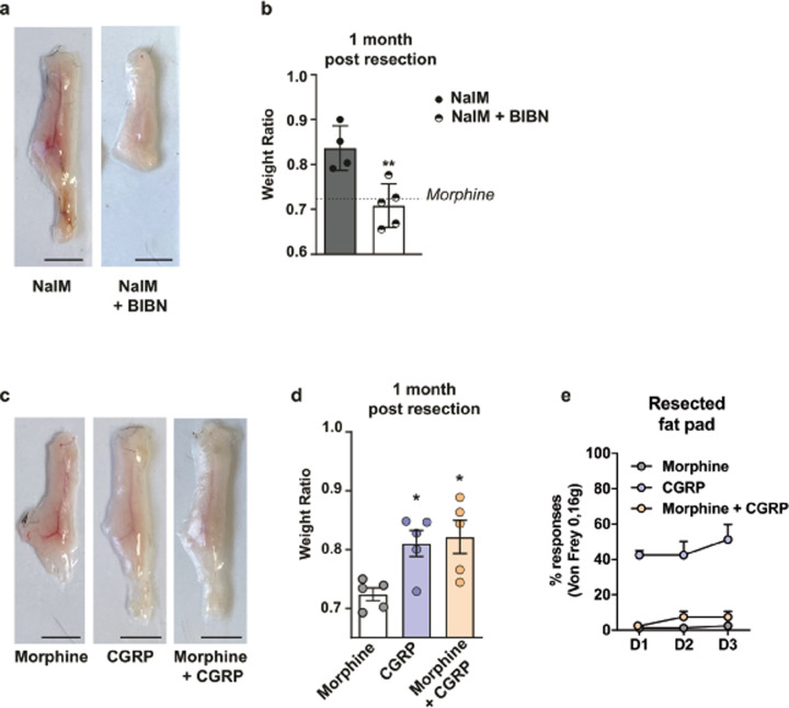
Tissue repair after injury in adult mammals, usually results in scarring and loss of function in contrast to lower vertebrates such as the newt and zebrafish that regenerate. Understanding the regulatory processes that guide the outcome of tissue repair is therefore a concerning challenge for regenerative medicine. In multiple regenerative animal species, the nerve dependence of regeneration is well established, but the nature of the innervation required for tissue regeneration remains largely undefined. Using our model of induced adipose tissue regeneration in adult mice, we demonstrate here that nociceptive nerves promote regeneration and their removal impairs tissue regeneration. We also show that blocking the receptor for the nociceptive neuropeptide calcitonin gene-related peptide (CGRP) inhibits regeneration, whereas CGRP administration induces regeneration. These findings reveal that peptidergic nociceptive neurons are required for adult mice tissue regeneration.


Conflict of interest statement

The authors declare no competing interests.

Figures

Fig. 1
Fig. 1. MORs mediate the deleterious effect of opioids on regenerative process.
a Representative pictures of scAT 1 month after resection, in C57Bl/6 mice treated with NaCl, morphine or NalM, and in MOR-KO mice (scale bars: 0.5 cm). b Quantification of regeneration. Weight ratio 1 month post-resection (n = 10–13 per group). c Quantification of nociceptive sensitivity using von Frey test. Mean frequency (±SEM) of withdrawal reflex after stimulation of the surgery area from day 1 (D1) to day 3 (D3) post-resection (n = 6–10 per group). d Representative pictures of pupils of C57Bl/6 mice treated with NalM or morphine, and of MOR-KO mice. e Quantification of nociceptive sensitivity using pupillary reflex test. Diameter ratio from day 1 (D1) to day 3 (D3) post-resection (n = 5–8 per group). NalM: naloxone methiodide. (*,$p < 0.05, **,$$p < 0.01. *: vs. NaCl, $: vs. morphine).
Fig. 2
Fig. 2. Selective denervation of nociceptive sensory neurons impairs tissue regeneration.
a Imaging of control or denervated scAT, 21 days after capsaicin injection, showing sensory fibres (CGRP, red), sympathetic fibres (TH, green) and nuclei (DAPI, blue) (scale bars: 0.5 cm). b Quantification of sensory (CGRP) and sympathetic fibres (TH) (n = 3). c Representative pictures of the stromal vascular fraction isolated from scAT 21 days after injection of capsaicin and placed in differentiation medium for 6 days (scale bars: 100 µm). d Quantification of triglycerides related to total proteins (nmol/µg) after 6 days of culture in differentiation medium (n = 2–3 per group). e, f Quantification of nociceptive sensitivity using von Frey test. Mean frequency (±SEM) of withdrawal reflex after stimulation of the surgery area (e) (n = 6–7 per group) or the paw (f) (n = 6–7 per group), from day 1 (D1) to day 3 (D3) post-resection, in mice treated with NalM and previously denervated with capsaicin or not. g Representative pictures of scAT 1 month after resection and morphine or NalM treatment in mice previously denervated with capsaicin or not (scale bar: 0.5 cm). h Quantification of regeneration. Weight ratio 1 month post-resection (n = 7–11 per group). CGRP: calcitonin gene-related peptide, TH: tyrosine hydroxylase, NalM: naloxone methiodide, Caps: capsaicin. (**p < 0.01, ***p < 0.001).
Fig. 3
Fig. 3. CGRP is sufficient to induce regeneration of the injured tissue and overcomes opioids’ scar healing effect.
a Representative pictures of scAT, 1 month after resection and NalM or NalM + BIBN treatment (scale bars: 0.5 cm). b Weight ratio quantification (n = 4–5 per group). The dotted line indicates the weight ratio in scar healing condition (morphine). c Representative pictures of scAT, 1 month after resection and morphine or CGRP or morphine + CGRP treatment (scale bars: 0.5 cm). d Weight ratio quantification (n = 5 per group). The dotted line indicates the weight ratio in scar healing condition (morphine). e Quantification of nociceptive sensitivity using von Frey test. Mean frequency (±SEM) of withdrawal reflex after stimulation of the surgery area from day 1 (D1) to day 3 (D3) post-resection (n = 5 per group). NalM: naloxone methiodide, BIBN: CGRP receptors antagonist BIBN-4096, CGRP: calcitonin gene-related peptide. (*p < 0.05, **p < 0.01).

References

    1. Eming SA, Martin P, Tomic-Canic M. Wound repair and regeneration: mechanisms, signaling, and translation. Sci. Transl. Med. 2014;6:265sr6. doi: 10.1126/scitranslmed.3009337. - DOI - PMC - PubMed
    1. Simoes MG, et al. Denervation impairs regeneration of amputated zebrafish fins. BMC Dev. Biol. 2014;14:49. doi: 10.1186/s12861-014-0049-2. - DOI - PMC - PubMed
    1. Todd T. On the process of reproduction of the members of the aquatic salamander. Q. J. Sci. Lit. Arts. 1823;16:84–96.
    1. Brockes JP. The nerve dependence of amphibian limb regeneration. J. Exp. Biol. 1987;132:79–91. doi: 10.1242/jeb.132.1.79. - DOI - PubMed
    1. Buckley G, Wong J, Metcalfe AD, Ferguson MW. Denervation affects regenerative responses in MRL/MpJ and repair in C57BL/6 ear wounds. J. Anat. 2012;220:3–12. doi: 10.1111/j.1469-7580.2011.01452.x. - DOI - PMC - PubMed