

Brain MRI in SARS-CoV-2 pneumonia patients with newly developed neurological manifestations suggestive of brain involvement
- PMID: 34650073
- PMCID: PMC8516985
- DOI: 10.1038/s41598-021-00064-5
Brain MRI in SARS-CoV-2 pneumonia patients with newly developed neurological manifestations suggestive of brain involvement
Abstract
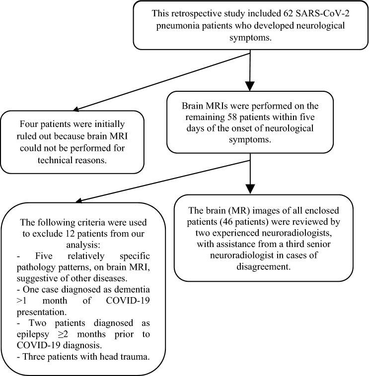
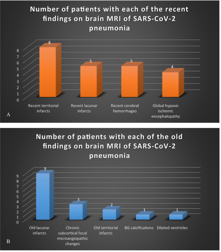
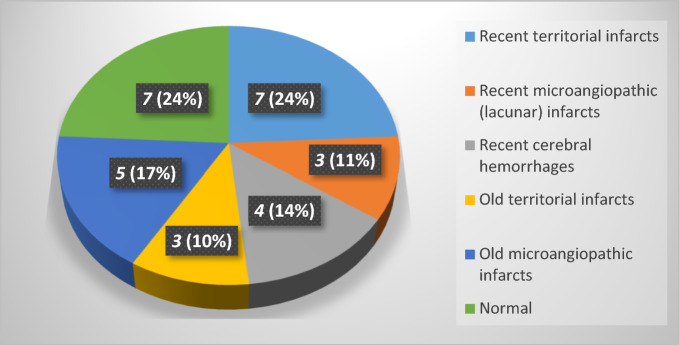
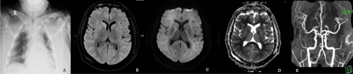
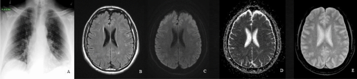
The increased frequency of neurological manifestations, including central nervous system (CNS) manifestations, in patients with coronavirus disease 2019 (COVID-19) pandemic is consistent with the virus's neurotropic nature. In most patients, brain magnetic resonance imaging (MRI) is a sensitive imaging modality in the diagnosis of viral encephalitides in the brain. The purpose of this study was to determine the frequency of brain lesion patterns on brain MRI in severe acute respiratory syndrome coronavirus 2 (SARS-CoV-2) pneumonia patients who developed focal and non-focal neurological manifestations. In addition, it will compare the impact of the Glasgow Coma Scale (GCS) as an index of deteriorating cerebral function on positive brain MRIs in both neurological manifestations. This retrospective study included an examination of SARS-CoV-2 pneumonia patients with real-time reverse transcription polymerase chain reaction (RT-PCR) confirmation, admitted with clinicoradiologic evidence of COVID-19 pneumonia, and who were candidates for brain MRI due to neurological manifestations suggesting brain involvement. Brain imaging acquired on a 3.0 T MRI system (Skyra; Siemens, Erlangen, Germany) with a 20-channel receive head coil. Brain MRI revealed lesions in 38 (82.6%) of the total 46 patients for analysis and was negative in the remaining eight (17.4%) of all finally enclosed patients with RT-PCR confirmed SARS-CoV-2 pneumonia. Twenty-nine (63%) patients had focal neurological manifestations, while the remaining 17 (37%) patients had non-focal neurological manifestations. The patients had a highly significant difference (p = 0.0006) in GCS, but no significant difference (p = 0.4) in the number of comorbidities they had. Brain MRI is a feasible and important imaging modality in patients with SARS-CoV-2 pneumonia who develop neurological manifestations suggestive of brain involvement, particularly in patients with non-focal manifestations and a decline in GCS.
© 2021. The Author(s).
Conflict of interest statement
The authors declare no competing interests.
Figures
Similar articles
-
Magnetic Resonance Imaging (MRI) and neurological manifestations in SARS-CoV-2 patients.Eur Rev Med Pharmacol Sci. 2021 Jan;25(2):1101-1108. doi: 10.26355/eurrev_202101_24681. Eur Rev Med Pharmacol Sci. 2021. PMID: 33577067 Review.
-
COVID-19 encephalopathy: Clinical and neurobiological features.J Med Virol. 2021 Jul;93(7):4374-4381. doi: 10.1002/jmv.26973. Epub 2021 Apr 23. J Med Virol. 2021. PMID: 33782993 Free PMC article.
-
Cerebrospinal Fluid Features in Patients With Coronavirus Disease 2019 and Neurological Manifestations: Correlation with Brain Magnetic Resonance Imaging Findings in 58 Patients.J Infect Dis. 2021 Feb 24;223(4):600-609. doi: 10.1093/infdis/jiaa745. J Infect Dis. 2021. PMID: 33249438 Free PMC article.
-
Neurological complications in COVID-19 - a diagnostic challenge.J Med Life. 2021 Mar-Apr;14(2):216-224. doi: 10.25122/jml-2021-0045. J Med Life. 2021. PMID: 34104245 Free PMC article.
-
Neurological symptoms, manifestations, and complications associated with severe acute respiratory syndrome coronavirus 2 (SARS-CoV-2) and coronavirus disease 19 (COVID-19).J Neurol. 2021 Sep;268(9):3059-3071. doi: 10.1007/s00415-021-10406-y. Epub 2021 Jan 23. J Neurol. 2021. PMID: 33486564 Free PMC article. Review.
Cited by
-
Significance of Persistent Systemic Support in the Clinical Course of Delayed Post-hypoxic Leukoencephalopathy Following Severe Coronavirus Disease 2019.Intern Med. 2024 Apr 15;63(8):1167-1172. doi: 10.2169/internalmedicine.1412-22. Epub 2024 Feb 1. Intern Med. 2024. PMID: 38296478 Free PMC article.
-
Clinical, Laboratory and Imaging Characteristics of Hospitalized COVID-19 Patients with Neurologic Involvement; a Cross-Sectional Study.Arch Acad Emerg Med. 2022 Jan 30;10(1):e10. doi: 10.22037/aaem.v10i1.1507. eCollection 2022. Arch Acad Emerg Med. 2022. PMID: 35402993 Free PMC article.
-
Melatonin: highlighting its use as a potential treatment for SARS-CoV-2 infection.Cell Mol Life Sci. 2022 Feb 20;79(3):143. doi: 10.1007/s00018-021-04102-3. Cell Mol Life Sci. 2022. PMID: 35187603 Free PMC article. Review.
-
Brain MRI findings in severe COVID-19 patients: a meta-analysis.Front Neurol. 2023 Oct 12;14:1258352. doi: 10.3389/fneur.2023.1258352. eCollection 2023. Front Neurol. 2023. PMID: 37900601 Free PMC article.
-
Brain resident memory T cells rapidly expand and initiate neuroinflammatory responses following CNS viral infection.Brain Behav Immun. 2023 Aug;112:51-76. doi: 10.1016/j.bbi.2023.05.009. Epub 2023 May 24. Brain Behav Immun. 2023. PMID: 37236326 Free PMC article.
References
-
- Moriguchi T, Harii N, Goto J, Harada D, Sugawara H, Takamino J, Ueno M, Sakata H, Kondo K, Myose N, Nakao A, Takeda M, Haro H, Inoue O, Suzuki-Inoue K, Kubokawa K, Ogihara S, Sasaki T, Kinouchi H, Kojin H, Ito M, Onishi H, Shimizu T, Sasaki Y, Enomoto N, Ishihara H, Furuya S, Yamamoto T, Shimada S. A first case of meningitis/encephalitis associated with SARS-Coronavirus-2. Int. J. Infect. Dis. 2020;94:55–58. doi: 10.1016/j.ijid.2020.03.062. - DOI - PMC - PubMed
Publication types
MeSH terms
LinkOut - more resources
Full Text Sources
Medical
Miscellaneous
