. 2021 Sep 28:15:712862.
doi: 10.3389/fnana.2021.712862. eCollection 2021.

Towards Deciphering the Fetal Foundation of Normal Cognition and Cognitive Symptoms From Sulcation of the Cortex

Affiliations

Towards Deciphering the Fetal Foundation of Normal Cognition and Cognitive Symptoms From Sulcation of the Cortex

Arnaud Cachia et al. Front Neuroanat. .

Abstract

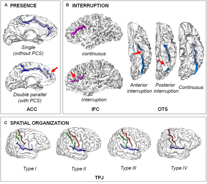
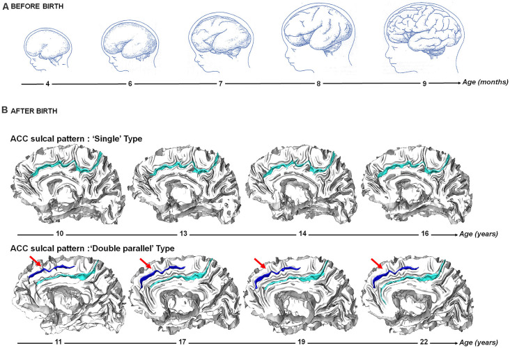
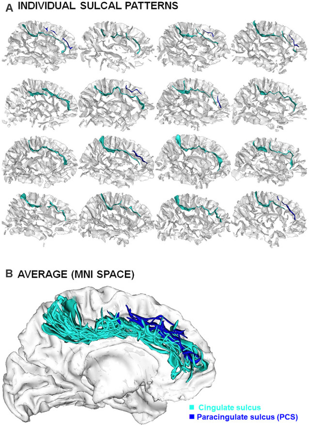
Growing evidence supports that prenatal processes play an important role for cognitive ability in normal and clinical conditions. In this context, several neuroimaging studies searched for features in postnatal life that could serve as a proxy for earlier developmental events. A very interesting candidate is the sulcal, or sulco-gyral, patterns, macroscopic features of the cortex anatomy related to the fold topology-e.g., continuous vs. interrupted/broken fold, present vs. absent fold-or their spatial organization. Indeed, as opposed to quantitative features of the cortical sheet (e.g., thickness, surface area or curvature) taking decades to reach the levels measured in adult, the qualitative sulcal patterns are mainly determined before birth and stable across the lifespan. The sulcal patterns therefore offer a window on the fetal constraints on specific brain areas on cognitive abilities and clinical symptoms that manifest later in life. After a global review of the cerebral cortex sulcation, its mechanisms, its ontogenesis along with methodological issues on how to measure the sulcal patterns, we present a selection of studies illustrating that analysis of the sulcal patterns can provide information on prenatal dispositions to cognition (with a focus on cognitive control and academic abilities) and cognitive symptoms (with a focus on schizophrenia and bipolar disorders). Finally, perspectives of sulcal studies are discussed.

Keywords: MRI; gyrification; neurodevelopment; psychiatry; psychology; sulcation.


Conflict of interest statement

The authors declare that the research was conducted in the absence of any commercial or financial relationships that could be construed as a potential conflict of interest.

Figures

Figure 1
Figure 1
Examples of different types of sulcal patterns. (A) Presence of the fold. The sulcal pattern of the anterior cingulate cortex (ACC) can be “single”, with only the cingulate sulcus, or “double parallel”, with an additional paracingulate sulcus (PCS). Adapted from Cachia et al. (2014). (B) Interruption of the fold. The sulcal pattern of the inferior frontal cortex (IFC) can be continuous or with an interruption (red arrow). The sulcal pattern of the lateral occipito-temporal sulcus (OTS) can be continuous or have an anterior or a posterior sulcal interruption. Adapted from Cachia et al. (2014); Borst et al. (2016); Cachia et al. (2018), and Tissier et al. (2018). (C) Spatial organization. The sulcal pattern of the temporo-parietal junction (TPJ) is characterized by the spatial organization of the posterior part of the right Sylvian fissure (pSF) including Sylvian fissure (in blue), post-central sulcus (green) and central sulcus (in red). Type I, has both a vertical branch (planum parietale) that ascends into the supramarginal gyrus and a horizontal branch (planum temporale) that forms the superior surface of the superior temporal gyrus. In Type II, the Sylvian fissure lacks a vertical branch. In Type III, the horizontal branch extends posteriorly to the supramarginal gyrus into the angular gyrus. In Type IV, the vertical branch connects the post-central sulcus anterior to the supramarginal gyrus, the horizontal branch is therefore absent. Adapted from Steinmetz et al. (1990) and Plaze et al. (2015).
Figure 2
Figure 2
Sulcogenesis before and after birth. (A) Before birth. Cortical folding during from 4–9 months post-conception during fetal life. Adapted from Welker (1990). (B) After birth. Longitudinal stability of the sulcal pattern of the Anterior Cingulate Cortex (ACC) pattern from 10–22 years in a participant with a “single type” ACC and in a participant with a “double parallel type” ACC. Adapted from Cachia et al. (2016).
Figure 3
Figure 3
Inter-individual variability of the sulcal patterns. (A) Example of individual ACC sulcal patterns in 12 healthy subjects. (B) Superimposition of individual sulcal patterns in a common reference space (MNI) after linear spatial normalization.

References

    1. Alemán-Gómez Y., Janssen J., Schnack H., Balaban E., Pina-Camacho L., Alfaro-Almagro F., et al. . (2013). The human cerebral cortex flattens during adolescence. J. Neurosci. 33, 15004–15010. 10.1523/JNEUROSCI.1459-13.2013 - DOI - PMC - PubMed
    1. Amiez C., Sallet J., Hopkins W. D., Meguerditchian A., Hadj-Bouziane F., Ben Hamed S., et al. . (2019). Sulcal organization in the medial frontal cortex provides insights into primate brain evolution. Nat. Commun. 10:3437. 10.1038/s41467-019-11347-x - DOI - PMC - PubMed
    1. Amiez C., Wilson C. R. E., Procyk E. (2018). Variations of cingulate sulcal organization and link with cognitive performance. Sci. Rep. 8:13988. 10.1038/s41598-018-32088-9 - DOI - PMC - PubMed
    1. Ansorge M. S., Hen R., Gingrich J. A. (2007). Neurodevelopmental origins of depressive disorders. Curr. Opin. Pharmacol. 7, 8–17. 10.1016/j.coph.2006.11.006 - DOI - PubMed
    1. Armstrong E., Schleicher A., Omran H., Curtis M., Zilles K. (1995). The ontogeny of human gyrification. Cereb. Cortex 5, 56–63. 10.1093/cercor/5.1.56 - DOI - PubMed

LinkOut - more resources