. 2021 Sep 23:12:734351.
doi: 10.3389/fphar.2021.734351. eCollection 2021.

Exosome-Derived ADAM17 Promotes Liver Metastasis in Colorectal Cancer

Affiliations

Exosome-Derived ADAM17 Promotes Liver Metastasis in Colorectal Cancer

Jinbing Sun et al. Front Pharmacol. .

Abstract

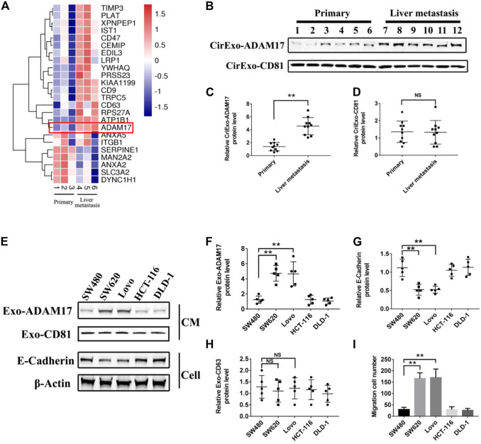
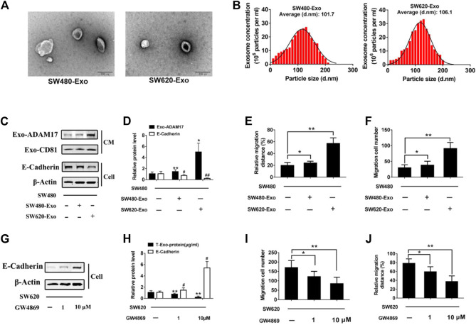
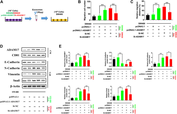
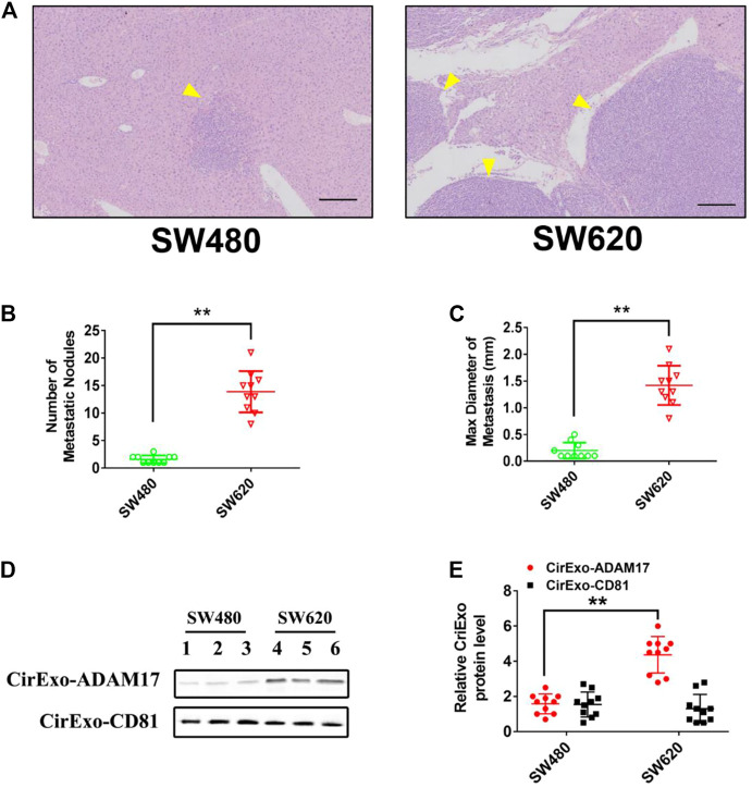
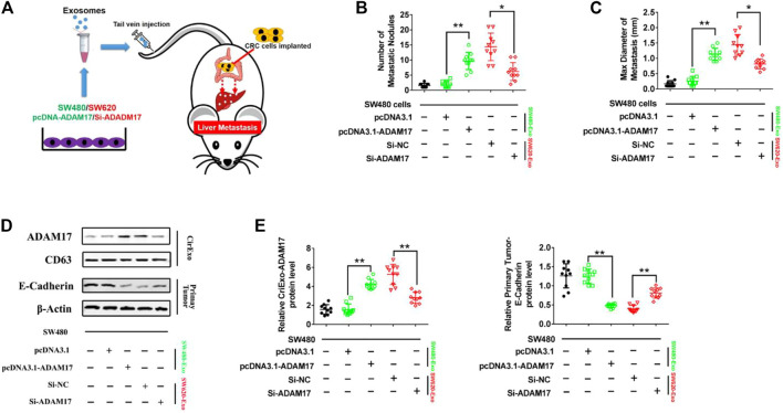
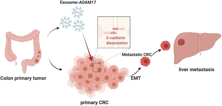
Exosomes derived from cancer cells are deemed important drivers of pre-metastatic niche formation at distant organs, but the underlying mechanisms of their effects remain largely unknow. Although the role of ADAM17 in cancer cells has been well studied, the secreted ADAM17 effects transported via exosomes are less understood. Herein, we show that the level of exosome-derived ADAM17 is elevated in the serum of patients with metastatic colorectal cancer as well as in metastatic colorectal cancer cells. Furthermore, exosomal ADAM17 was shown to promote the migratory ability of colorectal cancer cells by cleaving the E-cadherin junction. Moreover, exosomal ADAM17 overexpression as well as RNA interference results highlighted its function as a tumor metastasis-promoting factor in colorectal cancer in vitro and in vivo. Taken together, our current work suggests that exosomal ADAM17 is involved in pre-metastatic niche formation and may be utilized as a blood-based biomarker of colorectal cancer metastasis.

Keywords: ADAM17; E-cadherin; colorectal cancer; exosome; liver metastasis.


Conflict of interest statement

The authors declare that the research was conducted in the absence of any commercial or financial relationships that could be construed as a potential conflict of interest.

Figures

FIGURE 1
FIGURE 1
ADAM17 levels in exosomes from CRC patient sera and CRC cell lines. (A) Proteomic analysis of serum-derived exosomal protein differentially expressed between non-metastatic (primary) and liver metastastatic patients with CRC. A heatmap of differentially expressed exosomal proteins based on quantitative mass spectrometry values (technical triplicates, * false discovery rate < 0.05 via ANOVA). Hierarchical clustering (1—the sample Spearman’s rank correlation coefficient between observations) was performed on protein expression levels (n = 3). Exosomal ADAM17 was marked with a red frame as a differentially expressed protein. (B) The serum-derived exosomes of non-metastatic (primary) and liver metastatic patients with CRC were collected and lysed for analysis of ADAM17 and CD81 protein levels. (C,D) The relative protein levels were further calculated via gray analysis (the total exosomal protein was set as the internal control, n = 10). (E) SW480, SW620, Lovo, HCT-116, and DLD-1 exosomes and cells were subjected to analysis of ADAM17, CD81, and E-cadherin protein levels (CM, conditioned medium). (F–H) The relative protein levels were further calculated via gray analysis (the total exosomal protein was set as the internal control for exosomal protein analysis, ß-actin was set as the internal control for cells, n = 6). (I) Cell migration assessment of SW480, SW620, Lovo, HCT-116, and DLD-1 cells via transwell assays (n = 6). Data are expressed as means ± SDs. *, p < 0.05; **, p < 0.01.
FIGURE 2
FIGURE 2
Exosomal ADAM17 enhances the migratory properties of CRC cells. Characterization of CRC cell-derived exosomes. (A) Electron microscope images of exosomes isolated from the CM of cultured SW480 and SW620 cells. Scale bar, 200 nm. (B) Nanoparticle tracking analysis of isolated exosomes. (C,D) SW480 cells were treated with SW480-derived exosomes (SW480-Exo) and SW620-derived exosomes (SW620-Exo) at 50 µg (10 μg/ml) per 1×10⁵ cultured cells. Exosomes were collected, cells were lysed, and protein levels were further calculated via gray analysis (the total exosomal protein was set as the internal control for exosomal protein analysis, ß-actin was set as the internal control for cells, n = 6). (E,F) Changes in migration were measured via transwell and wound scratch assays using SW480 cells treated with SW480-Exo and SW620-Exo (n = 6). (G,H) SW620 cells were treated with different concentration GW4869, and the total exosomal protein as well as cellular E-cadherin levels were calculated (n = 6). (I,J) Changes in migration were measured via transwell and wound scratch assays using SW620 cells treated with GW4869 (n = 6). Data are expressed as means ± SDs. * or #, p < 0.05; ** or ##, p < 0.01.
FIGURE 3
FIGURE 3
Exosomal ADAM17 enhances the cleavage of E-cadherin. (A) Schematic description of the experimental design. The pcDNA3.1-, pcDNA3.1-ADAM17-transfected SW480-derived exosomes and Si-NC-, Si-ADAM17-transfected SW620-derived exosomes were isolated. 50 μg of exosomes were added to 5×10⁵ SW480 cells. (B,C) Changes in migration were measured via transwell and wound scratch assays using SW480 treated with pcDNA3.1-, pcDNA3.1-ADAM17-transfected SW480-derived exosomes as well as Si-NC-, Si-ADAM17-transfected SW620-derived exosomes (n = 6). (D,E) SW480 cells were treated with pcDNA3.1-, pcDNA3.1-ADAM17-transfected SW480-derived exosomes and Si-NC-, Si-ADAM17-transfected SW620-derived exosomes. Exosomes and lysed cells were collected for the assessment of protein levels via gray analysis (the total exosomal protein was set as the internal control for exosomal protein analysis, ß-actin was set as the internal control for cells, n = 6). Data are expressed as means ± SDs. *, p < 0.05; **, p < 0.01.
FIGURE 4
FIGURE 4
Exosomes derived from CRC cells promote liver metastasis. SW480 and SW620 cells were implanted into the mesentery at the tail end of the cecum. Autopsies were performed, and the presence of metastases was examined macroscopically 60 days after CRC implantation. (A) Representative H&E staining results of liver slices from SW480- and SW620-implanted mice. Arrows indicate the tumor nodules. Scale bar, 250 μm. (B,C) The number and maximal diameter of metastatic nodules in the livers of SW480- and SW620-implanted mice were calculated and analyzed (n = 10). (D,E) The serum-derived exosomes of SW480- and SW620-implanted mice were collected and lysed for analysis of ADAM17 and CD81 protein levels. The relative protein levels were further calculated via gray analysis (the total exosomal protein was set as the internal control, n = 10). Data are expressed as means ± SDs. *, p < 0.05; **, p < 0.01.
FIGURE 5
FIGURE 5
CRC cell-secreted exosomal ADAM17 promotes metastasis in vivo . (A) Schematic description of the experimental design. The pcDNA3.1-, pcDNA3.1-ADAM17-transfected SW480-derived exosomes and Si-NC-, Si-ADAM17-transfected SW620-derived exosomes were isolated. 10 μg exosomes were used to intravenously inject mice every 3 days for 2 months after the implantation of SW480 cells. (B,C) The number and maximal diameter of metastatic nodules in the livers of SW480-implanted mice treated with pcDNA3.1-, pcDNA3.1-ADAM17-transfected SW480-derived exosomes and Si-NC-, Si-ADAM17-transfected SW620-derived exosomes were calculated and analyzed (n = 10). (D,E) The serum-derived exosomes and primary tumors of SW480-implanted mice treated with pcDNA3.1-, pcDNA3.1-ADAM17-transfected SW480-derived exosomes and Si-NC-, Si-ADAM17-transfected SW620-derived exosomes were collected and lysed for the assessment of ADAM17, CD81, and E-cadherin protein levels. The relative protein levels were determined via gray analysis (the total exosomal protein was set as the internal control for exosomal protein analysis, ß-actin was set as the internal control for cells, n = 6). Data are expressed as means ± SDs. *, p < 0.05; **, p < 0.01.
FIGURE 6
FIGURE 6
Proposed mechanism for exosomal ADAM17-mediated promotion of CRC metastasis. The CRC-derived exosomal ADAM17 promoted the E-cadherin cleavage, enhancing the migratory properties of CRC cells, which in turn promoted hepatic metastasis in vivo.

Similar articles

Cited by

References

    1. Becker A., Thakur B. K., Weiss J. M., Kim H. S., Peinado H., Lyden D. (2016). Extracellular Vesicles in Cancer: Cell-To-Cell Mediators of Metastasis. Cancer Cell 30 (6), 836–848. 10.1016/j.ccell.2016.10.009 - DOI - PMC - PubMed
    1. Bollati V., Angelici L., Rizzo G., Pergoli L., Rota F., Hoxha M., et al. (2015). Microvesicle-associated microRNA Expression Is Altered upon Particulate Matter Exposure in Healthy Workers and in A549 Cells. J. Appl. Toxicol. 35 (1), 59–67. 10.1002/jat.2987 - DOI - PMC - PubMed
    1. Chafe S. C., Lou Y., Sceneay J., Vallejo M., Hamilton M. J., McDonald P. C., et al. (2015). Carbonic Anhydrase IX Promotes Myeloid-Derived Suppressor Cell Mobilization and Establishment of a Metastatic Niche by Stimulating G-CSF Production. Cancer Res. 75 (6), 996–1008. 10.1158/0008-5472.CAN-14-3000 - DOI - PubMed
    1. Chen C., Luo Y., He W., Zhao Y., Kong Y., Liu H., et al. (2020). Exosomal Long Noncoding RNA LNMAT2 Promotes Lymphatic Metastasis in Bladder Cancer. J. Clin. Invest. 130 (1), 404–421. 10.1172/JCI130892 - DOI - PMC - PubMed
    1. Cocucci E., Racchetti G., Meldolesi J. (2009). Shedding Microvesicles: Artefacts No More. Trends Cel Biol. 19 (2), 43–51. 10.1016/j.tcb.2008.11.003 - DOI - PubMed