. 2021 Sep 28:12:723972.
doi: 10.3389/fphys.2021.723972. eCollection 2021.

Aortic Stiffness Hysteresis in Isolated Mouse Aortic Segments Is Intensified by Contractile Stimuli, Attenuated by Age, and Reversed by Elastin Degradation

Affiliations

Aortic Stiffness Hysteresis in Isolated Mouse Aortic Segments Is Intensified by Contractile Stimuli, Attenuated by Age, and Reversed by Elastin Degradation

Sofie De Moudt et al. Front Physiol. .

Abstract

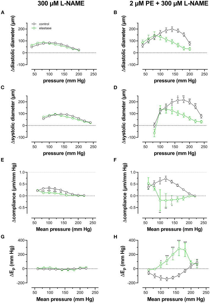
Aim: Cyclic stretch of vascular tissue at any given pressure reveals greater dimensions during unloading than during loading, which determines the cardiac beat-by-beat hysteresis loop on the pressure-diameter/volume relationship. The present study did not focus on hysteresis during a single stretch cycle but investigated whether aortic stiffness determined during continuous stretch at different pressures also displayed hysteresis phenomena. Methods: Aortic segments from C57Bl6 mice were mounted in the Rodent Oscillatory Set-up for Arterial Compliance (ROTSAC), where they were subjected to high frequency (10 Hz) cyclic stretch at alternating loads equivalent to a constant theoretical pulse pressure of 40 mm Hg. Diastolic and systolic diameter, compliance, and the Peterson elastic modulus (Ep), as a measure of aortic stiffness, was determined starting at cyclic stretch between alternating loads corresponding to 40 and 80 mm Hg, at each gradual load increase equivalent to 20 mm Hg, up to loads equivalent to pressures of 220 and 260 mm Hg (loading direction) and then repeated in the downward direction (unloading direction). This was performed in baseline conditions and following contraction by α1 adrenergic stimulation with phenylephrine or by depolarization with high extracellular K+ in aortas of young (5 months), aged (26 months) mice, and in segments treated with elastase. Results: In baseline conditions, diastolic/systolic diameters and compliance for a pulse pressure of 40 mm Hg were larger at any given pressure upon unloading (decreasing pressure) than loading (increasing pressure) of the aortic segments. The pressure-aortic stiffness (Ep) relationship was similar in the loading and unloading directions, and aortic hysteresis was absent. On the other hand, hysteresis was evident after activation of the VSMCs with the α1 adrenergic agonist phenylephrine and with depolarization by high extracellular K+, especially after inhibition of basal NO release with L-NAME. Aortic stiffness was significantly smaller in the unloading than in the loading direction. In comparison with young mice, old-mouse aortic segments also displayed contraction-dependent aortic hysteresis, but hysteresis was shifted to a lower pressure range. Elastase-treated segments showed higher stiffness upon unloading over nearly the whole pressure range. Conclusions: Mouse aortic segments display pressure- and contraction-dependent diameter, compliance, and stiffness hysteresis phenomena, which are modulated by age and VSMC-extracellular matrix interactions. This may have implications for aortic biomechanics in pathophysiological conditions and aging.

Keywords: aging; aortic stiffness; constriction; pressure-dependent hysteresis; viscosity.


Conflict of interest statement

The authors declare that the research was conducted in the absence of any commercial or financial relationships that could be construed as a potential conflict of interest.

Figures

Figure 1
Figure 1
Determination of the aortic segment extrapolated length and diameter. (A) Images of the aortic vessel segment at six different preloads between 10 and 60 mN; (B) length (l), width (w), and extrapolated diameter (D) as a function of preload; (C) relationship between length and extrapolated diameter (to obtain length at each calculated diameter to measure pressure).
Figure 2
Figure 2
(A) Test protocol applied to ROTSAC 1 to measure diameter, compliance, and Peterson modulus, Ep. Starting at diastolic and systolic preloads, according to 80 and 120 mm Hg, the preloads were adapted to obtain diastolic and systolic pressures of 40 and 80 mm Hg and subsequently increased by 20 mm Hg up to 200–240 mm Hg or higher (loading). Thereafter, the pressure was decreased back to 40–80 mm Hg (unloading) and, finally, to 80–120 mm Hg. In the insert, preload and calculated pressure are shown during the clamp of the segment at 10 Hz between 100 mm Hg (36.5 mN) and 140 mm Hg (54.4 mN). Similar protocols were applied to ROTSAC 2, 3, and 4 with the same pressure steps but other preloads. (B–D) Illustrate stretch-by-stretch diameter-pressure loops at 80–120 (black, red) and 160–200 (green, purple) during loading (up, black, green) and unloading (down, red, purple) in three experimental conditions: Krebs-Ringer (KR, B), 2 μM PE + 300-μM L-NAME (C), and 0.1 mg/ml elastase (D). The left y-axis refers to diastolic and systolic diameters at 80–120 mm Hg and the right axis to these parameters at 160–200 mm Hg. The y-axis scale is the same in all figures. The numbers along each loop indicate the Ep value in mm Hg.
Figure 3
Figure 3
Aortic segments displayed considerable pressure- and contraction-dependent hysteresis. Aortic segments were treated with 300-μM L-NAME in the absence (A,C,E,G) and presence (B,D,F,H) of 2 μM PE and subjected to cyclic stretch with an amplitude of 40 mm Hg, ranging from 40–80 mm Hg to 240–280 mm Hg (upward direction, open symbols, up) and back to 40–80 mm Hg (downward direction, closed symbols, down). Diastolic and systolic diameters (A–D), compliance (E,F), and Peterson's elastic modulus (Ep, G,H) were determined in aortic segments of 7 C57Bl6 mice (data points ± SEM). Data points for the upward pressure steps (open symbols, up) were compared at any given pressure with data points for the downward pressure steps (closed symbols, down) with two-way RM ANOVA with Sidak's multiple comparison test. *p < 0.05, **p < 0.01, and ***p < 0.001.
Figure 4
Figure 4
α1-Adrenergic stimulation and maximal depolarization of aortic segments cause different isometric and isobaric effects. Aortic segments were mounted in isometric (100 mm Hg, A) and isobaric (80–120 mm Hg, B) conditions and treated with 2 μM PE (circles) or depolarized with 50 mM K+ (squares) in the absence (open symbols) and presence (closed symbols) of 300-μM L-NAME. The increase of force (Δorce) and Ep (ΔEp) was determined in aortic segments of 5 (white bars) and 26 (blue bars) months old mice. Two-way RM ANOVA with Sidak's multiple comparison test. *p < 0.05, **p < 0.01, and ***p < 0.001.
Figure 5
Figure 5
Hysteresis is pressure, contraction and age dependent. Aortic segments of mice aged 5 months (black, n = 8) and 26 months (blue, n = 5) were subjected to upward (open symbols, up) and downward (closed symbols, down) pressure steps between 40 and 80 mm Hg to 200–240 mm Hg and back in control conditions (KR, A,D) in the presence of 2 μM PE (B,E) and 2 μM PE in the presence of 300-μM L-NAME to block endothelial NO release (C,F). Absolute values of Ep are shown in (A–C), whereas, in (D–F), data points for the upward pressure steps (open symbols, up) were compared at any given pressure, with data points for the downward pressure steps (closed symbols, down) (stiffness hysteresis). Two-way RM ANOVA with Sidak's multiple comparison test, *p < 0.05, **p < 0.01, ***p < 0.001.
Figure 6
Figure 6
Hysteresis is pressure, contraction, and age dependent. Aortic segments of mice aged 5 months (black, n = 8) and 26 months (blue, n = 5) were subjected to upward (open symbols, up) and downward (closed symbols, down) pressure steps between 40 and 80 mm Hg to 180–220 or 200–240 mm Hg and back in control conditions (KR, A,D) in the presence of 50 mM K+ (B,E) and 50 mMK+in the presence of 300-μM L-NAME to block endothelial NO release (C,F). Absolute values of Ep are shown in (A–C), whereas, in (D–F), data points for the upward pressure steps (open symbols, up) were compared at any given pressure with data points for the downward pressure steps (closed symbols, down) (stiffness hysteresis). Two-way RM ANOVA with Sidak's multiple comparison test (old vs. young mice), *p < 0.05, **p < 0.01, ***p < 0.001.
Figure 7
Figure 7
Pressure-dependent hysteresis is affected by the contractile stimulus and by age. Data from Figures 5F, 6F are combined in Figure 6A for mice of 5 months (n = 8, 5, black) and 26 months (n = 5, 26, blue). Contractions by 50 mM K+ (squares) and 2 μM PE (circles) were elicited in the presence of L-NAME. Three-way ANOVA: pressure: ***; age 0.12; stimulus *; pressure/age: **; pressure/stimulus ***; age/stimulus 0.73. For clarity, (A) was repeated in (B) (for 5 months) and (C) (for 26 months). Two-way RM ANOVA with Sidak's multiple comparison test, *p < 0.05, **p < 0.01, ***p < 0.001.
Figure 8
Figure 8
Diastolic and systolic diameters, compliance, and stiffness are elastin dependent. Diastolic diameter (A), systolic diameter (B), compliance (C), and Peterson modulus, Ep (D), were measured in aortic segments in control conditions (black symbols) or after treatment with 0.1 unit/ml elastase (green symbols). Data points for the upward pressure steps (open symbols, up) were compared at any given pressure with data points for the downward pressure steps (closed symbols, down). Two-way RM ANOVA with Sidak's multiple comparison test (elastase vs. control), *p < 0.05, **p < 0.01, ***p < 0.001, n = 5.
Figure 9
Figure 9
Diastolic and systolic diameters, compliance, and stiffness in the presence of PE are elastin dependent. Diastolic diameter (A), systolic diameter (B), compliance (C), and Peterson modulus, Ep (D), were measured in aortic segments in the presence of 2 μM PE and 300-μM L-NAME in control conditions (black symbols) or after treatment with 0.1 unit/ml elastase (green symbols). Two-way RM ANOVA with Sidak's multiple comparison test (elastase vs. control), *p < 0.05, **p < 0.01, ***p < 0.001, n = 5.
Figure 10
Figure 10
Pressure-dependent hysteresis induced by contraction with PE is elastin dependent. Diastolic (A,B) and systolic diameters (C,D), compliance (E,F), and Ep (G,H) were determined in untreated (black) and elastase-treated (0.1 unit/ml, green) aortic segments in KR (A,C,E,G) and in the presence of 2 μM PE/300-μM L-NAME (B,D,F,H). Data points for the upward pressure steps were compared at any given pressure with data points for the downward pressure steps (stiffness hysteresis). Hence, positive values for diameter and compliance refer to larger diameters and compliance for downward pressure steps. Negative values for Ep correspond to lower stiffness for the downward pressure steps (“de-stiffening”). Two-way RM ANOVA with Sidak's multiple comparison test, *p < 0.05, **p < 0.01, ***p < 0.001, n = 5.

Similar articles

Cited by

References

    1. Armentano R. L., Barra J. G., Levenson J., Simon A., Pichel R. H. (1995). Arterial wall mechanics in conscious dogs. assessment of viscous, inertial, and elastic moduli to characterize aortic wall behavior. Circ. Res. 76, 468–478. 10.1161/01.RES.76.3.468 - DOI - PubMed
    1. Barra J. G., Armentano R. L., Levenson J., Fischer E. I., Pichel R. H., Simon A. (1993). Assessment of smooth muscle contribution to descending thoracic aortic elastic mechanics in conscious dogs. Circ.Res. 73, 1040–1050. 10.1161/01.RES.73.6.1040 - DOI - PubMed
    1. Bauer R. D., Busse R., Schabert A., Summa Y., Wetterer E. (1979). Separate determination of the pulsatile elastic and viscous forces developed in the arterial wall in vivo. Pflugers Arch. 380, 221–226. 10.1007/BF00582900 - DOI - PubMed
    1. Bergel D. H. (1961a). The dynamic elastic properties of the arterial wall. J. Physiol. 156, 458–469. 10.1113/jphysiol.1961.sp006687 - DOI - PMC - PubMed
    1. Bergel D. H. (1961b). The static elastic properties of the arterial wall. J. Physiol. 156, 445–457. 10.1113/jphysiol.1961.sp006686 - DOI - PMC - PubMed

LinkOut - more resources