

Microplastics-Induced Eryptosis and Poikilocytosis in Early-Juvenile Nile Tilapia (Oreochromis niloticus)
- PMID: 34650449
- PMCID: PMC8507840
- DOI: 10.3389/fphys.2021.742922
Microplastics-Induced Eryptosis and Poikilocytosis in Early-Juvenile Nile Tilapia (Oreochromis niloticus)
Abstract
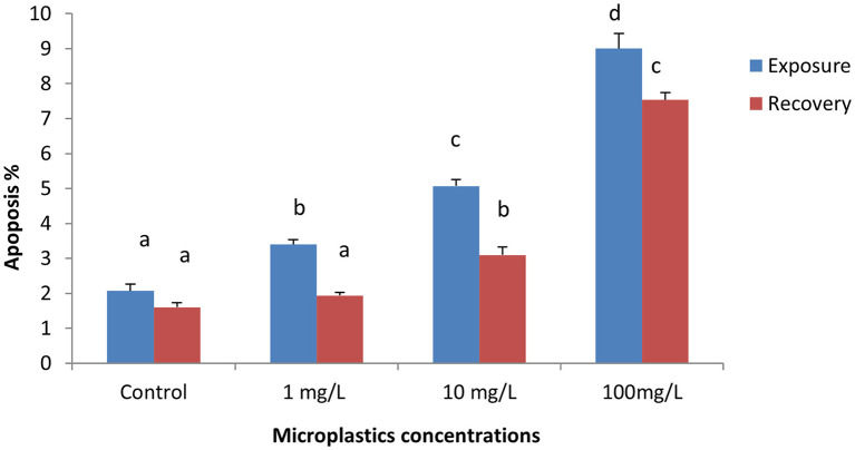
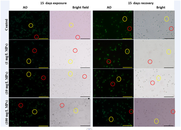
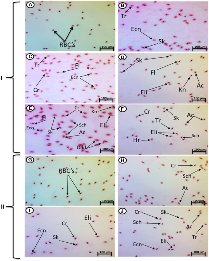
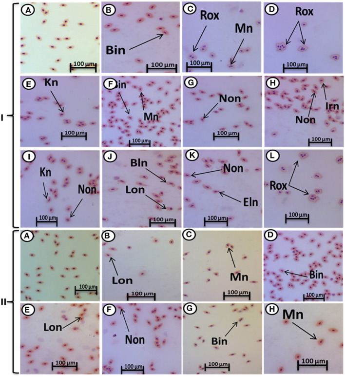
This study aims to assess the impact of microplastics (MPs) on erythrocytes using eryptosis (apoptosis) and an erythron profile (poikilocytosis and nuclear abnormalities), considered to be novel biomarkers in Nile tilapia (Oreochromis niloticus). In this study, four groups of fish were used: The first was the control group. In the second group, 1 mg/L of MPs was introduced to the samples. The third group was exposed to 10 mg/L of MPs. Finally, the fourth group was exposed to 100 mg/L of MPs for 15 days, following 15 days of recovery. The fish treated with MPs experienced an immense rise in the eryptosis percentage, poikilocytosis, and nuclear abnormalities of red blood cells (RBCs) compared with the control group in a concentration-dependent manner. Poikilocytosis of MP-exposed groups included sickle cell shape, schistocyte, elliptocyte, acanthocyte, and other shapes. Nuclear abnormalities of the MPs-exposed groups included micronuclei, binucleated erythrocytes, notched, lobed, blebbed, and hemolyzed nuclei. After the recovery period, a greater percentage of eryptosis, poikilocytotic cells, and nuclear abnormalities in RBCs were still evident in the groups exposed to MPs when crosschecked with the control group. The results show concerning facts regarding the toxicity of MPs in tilapia.
Keywords: Oreochromis niloticus; apoptosis; erythrocytes; microplastics; poikilocytosis; tilapia.
Copyright © 2021 Hamed, Osman, Badrey, Soliman and Sayed.
Conflict of interest statement
The authors declare that the research was conducted in the absence of any commercial or financial relationships that could be construed as a potential conflict of interest.
Figures
Similar articles
-
Assessment the effect of exposure to microplastics in Nile Tilapia (Oreochromis niloticus) early juvenile: I. blood biomarkers.Chemosphere. 2019 Aug;228:345-350. doi: 10.1016/j.chemosphere.2019.04.153. Epub 2019 Apr 22. Chemosphere. 2019. PMID: 31039541
-
Poikilocytosis and tissue damage as negative impacts of tramadol on juvenile of Tilapia (Oreochromis niloticus).Environ Toxicol Pharmacol. 2020 Aug;78:103383. doi: 10.1016/j.etap.2020.103383. Epub 2020 Apr 7. Environ Toxicol Pharmacol. 2020. PMID: 32305673
-
Single and combined toxicity of tadalafil (Cilais) and microplastic in Tilapia fish (Oreochromis niloticus).Sci Rep. 2024 Jun 25;14(1):14576. doi: 10.1038/s41598-024-64282-3. Sci Rep. 2024. PMID: 38914580 Free PMC article.
-
Impacts of microplastics on reproductive performance of male tilapia (Oreochromis niloticus) pre-fed on Amphora coffeaeformis.Environ Sci Pollut Res Int. 2021 Dec;28(48):68732-68744. doi: 10.1007/s11356-021-14984-2. Epub 2021 Jul 19. Environ Sci Pollut Res Int. 2021. PMID: 34279784
-
Antioxidants and molecular damage in Nile Tilapia (Oreochromis niloticus) after exposure to microplastics.Environ Sci Pollut Res Int. 2020 May;27(13):14581-14588. doi: 10.1007/s11356-020-07898-y. Epub 2020 Feb 11. Environ Sci Pollut Res Int. 2020. PMID: 32048193 Free PMC article.
Cited by
-
Hematological and Hematopoietic Analysis in Fish Toxicology-A Review.Animals (Basel). 2023 Aug 14;13(16):2625. doi: 10.3390/ani13162625. Animals (Basel). 2023. PMID: 37627416 Free PMC article. Review.
-
Assessing microplastic pollution vulnerability in a protected coastal lagoon in the Mediterranean Coast of Egypt using GIS modeling.Sci Rep. 2025 Apr 4;15(1):11557. doi: 10.1038/s41598-025-93329-2. Sci Rep. 2025. PMID: 40185773 Free PMC article.
-
Hemotoxic effects of polyethylene microplastics on mice.Front Physiol. 2023 Mar 8;14:1072797. doi: 10.3389/fphys.2023.1072797. eCollection 2023. Front Physiol. 2023. PMID: 36969612 Free PMC article.
-
Journey of micronanoplastics with blood components.RSC Adv. 2023 Oct 27;13(45):31435-31459. doi: 10.1039/d3ra05620a. eCollection 2023 Oct 26. RSC Adv. 2023. PMID: 37901269 Free PMC article. Review.
-
Toxicity of pharmaceutical micropollutants on common carp (Cyprinus carpio) using blood biomarkers.Sci Rep. 2025 May 28;15(1):18748. doi: 10.1038/s41598-025-01434-z. Sci Rep. 2025. PMID: 40436894 Free PMC article.
References
-
- Barboza L. G. A., Lopes C., Oliveira P., Bessa F., Otero V., Henriques B., et al. . (2020). Microplastics in wild fish from North East Atlantic Ocean and its potential for causing neurotoxic effects, lipid oxidative damage, and human health risks associated with ingestion exposure. Sci. Total Environ. 717:134625. 10.1016/j.scitotenv.2019.134625 - DOI - PubMed
LinkOut - more resources
Full Text Sources