

Peripheral Hybrid CB1R and iNOS Antagonist MRI-1867 Displays Anti-Fibrotic Efficacy in Bleomycin-Induced Skin Fibrosis
- PMID: 34650521
- PMCID: PMC8505776
- DOI: 10.3389/fendo.2021.744857
Peripheral Hybrid CB1R and iNOS Antagonist MRI-1867 Displays Anti-Fibrotic Efficacy in Bleomycin-Induced Skin Fibrosis
Abstract
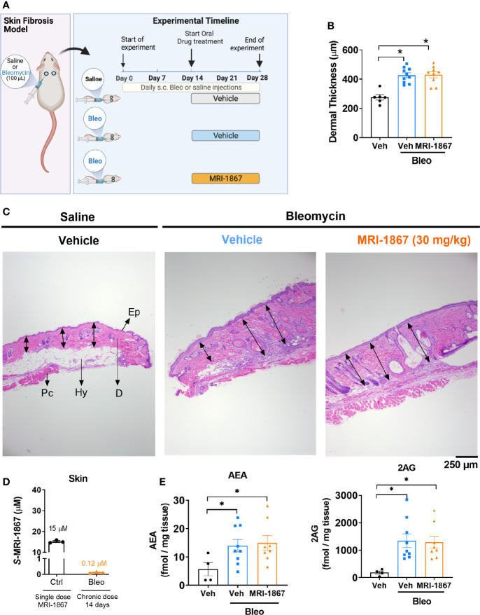
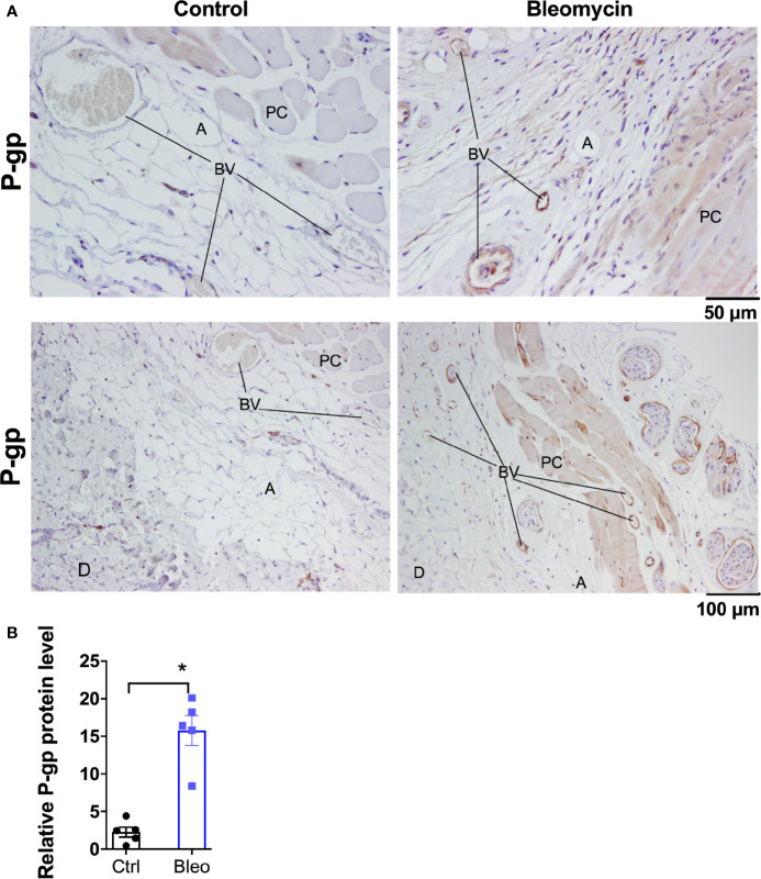
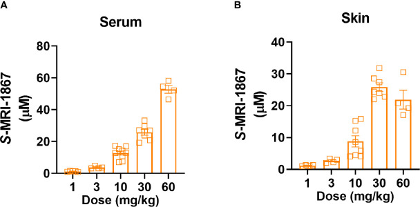
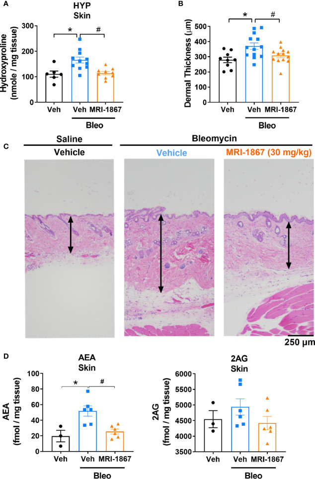
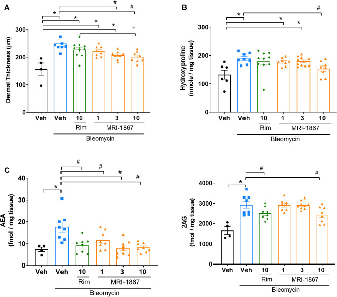
Scleroderma, or systemic sclerosis, is a multi-organ connective tissue disease resulting in fibrosis of the skin, heart, and lungs with no effective treatment. Endocannabinoids acting via cannabinoid-1 receptors (CB1R) and increased activity of inducible NO synthase (iNOS) promote tissue fibrosis including skin fibrosis, and joint targeting of these pathways may improve therapeutic efficacy. Recently, we showed that in mouse models of liver, lung and kidney fibrosis, treatment with a peripherally restricted hybrid CB1R/iNOS inhibitor (MRI-1867) yields greater anti-fibrotic efficacy than inhibiting either target alone. Here, we evaluated the therapeutic efficacy of MRI-1867 in bleomycin-induced skin fibrosis. Skin fibrosis was induced in C57BL/6J (B6) and Mdr1a/b-Bcrp triple knock-out (KO) mice by daily subcutaneous injections of bleomycin (2 IU/100 µL) for 28 days. Starting on day 15, mice were treated for 2 weeks with daily oral gavage of vehicle or MRI-1867. Skin levels of MRI-1867 and endocannabinoids were measured by mass spectrometry to assess target exposure and engagement by MRI-1867. Fibrosis was characterized histologically by dermal thickening and biochemically by hydroxyproline content. We also evaluated the potential increase of drug-efflux associated ABC transporters by bleomycin in skin fibrosis, which could affect target exposure to test compounds, as reported in bleomycin-induced lung fibrosis. Bleomycin-induced skin fibrosis was comparable in B6 and Mdr1a/b-Bcrp KO mice. However, the skin level of MRI-1867, an MDR1 substrate, was dramatically lower in B6 mice (0.023 µM) than in Mdr1a/b-Bcrp KO mice (8.8 µM) due to a bleomycin-induced increase in efflux activity of MDR1 in fibrotic skin. Furthermore, the endocannabinoids anandamide and 2-arachidonylglycerol were elevated 2-4-fold in the fibrotic vs. control skin in both mouse strains. MRI-1867 treatment attenuated bleomycin-induced established skin fibrosis and the associated increase in endocannabinoids in Mdr1a/b-Bcrp KO mice but not in B6 mice. We conclude that combined inhibition of CB1R and iNOS is an effective anti-fibrotic strategy for scleroderma. As bleomycin induces an artifact in testing antifibrotic drug candidates that are substrates of drug-efflux transporters, using Mdr1a/b-Bcrp KO mice for preclinical testing of such compounds avoids this pitfall.
Keywords: ATP-binding cassette transporters; P-gp (P-glycoprotein); bleomycin; cannabinoid (CB) receptor 1; endocannabinoids; peripheral CB1R antagonist; polypharmacology; skin fibrosis.
Copyright © 2021 Zawatsky, Park, Abdalla, Kunos, Iyer and Cinar.
Conflict of interest statement
This study received funding from SCOPUS BIOPHARMA INC., NY, USA. The funder was not involved in the study design, collection, analysis, interpretation of data, the writing of this article or the decision to submit it for publication. RC, GK, and MRI are listed as coinventors on a US patent covering MRI-1867 and related compounds (patent no. US 9,765,031 B2). The remaining authors declare that the research was conducted in the absence of any commercial or financial relationships that could be construed as a potential conflict of interest.
Figures
Similar articles
-
Targeting cannabinoid receptor 1 for antagonism in pro-fibrotic alveolar macrophages mitigates pulmonary fibrosis.JCI Insight. 2025 Jul 3;10(15):e187967. doi: 10.1172/jci.insight.187967. eCollection 2025 Aug 8. JCI Insight. 2025. PMID: 40608428 Free PMC article.
-
CB1 R and iNOS are distinct players promoting pulmonary fibrosis in Hermansky-Pudlak syndrome.Clin Transl Med. 2021 Jul;11(7):e471. doi: 10.1002/ctm2.471. Clin Transl Med. 2021. PMID: 34323400 Free PMC article.
-
Inactivation of fatty acid amide hydrolase exacerbates experimental fibrosis by enhanced endocannabinoid-mediated activation of CB1.Ann Rheum Dis. 2012 Dec;71(12):2051-4. doi: 10.1136/annrheumdis-2012-201823. Epub 2012 Aug 22. Ann Rheum Dis. 2012. PMID: 22915616
-
The therapeutic potential of second and third generation CB1R antagonists.Pharmacol Ther. 2020 Apr;208:107477. doi: 10.1016/j.pharmthera.2020.107477. Epub 2020 Jan 9. Pharmacol Ther. 2020. PMID: 31926199 Free PMC article. Review.
-
The Role of ABC Transporters in Skin Cells Exposed to UV Radiation.Int J Mol Sci. 2022 Dec 21;24(1):115. doi: 10.3390/ijms24010115. Int J Mol Sci. 2022. PMID: 36613554 Free PMC article. Review.
Cited by
-
Pharmacological Evaluation of Cannabinoid Receptor Modulators Using GRABeCB2.0 Sensor.Int J Mol Sci. 2024 May 3;25(9):5012. doi: 10.3390/ijms25095012. Int J Mol Sci. 2024. PMID: 38732230 Free PMC article.
-
An Integrative Multiomics Framework for Identification of Therapeutic Targets in Pulmonary Fibrosis.Adv Sci (Weinh). 2023 Jun;10(16):e2207454. doi: 10.1002/advs.202207454. Epub 2023 Apr 10. Adv Sci (Weinh). 2023. PMID: 37038090 Free PMC article.
-
Advances in targeted liquid chromatography-tandem mass spectrometry methods for endocannabinoid and N-acylethanolamine quantification in biological matrices: A systematic review.Mass Spectrom Rev. 2025 May-Jun;44(3):513-538. doi: 10.1002/mas.21897. Epub 2024 Jul 3. Mass Spectrom Rev. 2025. PMID: 38958096 Free PMC article.
-
Highly Selective Drug-Derived Fluorescent Probes for the Cannabinoid Receptor Type 1 (CB1R).J Med Chem. 2024 Jul 25;67(14):11841-11867. doi: 10.1021/acs.jmedchem.4c00465. Epub 2024 Jul 11. J Med Chem. 2024. PMID: 38990855 Free PMC article.
-
Targeting cannabinoid receptor 1 for antagonism in pro-fibrotic alveolar macrophages mitigates pulmonary fibrosis.JCI Insight. 2025 Jul 3;10(15):e187967. doi: 10.1172/jci.insight.187967. eCollection 2025 Aug 8. JCI Insight. 2025. PMID: 40608428 Free PMC article.
References
-
- Moinzadeh P, Aberer E, Ahmadi-Simab K, Blank N, Distler JH, Fierlbeck G, et al. . All Participating, Disease Progression in Systemic Sclerosis-Overlap Syndrome Is Significantly Different From Limited and Diffuse Cutaneous Systemic Sclerosis. Ann Rheum Dis (2015) 74:730–7. doi: 10.1136/annrheumdis-2013-204487 - DOI - PMC - PubMed
-
- de-Sa-Earp AP, do Nascimento AP, Carneiro SC, Porto LC, Monte-Alto-Costa A. Dermal Dendritic Cell Population and Blood Vessels Are Diminished in the Skin of Systemic Sclerosis Patients: Relationship With Fibrosis Degree and Disease Duration. Am J Dermatopathol (2013) 35:438–44. doi: 10.1097/DAD.0b013e3182712d1a - DOI - PubMed
Publication types
MeSH terms
Substances
LinkOut - more resources
Full Text Sources
Medical
Molecular Biology Databases
Research Materials