. 2021 Sep 28:12:737458.
doi: 10.3389/fmicb.2021.737458. eCollection 2021.

DexA70, the Truncated Form of a Self-Produced Dextranase, Effectively Disrupts Streptococcus mutans Biofilm

Affiliations

DexA70, the Truncated Form of a Self-Produced Dextranase, Effectively Disrupts Streptococcus mutans Biofilm

Nan Liu et al. Front Microbiol. .

Abstract

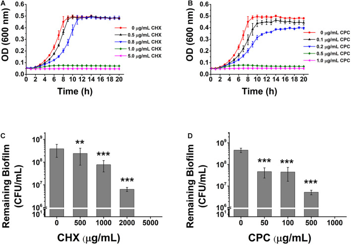
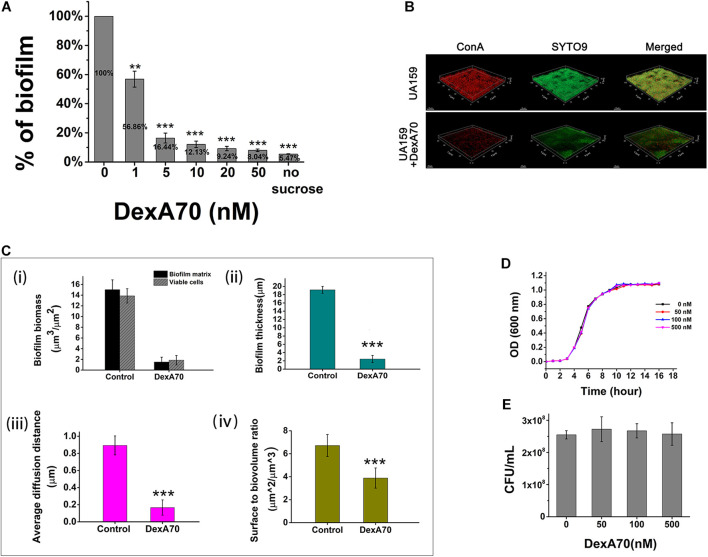
Billions of people suffer from dental caries every year in spite of the effort to reduce the prevalence over the past few decades. Streptococcus mutans is the leading member of a specific group of cariogenic bacteria that cause dental caries. S. mutans forms biofilm, which is highly resistant to harsh environment, host immunity, and antimicrobial treatments. In this study, we found that S. mutans biofilm is highly resistant to both antimicrobial agents and lysozyme. DexA70, the truncated form of DexA (amino acids 100-732), a dextranase in S. mutans, prevents S. mutans biofilm formation and disassembles existing biofilms within minutes at nanomolar concentrations when supplied exogenously. DexA70 treatment markedly enhances biofilm sensitivity to antimicrobial agents and lysozyme, indicating its great potential in combating biofilm-related dental caries.

Keywords: DexA70; Streptococcus mutans; biofilm; dental caries; lysozyme; polysaccharides.


Conflict of interest statement

The authors have filed a patent application on the use of DexA70.

Figures

FIGURE 1
FIGURE 1
The effects of CHX and CPC on S. mutans planktonic cells or biofilm. The planktonic cell growth curve and viable cells remaining in biofilms after treatment were tested. (A,B) The growth curves of S. mutans in BHI media containing different concentrations of CHX (A) or CPC (B). (C,D) CFU enumeration for remaining viable cells in biofilms treated with CHX (C) or CPC (D). Means ± SD from at least three independent experiments with technical triplicates are shown. Student’s t-test by SPSS 15 was used for data analysis. A p-value < 0.05 was considered statistically significant. **p < 0.01; ***p < 0.001.
FIGURE 2
FIGURE 2
Lysozyme inhibits S. mutans growth and biofilm formation. (A) The growth of S. mutans in BHI broth in various concentrations of lysozyme. (B) The quantification of biofilm formation in various concentrations of lysozyme. (C) Pre-formed biofilm treated with various concentrations of lysozyme. Statistical significance is indicated as compared with untreated biofilms using a t-test. All of the values shown represent the mean ± standard deviation of the results from three independent experiments. *p < 0.05; **p < 0.01; ***p < 0.001.
FIGURE 3
FIGURE 3
DexA70 inhibits biofilm formation without affecting bacterial growth. (A) DexA70 inhibited UA159 biofilm formation in a dose-dependent manner. Inhibition effect was calculated by the ratio of remaining biofilm biomass post DexA70 treatment to that of untreated control. (B) Exogenous DexA70 efficiently prevented the biofilm formation by UA159. Images were 1-day-old biofilm grown with/without DexA70 and corresponding CLSM z-stack images (dextran in red stained by ConA and viable cells in green stained by SYTO9). 63 × oil immersion magnification, scale bar = 15 μm. (C) Morphological parameters were evaluated by processing CLSM stack images of biofilms, cultivated with/without DexA70. (i). Biofilm biomass and viable cell biomass. (ii). biofilm thickness. (iii). Average diffusion distance of biofilm. (iv). Surface to biovolume ratio of biofilm. Data were calculated from three independent measurements and were reported as average. (D,E) The planktonic growth curve (D) and the colony-forming unit enumeration (E) of UA159 in BHI media with various concentrations of DexA70. The exogenous DexA70 does not affect cell growth. Statistical significance is indicated as compared with negative control using a t-test. **p < 0.01; ***p < 0.001.
FIGURE 4
FIGURE 4
Synergistic effect of DexA70 and lysozyme (LZM) on biofilm treatment. (A) The comparison between DexA and DexA70 in biofilm dispersion. Enzymes were added at 50, 100, 200, and 500 nM; the activity of DexA70 is much higher than DexA. Means ± SD are shown. (B) DexA70 disassembled 12 h biofilm in a dose-dependent manner with or without 2 mg/ml lysozyme. Means ± SD are shown. (C) The three-dimensional CLSM images for 12 h Streptococcus mutans biofilm treated with/without DexA70 and lysozyme (stained in red by ConA and green by SYTO9). (D) Quantitative analysis of biofilm structure in different conditions containing the biomass, mean thickness, thickness distribution, and surface-to-volume ratio. (i). Biofilm biomass and viable cell biomass. (ii). biofilm thickness. (iii). Average diffusion distance of biofilm. (iv). Surface to biovolume ratio of biofilm. Data were calculated from three independent measurements and were reported as average. Student’s t-test was used to conduct statistical analyses, and differences were considered significant when *p < 0.05. **p < 0.01; ***p < 0.001.
FIGURE 5
FIGURE 5
DexA70 and lysozyme make a dispersing and killing strategy. (A) The dry weight of UA159 biofilm treated with/without DexA70 and lysozyme. Co-administration of DexA70 and lysozyme efficiently reduced biofilm biomass. (B,C). CFU enumeration for remaining viable cells in biofilms treated with/without DexA70 and lysozyme. (B) Adding DexA70 and lysozyme (LZM) at the beginning of biofilm formation. (C) Adding enzymes after biofilm formation and incubating for 30 min. Experiments were performed in triplicate, and the results were shown as the mean ± SD. (D) LIVE/DEAD BacLight bacterial viability assay. Images show 12 h biofilm and cells prior to (0 min) and post 30 min of DexA70 and lysozyme treatment. Live bacteria are stained in green by SYTO 9 and dead bacteria are stained in red by propidium iodide. 63 × oil immersion magnification, scale bar = 15 μm. (E) Biomass (μm3/μm2) of alive and damaged cells was calculated by processing CLSM stack images. Experiments were performed in triplicate, and the results are shown as the mean ± SD. Statistical significance is indicated as compared with negative groups untreated with enzymes using a t-test. *p < 0.05; **p < 0.01.
FIGURE 6
FIGURE 6
DexA treatment improved biofilm sensitivity to Chlorhexidine and Cetylpyridinium Chloride. MBIC (A) and MBEC (B) of biofilm treated with (⚫) and without (■) DexA. Arrows indicate the corresponding values of MBIC and MBEC. (C) MBVEC of biofilm treated with CHX (left) or CPC (right) and with/without DexA70. Means ± SD are shown. t-test was performed for testing differences between groups. *p < 0.05; **p < 0.01; ***p < 0.001.

Similar articles

Cited by

References

    1. Aires A., Barreto A. S., Semedo-Lemsaddek T. (2020). Antimicrobial effects of essential oils on oral microbiota biofilms: the toothbrush in vitro model. Antibiotics (Basel) 10:21. 10.3390/antibiotics10010021 - DOI - PMC - PubMed
    1. Bowden G. H., Hamilton I. R. (1998). Survival of oral bacteria. Crit. Rev. Oral Biol. Med. 9 54–85. 10.1177/10454411980090010401 - DOI - PubMed
    1. Bowen W. H., Burne R. A., Wu H., Koo H. (2018). Oral biofilms: pathogens, matrix, and polymicrobial interactions in microenvironments. Trends Microbiol. 26 229–242. 10.1016/j.tim.2017.09.008 - DOI - PMC - PubMed
    1. Branda S. S., Vik S., Friedman L., Kolter R. (2005). Biofilms: the matrix revisited. Trends Microbiol. 13 20–26. 10.1016/j.tim.2004.11.006 - DOI - PubMed
    1. Brown M. R. W., Gilbert P. (1993). Sensitivity of biofilms to antimicrobial agents. J. Appl. Bacteriol. 74 S87–S97. 10.1111/j.1365-2672.1993.tb04345.x - DOI - PubMed