

Tumor-Derived Exosomal Protein Tyrosine Phosphatase Receptor Type O Polarizes Macrophage to Suppress Breast Tumor Cell Invasion and Migration
- PMID: 34650968
- PMCID: PMC8505750
- DOI: 10.3389/fcell.2021.703537
Tumor-Derived Exosomal Protein Tyrosine Phosphatase Receptor Type O Polarizes Macrophage to Suppress Breast Tumor Cell Invasion and Migration
Abstract
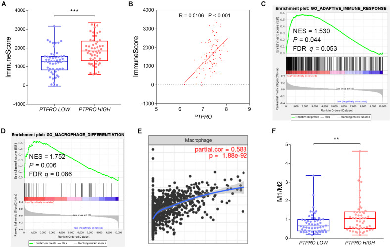
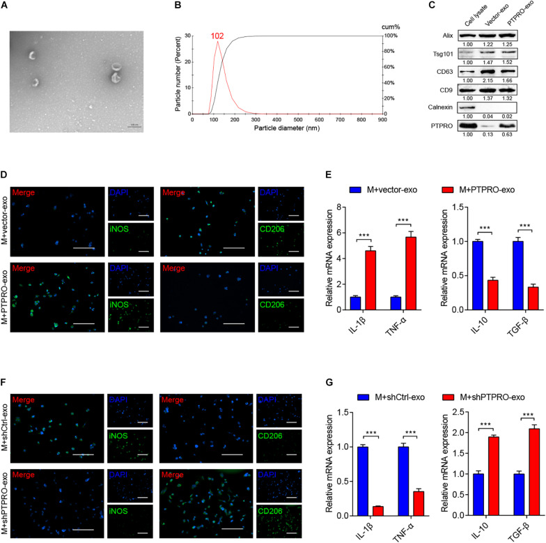
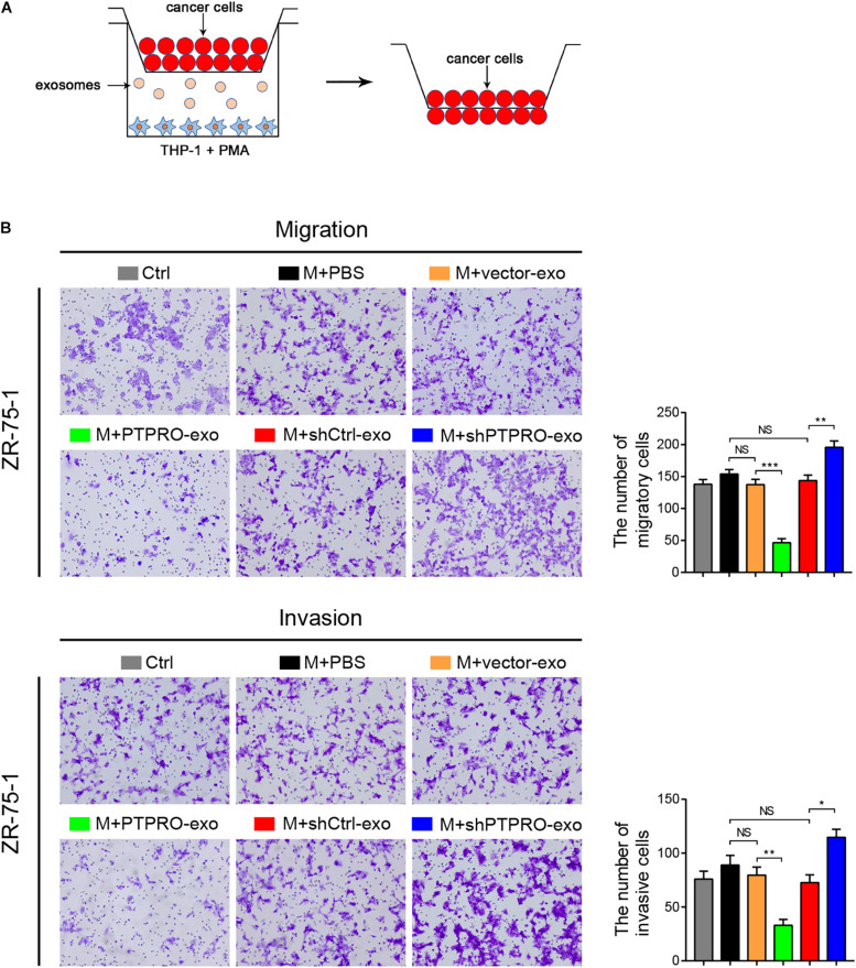
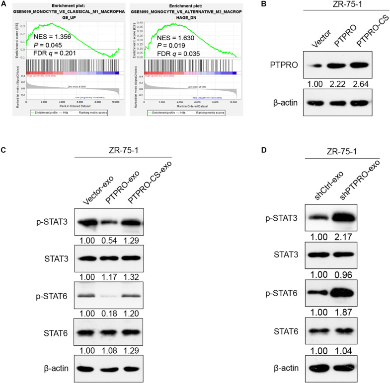
Tumor-derived exosomes, containing multiple nucleic acids and proteins, have been implicated to participate in the interaction between tumor cells and microenvironment. However, the functional involvement of phosphatases in tumor-derived exosomes is not fully understood. We and others previously demonstrated that protein tyrosine phosphatase receptor type O (PTPRO) acts as a tumor suppressor in multiple cancer types. In addition, its role in tumor immune microenvironment remains elusive. Bioinformatical analyses revealed that PTPRO was closely associated with immune infiltration, and positively correlated to M1-like macrophages, but negatively correlated to M2-like macrophages in breast cancer tissues. Co-cultured with PTPRO-overexpressing breast cancer cells increased the proportion of M1-like tumor-associated macrophages (TAMs) while decreased that of M2-like TAMs. Further, we observed that tumor-derived exosomal PTPRO induced M1-like macrophage polarization, and regulated the corresponding functional phenotypes. Moreover, tumor cell-derived exosomal PTPRO inhibited breast cancer cell invasion and migration, and inactivated STAT signaling in macrophages. Our data suggested that exosomal PTPRO inhibited breast cancer invasion and migration by modulating macrophage polarization. Anti-tumoral effect of exosomal PTPRO was mediated by inactivating STAT family in macrophages. These findings highlight a novel mechanism of tumor invasion regulated by tumor-derived exosomal tyrosine phosphatase, which is of translational potential for the therapeutic strategy against breast cancer.
Keywords: breast cancer; invasion and migration; macrophage polarization; protein tyrosine phosphatase receptor type O; tumor-derived exosomes.
Copyright © 2021 Dong, Xie, Jiang, Li, Lin, Pang, Xiong, Zheng, Ke, Chen, Li and Zhang.
Conflict of interest statement
The authors declare that the research was conducted in the absence of any commercial or financial relationships that could be construed as a potential conflict of interest.
Figures

Similar articles
-
Tumor-derived exosomal miR-148b-3p mediates M2 macrophage polarization via TSC2/mTORC1 to promote breast cancer migration and invasion.Thorac Cancer. 2023 Jun;14(16):1477-1491. doi: 10.1111/1759-7714.14891. Epub 2023 May 5. Thorac Cancer. 2023. PMID: 37144254 Free PMC article.
-
Tumor-derived Exosomal ENO2 Modulates Polarization of Tumor-associated Macrophages through Reprogramming Glycolysis to Promote Progression of Diffuse Large B-cell Lymphoma.Int J Biol Sci. 2024 Jan 1;20(3):848-863. doi: 10.7150/ijbs.91154. eCollection 2024. Int J Biol Sci. 2024. PMID: 38250157 Free PMC article.
-
Cancer-derived exosomal miR-138-5p modulates polarization of tumor-associated macrophages through inhibition of KDM6B.Theranostics. 2021 May 3;11(14):6847-6859. doi: 10.7150/thno.51864. eCollection 2021. Theranostics. 2021. PMID: 34093857 Free PMC article.
-
Exosomal ncRNAs facilitate interactive 'dialogue' between tumor cells and tumor-associated macrophages.Cancer Lett. 2023 Jan 1;552:215975. doi: 10.1016/j.canlet.2022.215975. Epub 2022 Oct 25. Cancer Lett. 2023. PMID: 36306940 Review.
-
Exosomal MicroRNAs as Mediators of Cellular Interactions Between Cancer Cells and Macrophages.Front Immunol. 2020 Jun 11;11:1167. doi: 10.3389/fimmu.2020.01167. eCollection 2020. Front Immunol. 2020. PMID: 32595638 Free PMC article. Review.
Cited by
-
Regulatory Functions of Protein Tyrosine Phosphatase Receptor Type O in Immune Cells.Front Immunol. 2021 Nov 22;12:783370. doi: 10.3389/fimmu.2021.783370. eCollection 2021. Front Immunol. 2021. PMID: 34880876 Free PMC article. Review.
-
Decoding the Tumor Microenvironment: Exosome-Mediated Macrophage Polarization and Therapeutic Frontiers.Int J Biol Sci. 2025 Jun 20;21(9):4187-4214. doi: 10.7150/ijbs.114222. eCollection 2025. Int J Biol Sci. 2025. PMID: 40612677 Free PMC article. Review.
-
Exosomes derived from natural killer cells: transforming immunotherapy for aggressive breast cancer.Med Oncol. 2025 Mar 18;42(4):114. doi: 10.1007/s12032-025-02647-y. Med Oncol. 2025. PMID: 40100465 Review.
-
Tumor cell-derived exosomes regulate macrophage polarization: Emerging directions in the study of tumor genesis and development.Heliyon. 2023 Aug 22;9(9):e19296. doi: 10.1016/j.heliyon.2023.e19296. eCollection 2023 Sep. Heliyon. 2023. PMID: 37662730 Free PMC article. Review.
-
Tumor-derived extracellular vesicles modulate innate immune responses to affect tumor progression.Front Immunol. 2022 Nov 2;13:1045624. doi: 10.3389/fimmu.2022.1045624. eCollection 2022. Front Immunol. 2022. PMID: 36405712 Free PMC article. Review.
References
LinkOut - more resources
Full Text Sources