Increased Nuclear Transporter Importin 7 Contributes to the Tumor Growth and Correlates With CD8 T Cell Infiltration in Cervical Cancer
- PMID: 34650978
- PMCID: PMC8505702
- DOI: 10.3389/fcell.2021.732786
Increased Nuclear Transporter Importin 7 Contributes to the Tumor Growth and Correlates With CD8 T Cell Infiltration in Cervical Cancer
Abstract
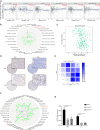
Background: Importin 7 (IPO7), a karyopherin-β protein, is involved in various tumorigenesis and progression abilities by mediating the nuclear import of oncoproteins. However, the exact biological functions of IPO7 remain to be further elucidated. Materials and Methods: TCGA and GEO datasets were used to identify dysregulated expression of IPO7 in various cancers. Gain-of-function and loss-of-function analyses were used to identify the oncogenic functions of IPO7 in vitro and in vivo. Moreover, LC-MS/MS and parallel reaction monitoring analysis were used to comparatively profiled IPO7-related proteomics and potential molecular machinery. Results: Our works demonstrated that the expression of IPO7 was upregulated and was correlated with a poor prognosis in cervical cancer. In vitro and in vivo experiments demonstrated that knockdown of IPO7 inhibited the proliferation of HeLa and C-4 I cells. LC-MS/MS analysis showed that IPO7-related cargo proteins mainly were enriched in gene transcription regulation. Then independent PRM analysis for the first time demonstrated that 32 novel IPO7 cargo proteins, such as GTF2I, RORC1, PSPC1, and RBM25. Moreover, IPO7 contributed to activating the PI3K/AKT-mTOR pathway by mediating the nuclear import of GTF2I in cervical cancer cells. Intriguingly, we found that the IPO7 expression was negatively correlated with CD8 T cell infiltration via regulating the expression of CD276 in cervical cancer. Conclusion: This study enhances our understanding of IPO7 nuclear-cytoplasmic translocation and might reveal novel potential therapeutic targets. The results of a negative correlation between the IPO7 and CD8 T cell infiltration indicate that the IPO7 might play an important impact on the immune microenvironment of cervical cancer.
Keywords: IPO7; cervical cancer; immune infiltration; mass spectrometry; proteome.
Copyright © 2021 Chen, Hu, Teng and Yang.
Conflict of interest statement
The authors declare that the research was conducted in the absence of any commercial or financial relationships that could be construed as a potential conflict of interest.
Figures


Similar articles
-
Forkhead Box M1 Is Essential for Nuclear Localization of Glioma-associated Oncogene Homolog 1 in Glioblastoma Multiforme Cells by Promoting Importin-7 Expression.J Biol Chem. 2015 Jul 24;290(30):18662-70. doi: 10.1074/jbc.M115.662882. Epub 2015 Jun 17. J Biol Chem. 2015. PMID: 26085085 Free PMC article.
-
IPO7 promotes pancreatic cancer progression via regulating ERBB pathway.Clinics (Sao Paulo). 2022 May 16;77:100044. doi: 10.1016/j.clinsp.2022.100044. eCollection 2022. Clinics (Sao Paulo). 2022. PMID: 35588577 Free PMC article.
-
SWATH-MS based quantitative proteomics analysis reveals that curcumin alters the metabolic enzyme profile of CML cells by affecting the activity of miR-22/IPO7/HIF-1α axis.J Exp Clin Cancer Res. 2018 Jul 25;37(1):170. doi: 10.1186/s13046-018-0843-y. J Exp Clin Cancer Res. 2018. PMID: 30045750 Free PMC article.
-
Identification of ZNF217, hnRNP-K, VEGF-A and IPO7 as targets for microRNAs that are downregulated in prostate carcinoma.Int J Cancer. 2013 Feb 15;132(4):775-84. doi: 10.1002/ijc.27731. Epub 2012 Aug 3. Int J Cancer. 2013. PMID: 22815235
-
FAM83H overexpression predicts worse prognosis and correlates with less CD8+ T cells infiltration and Ras-PI3K-Akt-mTOR signaling pathway in pancreatic cancer.Clin Transl Oncol. 2020 Dec;22(12):2244-2252. doi: 10.1007/s12094-020-02365-z. Epub 2020 May 18. Clin Transl Oncol. 2020. PMID: 32424701
Cited by
-
Investigation and development of natural products that target chemotherapy resistance factors in cancer cells.J Nat Med. 2025 Sep;79(5):1005-1016. doi: 10.1007/s11418-025-01942-2. Epub 2025 Aug 8. J Nat Med. 2025. PMID: 40779221 Free PMC article. Review.
-
Nuclear localization signal-tagged systems: relevant nuclear import principles in the context of current therapeutic design.Chem Soc Rev. 2024 Jan 2;53(1):204-226. doi: 10.1039/d1cs00269d. Chem Soc Rev. 2024. PMID: 38031452 Free PMC article. Review.
-
Cervical cancer immune infiltration microenvironment identification, construction of immune scores, assisting patient prognosis and immunotherapy.Front Immunol. 2023 Mar 10;14:1135657. doi: 10.3389/fimmu.2023.1135657. eCollection 2023. Front Immunol. 2023. PMID: 36969161 Free PMC article.
-
Nuclear transport maintenance of USP22-AR by Importin-7 promotes breast cancer progression.Cell Death Discov. 2023 Jul 1;9(1):211. doi: 10.1038/s41420-023-01525-8. Cell Death Discov. 2023. PMID: 37391429 Free PMC article.
-
Protein Signature Differentiating Neutrophils and Myeloid-Derived Suppressor Cells Determined Using a Human Isogenic Cell Line Model and Protein Profiling.Cells. 2024 May 7;13(10):795. doi: 10.3390/cells13100795. Cells. 2024. PMID: 38786019 Free PMC article.
References
LinkOut - more resources
Full Text Sources
Research Materials
Miscellaneous

