. 2021 Sep 3;4(5):1514-1527.
doi: 10.1021/acsptsci.1c00080. eCollection 2021 Oct 8.

Inhibition of Severe Acute Respiratory Syndrome Coronavirus 2 Replication by Hypertonic Saline Solution in Lung and Kidney Epithelial Cells

Affiliations

Inhibition of Severe Acute Respiratory Syndrome Coronavirus 2 Replication by Hypertonic Saline Solution in Lung and Kidney Epithelial Cells

Rafael R G Machado et al. ACS Pharmacol Transl Sci. .

Abstract

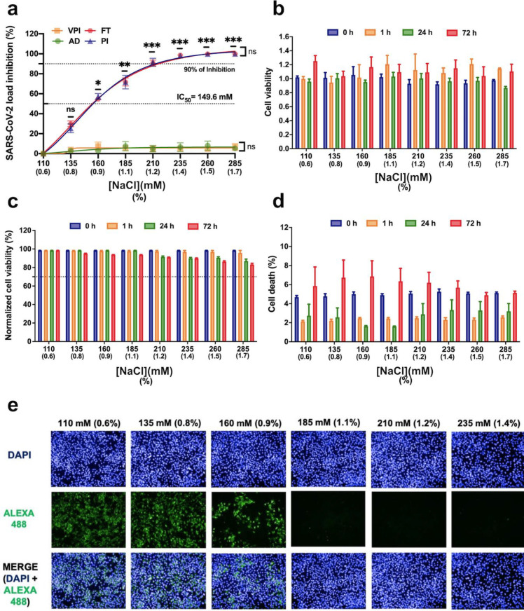
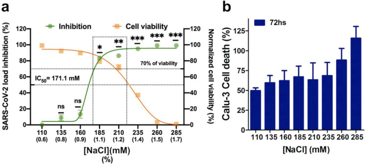
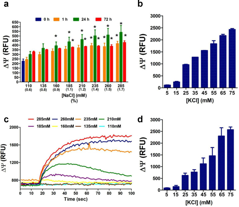
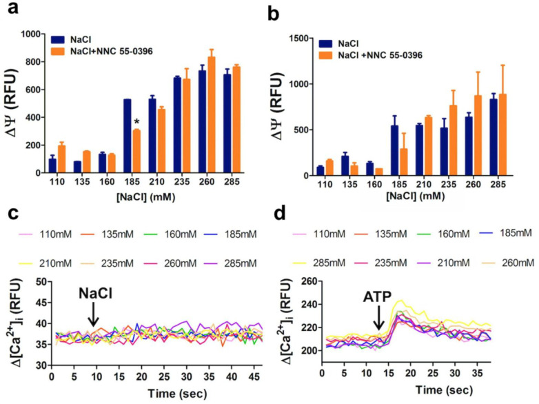
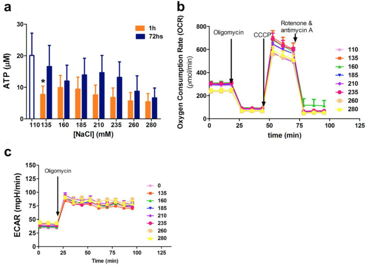
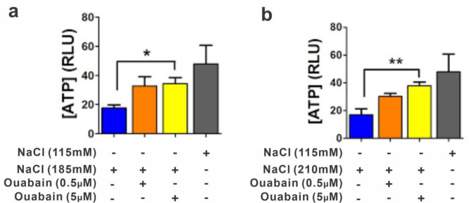
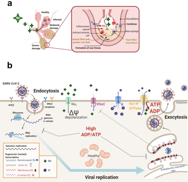
An unprecedented global health crisis has been caused by a new virus called severe acute respiratory syndrome coronavirus 2 (SARS-CoV-2). We performed experiments to test if a hypertonic saline solution was capable of inhibiting virus replication. Our data show that 1.2% NaCl inhibited virus replication by 90%, achieving 100% of inhibition at 1.5% in the nonhuman primate kidney cell line Vero, and 1.1% of NaCl was sufficient to inhibit the virus replication by 88% in human epithelial lung cell line Calu-3. Furthermore, our results indicate that the inhibition is due to an intracellular mechanism and not to the dissociation of the spike SARS-CoV-2 protein and its human receptor. NaCl depolarizes the plasma membrane causing a low energy state (high ADP/ATP concentration ratio) without impairing mitochondrial function, supposedly associated with the inhibition of the SARS-CoV-2 life cycle. Membrane depolarization and intracellular energy deprivation are possible mechanisms by which the hypertonic saline solution efficiently prevents virus replication in vitro assays.


Conflict of interest statement

The authors declare no competing financial interest.

Figures

Figure 1
Figure 1
Antiviral activity of NaCl against SARS-CoV-2 in vitro assay. (a) Y axis labeling of the graph represents percentage inhibition of virus load in cellular supernatant by increasing NaCl concentration. Four different times of NaCl addition were evaluated, comprising the virus preincubation (VPI, orange line), absorption (AD, green line), postinfection (PI, blue line), and adsorption plus postinfection (FT, red line). Error bars indicate the standard error of the mean of three independent experiments with each one carried out in triplicate. *p < 0.05, **p < 0.005, and ***p < 0.0005 when compared to 110 mM NaCl. Vero CCL-81 cell viability was not significantly impaired in the presence of increasing concentrations of NaCl (110 mM up to 285 mM). (b) Cell viability was determined following 0, 1, 24, and 72 h post-treatment with different concentrations of NaCl using AlamarBlue Cell Viability Reagent (Thermo Fisher Scientific). (c) Cell viability was also determined by quantification of LDH released into the culture supernatant from cells with damaged membranes, using the CytoTox 96 Non-Radioactive Cytotoxicity Assay (Promega Corp., Madison, WI). Cell viability was normalized to that determined with cells kept at a physiological NaCl concentration (110 mM NaCl). (d) Percentage of dead cells determined by propidium iodide/Hoechst 33342 staining. Viability below 70% (cell death rates of more than 30%) was considered as evidence of cytotoxicity. Error bars represent the mean ± SEM of three independent experiments carried out in triplicate. (e) Infected cells by SARS-CoV-2 were observed by indirect immunofluorescence (IIF) assay. SARS-CoV-infected and noninfected Vero cells were stained with a convalescent serum, followed by incubation with the Alexa488-conjugated goat antihuman IgG antibody (green). Cells were counterstained with DAPI for nuclear staining (blue). Positive (infected nontreated cells) and negative (noninfected cells) controls are represented in the bottom of the image. Representative images were captured with a 20× objective using the Operetta High Content Imaging System (PerkinElmer).
Figure 2
Figure 2
Effects of increasing NaCl concentrations on Calu-3 cells (lung epithelial cells). (a) The left Y-axis of the graph represents percentage inhibition of virus load in postinfection cellular supernatant. Error bars indicate the standard error of the mean of three independent experiments, each of them carried out in triplicate. *p < 0.05, **p < 0.005, and ***p < 0.0005 when compared to 110 mM NaCl. Cells were treated with increasing concentrations of NaCl (110 mM up to 285 mM). Then, 72 h post-treatment cellular viability was determined. The dotted rectangular area (185 to 210 mM NaCl) shows the concentrations of NaCl that significantly inhibit SARS-CoV-2 replication, and more than 70% of the cells are viable. (b) Percentage of dead cells determined via propidium iodide/Hoechst 33342 staining. Viability below 70% (cell death more than 30%) was considered as evidence of cytotoxicity. Error bars represent the mean ± SEM of three independent experiments carried out in triplicate.
Figure 3
Figure 3
Vero and Calu-3 cell membrane depolarization by increasing concentrations of NaCl and KCl. (a) Membrane potential indicated as relative fluorescence units (RFU) in Vero cells after 0, 1, 24, and 72 h post-treatment with increasing concentrations of NaCl (110 up to 285 mM) as determined by microfluorimetry. Increasing NaCl concentration caused membrane depolarization that was maximal after 24 h and tended to return to control values after 72 h of treatment. (b) Increasing KCl concentrations caused immediate membrane depolarization at concentrations above 25 mM in Vero cells as determined by real time fluorescence measurements. (c) The increasing NaCl concentrations caused an immediate membrane depolarization at concentrations above 160 mM. Membrane depolarization of cells treated with up to 210 mM NaCl returned to the resting point after 70 s. (d) Increasing KCl concentrations caused immediate membrane depolarization at concentrations above 25 mM in Calu-3 cells as determined by real time fluorescence measurements. Data are representative of three independent experiments and shown as mean values ± SEM; one-way ANOVA (*p < 0.05).
Figure 4
Figure 4
Increasing concentrations of NaCl affect ATP homeostasis independently of voltage-gated calcium channels. Membrane potential indicated as relative fluorescence units (RFU) in Vero cells (a) and Calu-3 cells (b) 3 min after the addition of increasing concentrations of NaCl (110 up to 285 mM) as determined by microfluorimetry. NN55-0396 (1 μM), a blocker of Cav Ca2+ channels, was incubated 5 min prior to NaCl challenge. (c, d) Variations in intracellular free calcium [Ca2+]i concentration, indicated as relative fluorescence units (RFU) in Vero cells exposed to increasing concentrations of NaCl (110 up to 285 mM) (c) and upon addition of 10 μM ATP (shown by arrows) (d), as determined by microfluorimetry. The data are representative of three independent experiments and shown as mean values ± SEM; two-way ANOVA (*p ≤ 0.05).
Figure 5
Figure 5
NaCl-induced depolarization affects the cell energetic state without impairment of mitochondrial function. (a) Total cellular ATP quantification of Vero cells treated for 1 h or 72 h with increasing NaCl concentrations by the luciferase assay. The oxygen consumption rate, OCR, (b) and the extracellular acidification rate, ECAR, (c) were measured in cultured Vero cells (40 000 cells/well) after 72 h of NaCl incubation, using Seahorse technology with the Mito Stress Test kit. Sequential injections of oligomycin, carbonyl cyanide 3-chlorophenylhydrazone (CCCP), and rotenone plus antimycin A are indicated. Mitochondrial respiration was evaluated under four different conditions: basal respiration, corresponding to the cell basal consumed oxygen; proton leak, after oligomycin (ATP-synthase blocker) addition; uncoupled, after addition of the respiratory chain uncoupler CCCP, where the oxygen consumed reflects the maximal respiration rate (irreversibly uncoupling from ATP synthesis); and inhibited, through complex I and complex III total inhibition by rotenone and antimycin A, respectively. The ECAR rate was verified by application of the glycolysis stress test kit (Agilent Technologies). The data are representative of three independent experiments and shown as mean values ± SEM; two-way ANOVA (*p ≤ 0.05).
Figure 6
Figure 6
Ouabain treatment abolished the cell energetic state impairment caused by NaCl. Relative quantification of cellular ATP concentrations of Vero cells treated for 1 h with DMEM cell culture medium, containing 115 mM NaCl (resting conditions) or with DMEM with NaCl supplemented to 185 or to 210 mM final NaCl concentrations, as shown in panels a and b, respectively. Relative ATP concentrations were measured by a luciferase assay. The data are representative of three independent experiments and shown as mean values ± SEM; one-way ANOVA (*p ≤ 0.05, **p ≤ 0.01).
Figure 7
Figure 7
Confirmed and suggested mechanisms of NaCl hyperosmotic treatment resulting in SARS-CoV-2 replication inhibition. (a) Illustration of SARS-CoV-2 alveoli infection, which can lead to pulmonary edema, high levels of cytokines, and tissue damage. (b) Hypothesis for a possible mechanism involving the effect of NaCl treatment in the inhibition of SARS-CoV-2 replication in Vero and Calu-3 cells. Hypertonic saline solution causes membrane depolarization and an overflow of Na+ in cells, causing a low energy state (high ADP/ATP concentration ratio), leading to impaired virus replication. Hyperosmotic extracellular NaCl concentrations probably activate sodium-sensitive (but not voltage-sensitive) Nax channels, which are critically involved in body-fluid homeostasis. High cytoplasmic Na+ concentrations can recruit epithelial sodium (Na+) channels (ENaC), further increasing intracellular Na+ concentration, causing cell membranes to depolarize. The activation of K+ channels may also happen to pump out K+. Na+/K+ ATPase activation for restoring resting membrane potential results in increased ADP/ATP ratio. This low energetic state would be detrimental for viral replication. Thus, the imbalance of intracellular K+ concentration may also affect the functioning of different potassium channels that may be important for the virus life cycle. The illustration was created with the web-based tool BioRender (https://biorender.com).

Similar articles

Cited by

References

    1. Zhu N.; Zhang D.; Wang W.; Li X.; Yang B.; Song J.; Zhao X.; Huang B.; Shi W.; Lu R.; Niu P.; Zhan F.; Ma X.; Wang D.; Xu W.; Wu G.; Gao G. F.; Tan W. China Novel Coronavirus Investigating and Research Team. A Novel Coronavirus from Patients with Pneumonia in China, 2019. N. Engl. J. Med. 2020, 382 (8), 727–733. 10.1056/NEJMoa2001017. - DOI - PMC - PubMed
    1. Karim S. S. A. Vaccines and SARS-CoV-2 Variants: The Urgent Need for a Correlate of Protection. Lancet 2021, 397 (10281), 1263–1264. 10.1016/S0140-6736(21)00468-2. - DOI - PMC - PubMed
    1. Abdool Karim S. S.; de Oliveira T. New SARS-CoV-2 Variants — Clinical, Public Health, and Vaccine Implications. N. Engl. J. Med. 2021, 384, 1866–1868. 10.1056/NEJMc2100362. - DOI - PMC - PubMed
    1. Letko M.; Marzi A.; Munster V. Functional Assessment of Cell Entry and Receptor Usage for SARS-CoV-2 and Other Lineage B Betacoronaviruses. Nat. Microbiol 2020, 5 (4), 562–569. 10.1038/s41564-020-0688-y. - DOI - PMC - PubMed
    1. Wang K.; Chen W.; Zhang Z.; Deng Y.; Lian J.-Q.; Du P.; Wei D.; Zhang Y.; Sun X.-X.; Gong L.; Yang X.; He L.; Zhang L.; Yang Z.; Geng J.-J.; Chen R.; Zhang H.; Wang B.; Zhu Y.-M.; Nan G.; Jiang J.-L.; Li L.; Wu J.; Lin P.; Huang W.; Xie L.; Zheng Z.-H.; Zhang K.; Miao J.-L.; Cui H.-Y.; Huang M.; Zhang J.; Fu L.; Yang X.-M.; Zhao Z.; Sun S.; Gu H.; Wang Z.; Wang C.-F.; Lu Y.; Liu Y.-Y.; Wang Q.-Y.; Bian H.; Zhu P.; Chen Z.-N. CD147-Spike Protein Is a Novel Route for SARS-CoV-2 Infection to Host Cells. Signal Transduct Target Ther 2020, 5 (1), 283. 10.1038/s41392-020-00426-x. - DOI - PMC - PubMed

LinkOut - more resources