

Predicting the nature of pleural effusion in patients with lung adenocarcinoma based on 18F-FDG PET/CT
- PMID: 34652524
- PMCID: PMC8519982
- DOI: 10.1186/s13550-021-00850-2
Predicting the nature of pleural effusion in patients with lung adenocarcinoma based on 18F-FDG PET/CT
Abstract
Background: This study aims to establish a predictive model on the basis of 18F-FDG PET/CT for diagnosing the nature of pleural effusion (PE) in patients with lung adenocarcinoma.
Methods: Lung adenocarcinoma patients with PE who underwent 18F-FDG PET/CT were collected and divided into training and test cohorts. PET/CT parameters and clinical information in the training cohort were collected to estimate the independent predictive factors of malignant pleural effusion (MPE) and to establish a predictive model. This model was then applied to the test cohort to evaluate the diagnostic efficacy.
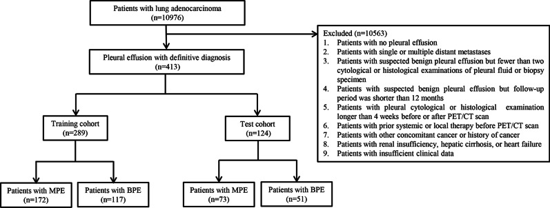
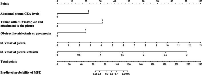
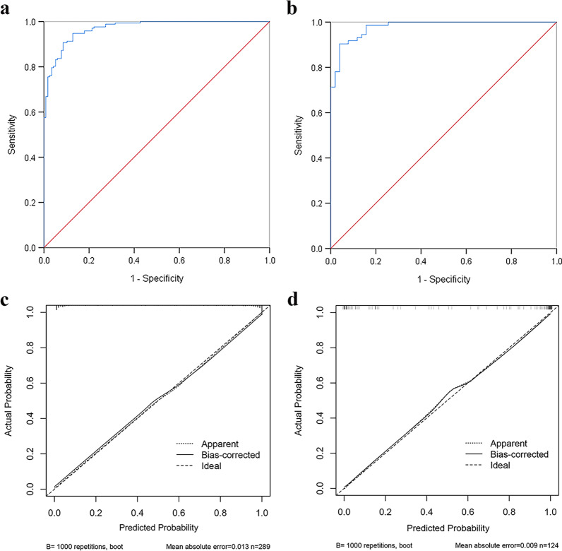
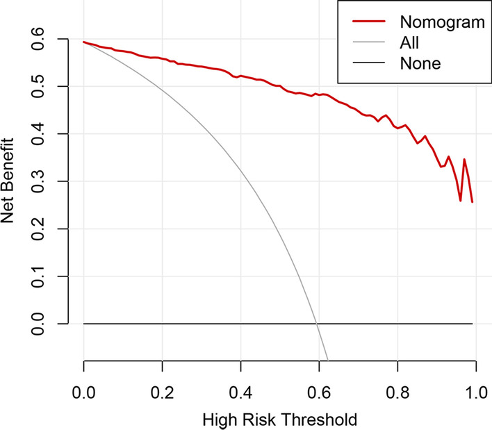
Results: A total of 413 lung adenocarcinoma patients with PE were enrolled in this study, including 245 patients with MPE and 168 patients with benign PE (BPE). The patients were divided into training (289 patients) and test (124 patients) cohorts. CEA, SUVmax of tumor and attachment to the pleura, obstructive atelectasis or pneumonia, SUVmax of pleura, and SUVmax of PE were identified as independent significant factors of MPE and were used to construct a predictive model, which was graphically represented as a nomogram. This predictive model showed good discrimination with the area under the curve (AUC) of 0.970 (95% CI 0.954-0.986) and good calibration. Application of the nomogram in the test cohort still gave good discrimination with AUC of 0.979 (95% CI 0.961-0.998) and good calibration. Decision curve analysis demonstrated that this nomogram was clinically useful.
Conclusions: Our predictive model based on 18F-FDG PET/CT showed good diagnostic performance for PE, which was helpful to differentiate MPE from BPE in patients with lung adenocarcinoma.
Keywords: 18F-FDG; Lung adenocarcinoma; PET/CT; Pleural effusion; Predictive model.
© 2021. The Author(s).
Conflict of interest statement
The authors declare that they have no competing interests.
Figures
References
-
- Rodríguez PF. Diagnosis and treatment of malignant pleural mesothelioma. Arch Bronconeumol. 2015;51:177–184. - PubMed
Grants and funding
LinkOut - more resources
Full Text Sources
