. 2021 Dec;140(12):1709-1731.
doi: 10.1007/s00439-021-02379-9. Epub 2021 Oct 15.

TUBB3 Arg262His causes a recognizable syndrome including CFEOM3, facial palsy, joint contractures, and early-onset peripheral neuropathy

Affiliations

TUBB3 Arg262His causes a recognizable syndrome including CFEOM3, facial palsy, joint contractures, and early-onset peripheral neuropathy

Mary C Whitman et al. Hum Genet. 2021 Dec.

Abstract

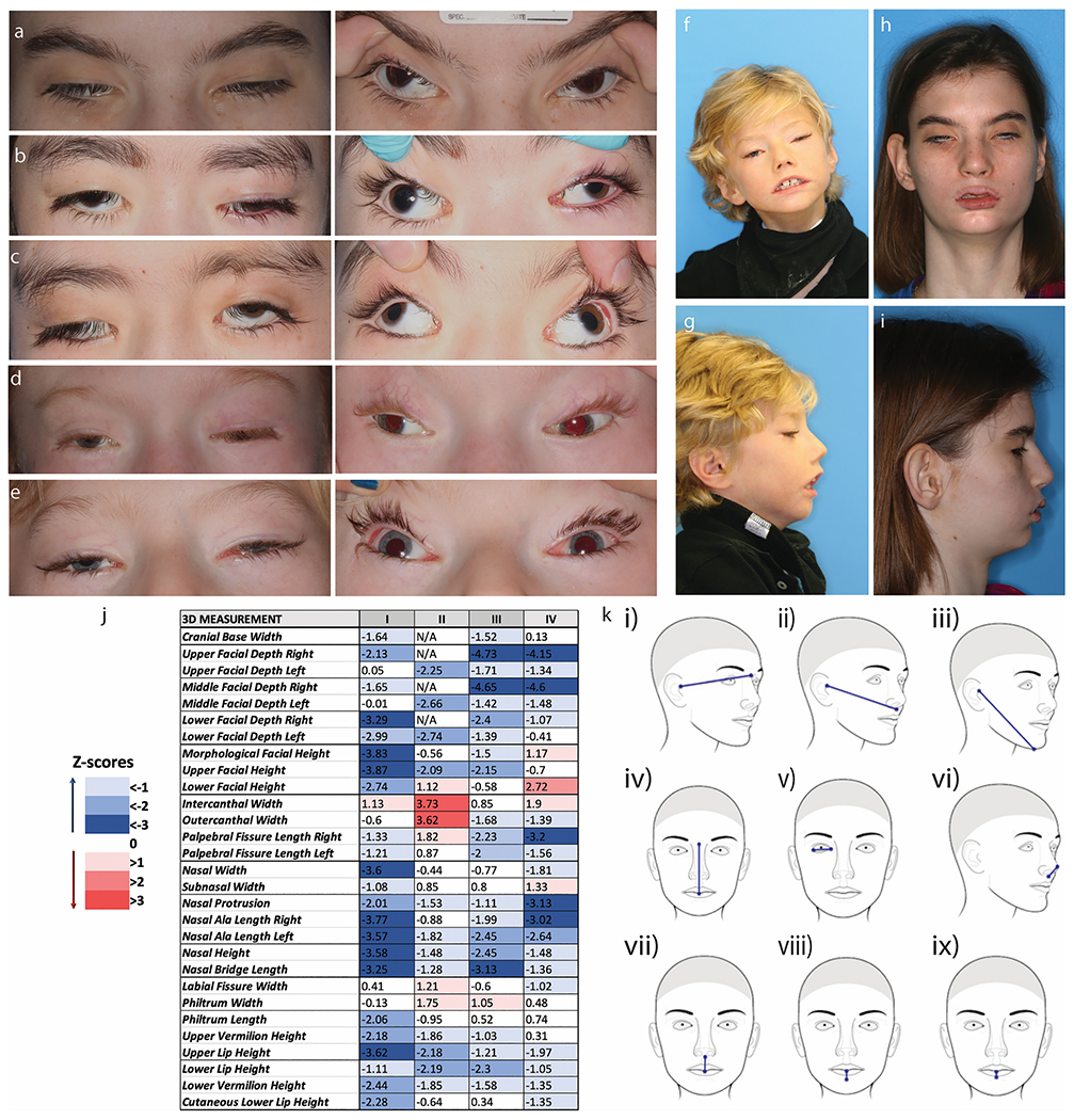
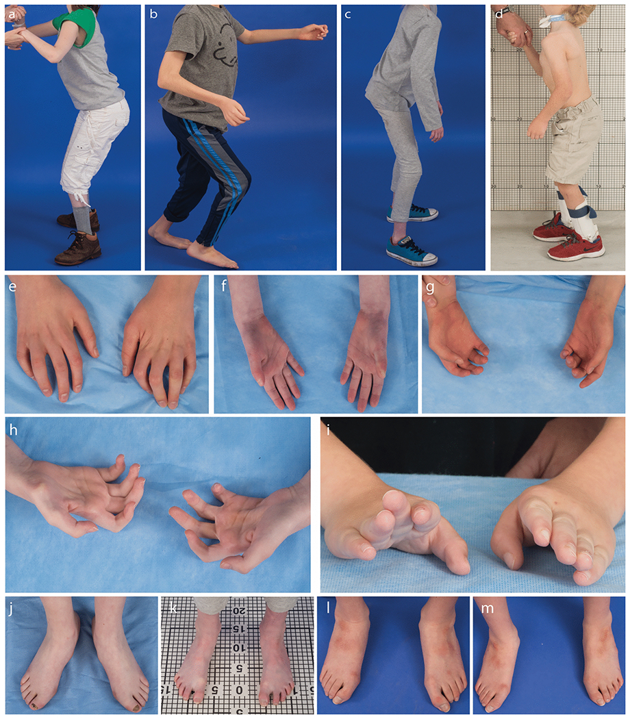
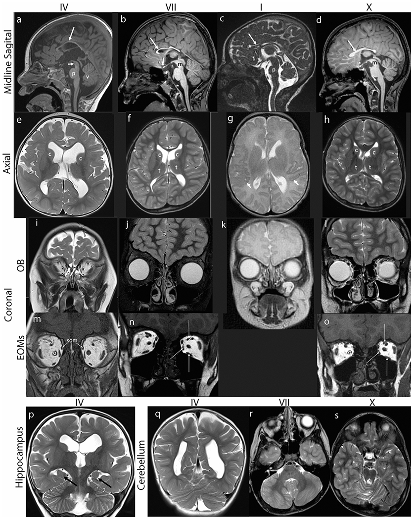
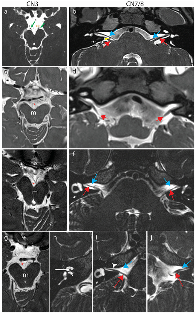
Microtubules are formed from heterodimers of alpha- and beta-tubulin, each of which has multiple isoforms encoded by separate genes. Pathogenic missense variants in multiple different tubulin isoforms cause brain malformations. Missense mutations in TUBB3, which encodes the neuron-specific beta-tubulin isotype, can cause congenital fibrosis of the extraocular muscles type 3 (CFEOM3) and/or malformations of cortical development, with distinct genotype-phenotype correlations. Here, we report fourteen individuals from thirteen unrelated families, each of whom harbors the identical NM_006086.4 (TUBB3):c.785G>A (p.Arg262His) variant resulting in a phenotype we refer to as the TUBB3 R262H syndrome. The affected individuals present at birth with ptosis, ophthalmoplegia, exotropia, facial weakness, facial dysmorphisms, and, in most cases, distal congenital joint contractures, and subsequently develop intellectual disabilities, gait disorders with proximal joint contractures, Kallmann syndrome (hypogonadotropic hypogonadism and anosmia), and a progressive peripheral neuropathy during the first decade of life. Subsets may also have vocal cord paralysis, auditory dysfunction, cyclic vomiting, and/or tachycardia at rest. All fourteen subjects share a recognizable set of brain malformations, including hypoplasia of the corpus callosum and anterior commissure, basal ganglia malformations, absent olfactory bulbs and sulci, and subtle cerebellar malformations. While similar, individuals with the TUBB3 R262H syndrome can be distinguished from individuals with the TUBB3 E410K syndrome by the presence of congenital and acquired joint contractures, an earlier onset peripheral neuropathy, impaired gait, and basal ganglia malformations.


Conflict of interest statement

Conflicts of interest: None of the authors have any relevant conflicts of interest.

Figures

Fig. 1
Fig. 1. Ophthalmological and Craniofacial Evaluation.
All subjects have severe ptosis, extremely limited eye movements, and severe exotropia. a-e Ocular photographs of the five subjects evaluated at NIH (XIII, X, XI, XII, VII). Panels on the left show the severe ptosis. Panels on the right, with the lids artificially raised, display the severe exotropia. Subjects have almost no voluntary eye movements. F-I. Frontal and profile photos of subjects VII (f&g) and XIII (h&i). j. Color scale table depicting the deviation of the soft-tissue measurements in four TUBB3 R262H subjects, expressed in the form of Z-scores, which were computed from the normal values provided by FaceBase. k. Schematic representation of the most negatively affected linear 3D measurements in this cohort. In general, most values are lower than the average normal values, particularly the facial depth measures (i, ii, iii), the upper facial height (iv), the palpebral fissure lengths (v), the protrusion of the nose (vi), and the vertical dimensions of the lips (vii, viii, ix). The only measure with increased or normal values in all four subjects is the intercanthal width.
Fig. 2
Fig. 2. The combination of joint contractures and peripheral neuropathy limit mobility.
Standing photographs of subjects XIII (a), XI (b), X (c), and VII (d) show the wide based, hinged stance subjects require to stand. Representative images of hand (e-i) and feet (h-m) contractures from subjects VII (i), X (f,k), XI (g,m), XII (e,l), and XIII (h,j).
Fig. 3
Fig. 3. Brain MR imaging.
Hypoplasia of corpus callosum, dysmorphic basal ganglia, absence of olfactory bulbs, small extraocular muscles, incomplete hippocampal inversion, and cerebellar abnormalities in subjects IV (a,e,i,m,p,q), VII (b,f,j,n,r), I (c,g,k) and X (d,h,l,o,s). (a-d) Midline sagittal T1 weighted (a, b, d) or FIESTA (c) images demonstrate marked thinning of the corpus callosum (long arrows). (a) The pons (p) appears mildly shortened in height and narrowed in AP diameter. The tegmentum of the midbrain (short arrow) appears slightly thinned and elongated. There is slight uplifting of the vermis (v). (b) The pons is minimally narrowed in AP diameter with slightly accentuated dorsal indentation and the distal midbrain (m) tegmentum is thinned. (c) The pons (p) is hypoplastic, appearing shortened in height and anteroposterior diameter with accentuated dorsal indentation. The vermis is slightly uplifted but normal in size. (d) There is mild thinning of the distal midbrain (m) and the pons is minimally shortened in height with accentuated dorsal indentation. (e-h) Axial T2 weighted MR images of the brain demonstrate asymmetry of the lateral ventricles and dysmorphic basal ganglia. (e) The caudate nuclei (c) and basal ganglia are asymmetric in shape and position, with the left caudate head more dorsally positioned than the right. The white matter appears mildly decreased in thickness. The lateral ventricles are prominent. (f) The caudate heads (c) are asymmetric with the left appearing shorter and wider. Mildly asymmetric shape and positioning of the thalami (t). The Sylvian fissures are asymmetric with the left shorter in length than right. The parietal and right frontal white matter appears mildly decreased in thickness with a small focus of high signal intensity adjacent to the right lateral ventricular atrium. (g) The perisylvian cortex has a serrated appearance raising concern for polymicrogyria (short arrows). (h) The lateral ventricles, caudate heads (c) and thalami (t) are asymmetric in shape. Mildly decreased thickness of right frontal and left > right parietal periventricular white matter. (i-l) Coronal MR images at the level of the cribriform plates (white arrows in I) reveal absent olfactory bulbs and sulci with only small vessels visible. (m-o) Coronal T1 weighted MR images show small medial, superior, and inferior rectus muscles (long white arrows in N and O). The superior oblique muscles (som, arrows, M) are also small and inferomedially positioned. The optic nerves (o) appear medialized. (p-q) Coronal T2 weighted MR images of the brain show (P) incomplete hippocampal inversion bilaterally (black arrows). The 3rd and lateral ventricles are moderately dilated with the left lateral ventricle appearing larger than the right. The body of the left caudate nucleus appears smaller than the right (c), and more inferolaterally positioned. (q) The cerebellum appears mildly rotated. (r) Axial T2 weighted MR image at the level of the posterior fossa shows asymmetric architecture of the cerebellar hemispheres with generalized mild dysmorphism on the right. There is minimal disorganization of the superior cerebellar vermian folia. (s) Axial T2 weighted images show minimal dysmorphism of the superior cerebellar vermis.
Fig. 4
Fig. 4. Cranial nerve MR imaging.
Oculomotor nerve (a,c,e,g) and facial nerve (b,d,f,i,j) and cochlear nerve (h) imaging in comparison subjects (a,b), Subjects IV (c,d), X (e,f), and VII (g-j). (a) Normal CN III. 3 Tesla Axial constructive interference in steady-state (CISS) image through the midbrain at the level of the interpeduncular cistern showing normal CNs III emerging from the ventral aspect of the cerebral peduncles (green arrows). The oculomotor nerves course inferolaterally to the cavernous sinuses in close proximity to the posterior cerebral artery (PCA) on each side (orange asterisk left PCA). (b) Normal CN VII and VIII. 3 Tesla Axial sampling perfection with application optimized contrasts using different flip angle evolution (SPACE) image demonstrating normal anatomy of CN VII (blue arrows) coursing parallel and ventral to CN VIII (red arrows) on each side. The anterior inferior cerebellar artery flow void is seen between the cranial nerves on the right (yellow arrowhead). (c,e,g) Axial sampling perfection with application optimized contrasts using different flip angle evolution (SPACE) images (c) or Axial T2 weighted (e,g) images at the level of the interpeduncular cistern (red asterisk) show apparent absence of the cisternal segments of cranial nerves III which usually course anterolaterally from the ventral aspect of the midbrain (m) on each side. (d,f,i,j) At the level of the superior internal auditory meati, the cisternal segments of cranial nerve VIII are well seen bilaterally (red arrows, d [SPACE], f, i, j [axial T2 weighted]). Cranial nerves VII, which ordinarily course anterior and parallel to VIII are either not seen on either side (d) or appear markedly hypoplastic (blue arrows in f, i, j) and are faintly visualized coursing ventral and parallel to the cranial nerves VIII (red arrows). The right internal auditory meatus is asymmetrically narrowed (i, arrowhead). (h) In subject VII, who has right-sided hearing loss, the cochlear modiolus is thickened and misshapen (longer white arrow) with severe stenosis/atresia of the cochlear aperture (short white arrow). The cochlear division of the right CN VIII, which usually courses through the cochlear aperture, is not visible.

References

    1. Achenbach TM, Ruffle TM (2000) The Child Behavior Checklist and related forms for assessing behavioral/emotional problems and competencies. Pediatr Rev 21: 265–71. doi: 10.1542/pir.21-8-265 - DOI - PubMed
    1. Bahi-Buisson N, Poirier K, Fourniol F, Saillour Y, Valence S, Lebrun N, Hully M, Bianco CF, Boddaert N, Elie C, Lascelles K, Souville I, Consortium LI-T, Beldjord C, Chelly J (2014) The wide spectrum of tubulinopathies: what are the key features for the diagnosis? Brain 137: 1676–700. doi: 10.1093/brain/awu082 - DOI - PubMed
    1. Bonomi M, Vezzoli V, Krausz C, Guizzardi F, Vezzani S, Simoni M, Bassi I, Duminuco P, Di lorgi N, Giavoli C, Pizzocaro A, Russo G, Moro M, Fatti L, Ferlin A, Mazzanti L, Zatelli MC, Cannavo S, Isidori AM, Pincelli AI, Prodam F, Mancini A, Limone P, Tanda ML, Gaudino R, Salerno M, Francesca P, Maghnie M, Maggi M, Persani L, Italian Network on Central H, Italian Network on Central H (2018) Characteristics of a nationwide cohort of patients presenting with isolated hypogonadotropic hypogonadism (IHH). Eur J Endocrinol 178: 23–32. doi: 10.1530/EJE-17-0065 - DOI - PubMed
    1. Brinkley JF, Fisher S, Harris MP, Holmes G, Hooper JE, Jabs EW, Jones KL, Kesselman C, Klein OD, Maas RL, Marazita ML, Selleri L, Spritz RA, van Bakel H, Visel A, Williams TJ, Wysocka J, FaceBase C, Chai Y (2016) The FaceBase Consortium: a comprehensive resource for craniofacial researchers. Development 143: 2677–88. doi: 10.1242/dev.135434 - DOI - PMC - PubMed
    1. Burns TG, King TZ, Spencer KS (2013) Mullen scales of early learning: the utility in assessing children diagnosed with autism spectrum disorders, cerebral palsy, and epilepsy. Appl Neuropsychol Child 2: 33–42. doi: 10.1080/21622965.2012.682852 - DOI - PubMed

MeSH terms

Supplementary concepts