. 2022 Jan:347:113895.
doi: 10.1016/j.expneurol.2021.113895. Epub 2021 Oct 13.

Exosomes derived from bone marrow mesenchymal stromal cells promote remyelination and reduce neuroinflammation in the demyelinating central nervous system

Affiliations

Exosomes derived from bone marrow mesenchymal stromal cells promote remyelination and reduce neuroinflammation in the demyelinating central nervous system

Jing Zhang et al. Exp Neurol. 2022 Jan.

Abstract

Injury of oligodendrocytes (OLs) induces demyelination, and patients with neurodegenerative diseases exhibit demyelination concomitantly with neurological deficit and cognitive impairment. Oligodendrocyte progenitor cells (OPCs) are present in the adult central nervous system (CNS), and they can proliferate, differentiate, and remyelinate axons after damage. However, remyelination therapies are not in clinical use. Multiple sclerosis (MS) is a major demyelinating disease in the CNS. Mesenchymal stromal cells (MSCs) have demonstrated therapeutic promise in animal models and in clinical trials of MS. Exosomes are nanoparticles generated by nearly all cells and they mediate cell-cell communication by transferring cargo biomaterials. Here, we hypothesize that exosomes harvested from MSCs have a similar therapeutic effect on enhancement of remyelination as that of MSCs. In the present study we employed exosomes derived from rhesus monkey MSCs (MSC-Exo). Two mouse models of demyelination were employed: 1) experimental autoimmune encephalomyelitis (EAE), an animal model of MS; and 2) cuprizone (CPZ) diet model, a toxic demyelination model. MSC-Exo or PBS were intravenously injected twice a week for 4 weeks, starting on day 10 post immunization in EAE mice, or once a week for 2 weeks starting on the day of CPZ diet withdrawal. Neurological and cognitive function were tested, OPC differentiation and remyelination, neuroinflammation and the potential underlying mechanisms were investigated using immunofluorescent staining, transmission electron microscopy and Western blot. Data generated from the EAE model revealed that MSC-Exo cross the blood brain barrier (BBB) and target neural cells. Compared with the controls (p < 0.05), treatment with MSC-Exo: 1) significantly improved neurological outcome; 2) significantly increased the numbers of newly generated OLs (BrdU+/APC+) and mature OLs (APC+), and the level of myelin basic protein (MBP); 3) decreased amyloid-β precursor protein (APP)+ density; 4) decreased neuroinflammation by increasing the M2 phenotype and decreasing the M1 phenotype of microglia, as well as their related cytokines; 5) inhibited the TLR2/IRAK1/NFκB pathway. Furthermore, we confirmed that the MSC-Exo treatment significantly improved cognitive function, promoted remyelination, increased polarization of M2 phenotype and blocked TLR2 signaling in the CPZ model. Collectively, MSC-Exo treatment promotes remyelination by both directly acting on OPCs and indirectly by acting on microglia in the demyelinating CNS. This study provides the cellular and molecular basis for this cell-free exosome therapy on remyelination and modulation of neuroinflammation in the CNS, with great potential for treatment of demyelinating and neurodegenerative disorders.

Keywords: Exosome; Mesenchymal stem cell; Microglia; Neuroinflammation; Oligodendrocyte; Oligodendrocyte progenitor cells; Remyelination.


Conflict of interest statement

Declaration of Competing Interest

None.

Figures

Fig. 1.
Fig. 1.
A. qNano shows distribution of MSC-Exo particle size. B. TEM image of MSC-Exo. C. Western blot analysis shows the exosomal proteins CD63 and Alix in MSC-Exo. D. MSCs transfected with CD63-GFP (green). E. Orthogonal view of a confocal Z-stack image shows that Exo/CD63-GFP (green) were internalized by neural cells in the corpus callosum after IV injection. F. Double immunostaining show that Exo/CD63-GFP (green) colocalized to PDGFRa+ OPCs (red), Scale bar in E = 10 μm, F = 25 μm. (For interpretation of the references to colour in this figure legend, the reader is referred to the web version of this article.)
Fig. 2.
Fig. 2.
A. The schema of treatments in the EAE model. B. Average clinical daily score show that neurological function significantly improved in the MOG-EAE mice treated with MSC-Exo compared with the Exo-con and the PBS control groups. Green arrows indicate the treatment time points. *p < 0.05 vs MSC-Exo group. C. The schema of experiments in the CPZ model. D. Social behavior test in the CPZ mice treated with MSC-Exo and control. The duration of time spent with empty column and stranger 1 (Session I), *p < 0.05, p < 0.01 vs Empty, the duration of time spent with stranger 1 and stranger 2 (Session II), *p < 0.05 vs Stranger 1. N = 10 per group. (For interpretation of the references to colour in this figure legend, the reader is referred to the web version of this article.)
Fig. 3.
Fig. 3.
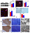
A. Western blot results show that the protein level of MBP significantly increased in the spinal cord of the MSC-Exo group compared with the control group. B ~ C. Representative images of immunofluorescent stainings of BrdU-NG2 (arrow) and BrdU-APC (arrow), respectively. Quantitative data show that proliferation of OPCs (BrdU-NG2), differentiation of OPCs (BrdU-APC) and the number of APC+ cells significantly increased in the spinal cord of the MSC-Exo group compared with the control group. D. Immunostaining data show that APP+ density significantly decreased in the spinal cord after MSC-Exo treatment compared with PBS treatment. E. MBP immunostaining in the dorsal column of spinal cords. F. Enlarged area of red square in E by Paragon staining. G. Enlarged area of red square in F scanned by TEM. TEM images show significant demyelination and axonal damage in the spinal cord of the control mice, and more intact axons with thinner myelin (arrows) in the MSC-Exo group at day 35p.i. *p < 0.05 vs control. Scale bar =25 μm in B and C, 50 μm D, 25 μm in F, and 2 μm in G. (For interpretation of the references to colour in this figure legend, the reader is referred to the web version of this article.)
Fig. 4.
Fig. 4.
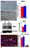
A. Immunostaining results show MBP+ density (myelin area); B. Western blot results of MBP protein level; C. Double immunostaining of mature OLs (APC, red) and proliferating cells (BrdU, green) in the corpus callosum. MSC-Exo treatment significantly increased level of MBP, numbers of APC+ OLs and BrdU+-APC+ (OPC differentiation, arrow) compared with the control group after 2 weeks of treatments. *p < 0.05 vs control. Scale bar in A and C = 25um. (For interpretation of the references to colour in this figure legend, the reader is referred to the web version of this article.)
Fig. 5.
Fig. 5.
A. Western blot results show that there was no significant difference of IBA1 protein level in the EAE model between the MSC-Exo group and the control group, while YM1 protein level significantly increased and iNOS protein level significantly decreased in the spinal cord of the MSC-Exo group compared with the control group. B. Analysis of Western blots show the protein levels of pro-and anti-inflammatory cytokines in the spinal cord of EAE mice treated with MSC-Exo and PBS control on day 35p.i.. C. Immunostaining results show that there was no significant difference of the number of IBA1+ cells, while the number of YM1+ cells significantly increased in the corpus callosum of CPZ mice treated with MSC-Exo compared with that in the control group. Scale bar = 25 μm. D. Western blot results show that compared with the control, MSC-Exo did not significantly change IBA1 protein level, however, it significantly increased YM1 protein level and decreased iNOS protein level in the corpus callosum on Week 7. E. Analysis of Western blots show the protein levels of pro-and anti-inflammatory cytokines in the corpus callosum of CPZ mice treated with MSC-Exo and PBS on Week 7. *p < 0.05, **p < 0.01 vs control. N = 6 per group.
Fig. 6.
Fig. 6.
A. Analysis of Western blots show the protein levels of TLR2, IRAK1 and pNF-kB significantly decreased in the spinal cord of the EAE mice treated with MSC-Exo compared with the PBS controls at day 35 p.i. B. Western blot data show that MSC-Exo significantly decreased protein level of TLR2 and pNF-kB in the corpus callosum at Week 7 compared with the controls (*p < 0.05).
Fig. 7.
Fig. 7.
The sketch of the potential mechanisms of MSC-Exo actions.

References

    1. Adams KL, Gallo V, 2018. The diversity and disparity of the glial scar. Nat. Neurosci 21, 9–15. - PMC - PubMed
    1. Aharoni R, Herschkovitz A, Eilam R, Blumberg-Hazan M, Sela M, Bruck W, Arnon R, 2008. Demyelination arrest and remyelination induced by glatiramer acetate treatment of experimental autoimmune encephalomyelitis. Proc. Natl. Acad. Sci. U. S. A 105, 11358–11363. - PMC - PubMed
    1. Alvarez-Erviti L, Seow Y, Yin H, Betts C, Lakhal S, Wood MJ, 2011. Delivery of siRNA to the mouse brain by systemic injection of targeted exosomes. Nat. Biotechnol 29, 341–345. - PubMed
    1. Asgarpour K, Shojaei Z, Amiri F, Ai J, Mahjoubin-Tehran M, Ghasemi F, ArefNezhad R, Hamblin MR, Mirzaei H, 2020. Exosomal microRNAs derived from mesenchymal stem cells: cell-to-cell messages. Cell Commun. Signal 18, 149. - PMC - PubMed
    1. Back SA, Tuohy TM, Chen H, Wallingford N, Craig A, Struve J, Luo NL, Banine F, Liu Y, Chang A, Trapp BD, Bebo BF Jr., Rao MS, Sherman LS, 2005. Hyaluronan accumulates in demyelinated lesions and inhibits oligodendrocyte progenitor maturation. Nat. Med 11, 966–972. - PubMed