. 2022 Apr 12;6(7):2361-2372.
doi: 10.1182/bloodadvances.2021006034.

Diffuse large B-cell lymphomas in adults with aberrant coexpression of CD10, BCL6, and MUM1 are enriched in IRF4 rearrangements

Affiliations

Diffuse large B-cell lymphomas in adults with aberrant coexpression of CD10, BCL6, and MUM1 are enriched in IRF4 rearrangements

Leonie Frauenfeld et al. Blood Adv. .

Abstract

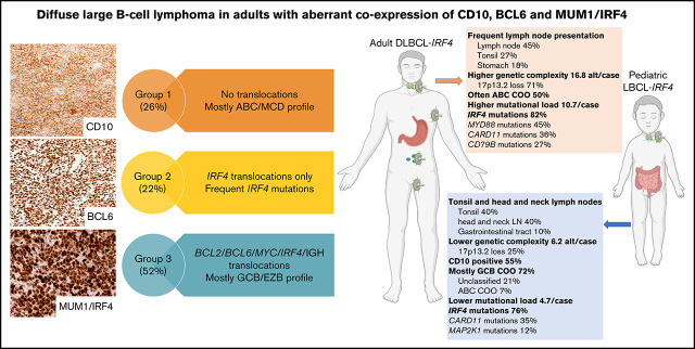
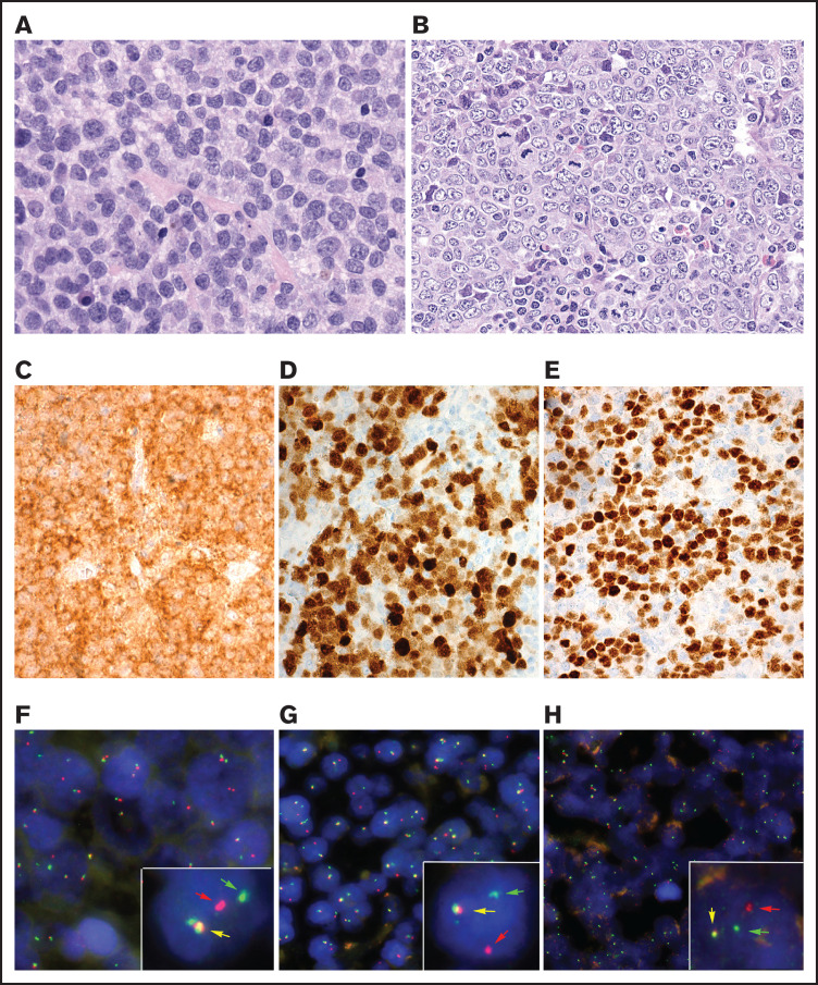
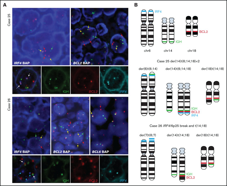
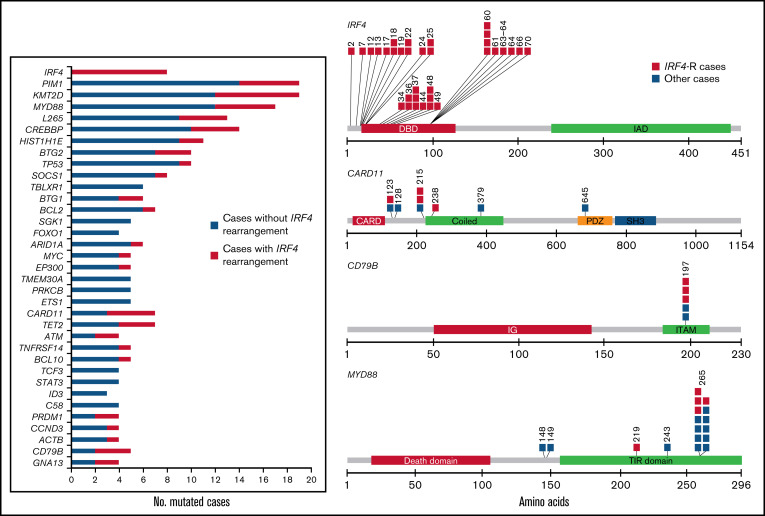
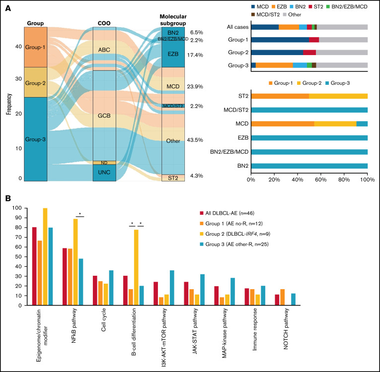
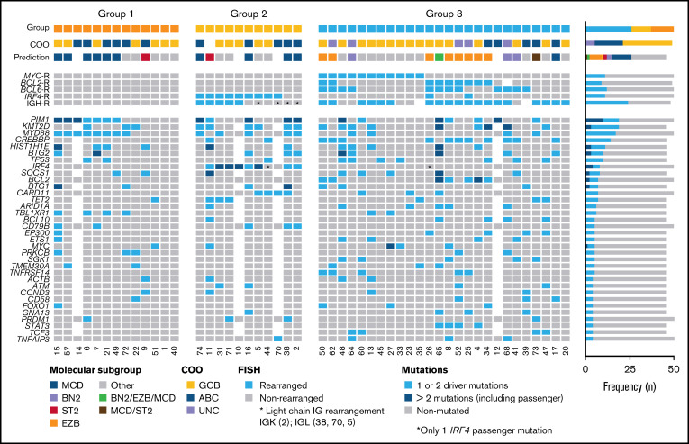
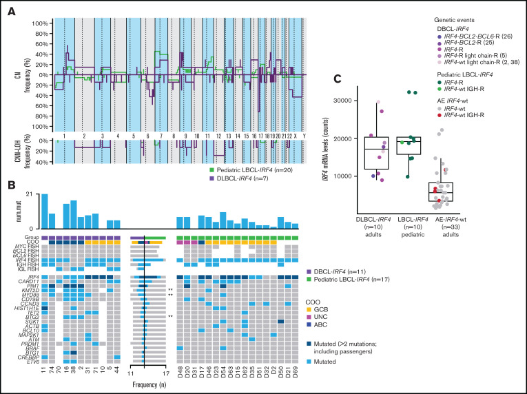
Diffuse large B-cell lymphoma (DLBCL) with aberrant coexpression of CD10+BCL6+MUM1+ (DLBCL-AE), classified as germinal center B cell (GCB) type by the Hans algorithm (HA), was genetically characterized. To capture the complexity of DLBCL-AE, we used an integrated approach that included gene expression profiling (GEP), fluorescence in situ hybridization, targeted gene sequencing, and copy number (CN) arrays. According to GEP, 32/54 (59%) cases were classified as GCB-DLBCL, 16/54 (30%) as activated B-cell (ABC) DLBCL, and 6/54 (11%) as unclassifiable. The discrepancy between HA and GEP was 41%. Three genetic subgroups were identified. Group 1 included 13/50 (26%) cases without translocations and mainly showing and ABC/MCD molecular profile. Group 2 comprised 11/50 (22%) cases with IRF4 alterations (DLBCL-IRF4), frequent mutations in IRF4 (82%) and NF-κB pathway genes (MYD88, CARD11, and CD79B), and losses of 17p13.2. Five cases each were classified as GCB- or ABC-type. Group 3 included 26/50 (52%) cases with 1 or several translocations in BCL2/BCL6/MYC/IGH, and GCB/EZB molecular profile predominated. Two cases in this latter group showed complex BCL2/BCL6/IRF4 translocations. DLBCL-IRF4 in adults showed a similar copy number profile and shared recurrent CARD11 and CD79B mutations when compared with LBCL-IRF4 in the pediatric population. However, adult cases showed higher genetic complexity, higher mutational load with frequent MYD88 and KMT2D mutations, and more ABC GEP. IRF4 mutations were identified only in IRF4-rearranged cases, indicating its potential use in the diagnostic setting. In conclusion, DLBCL-AE is genetically heterogeneous and enriched in cases with IRF4 alterations. DLBCL-IRF4 in adults has many similarities to the pediatric counterpart.


Figures

None
Graphical abstract
Figure 1.
Figure 1.
Morphological, immunophenotypic, and genetic features of triple-positive DLBCLs. (A) B-cell lymphoma with blastic morphology characterized by medium- to large-sized cells with irregular nuclei, fine chromatin, inconspicuous nucleolus, and scant cytoplasm (case 10), (original magnification ×400; hematoxylin and eosin stain). (B-G) Case 5 shows a characteristic centroblastic morphology (original magnification ×400; hematoxylin and eosin stain). The tumor cells are CD10+ (C), MUM1/IRF4+ (D), and BCL6+ (E) (original magnification ×400; immunostaining). FISH demonstrates an IRF4 (F) and IGL (G) break with 1 colocalized signal (yellow arrow) and 1 split signal (green and red arrows) consistent with gene rearrangement. (H) In case 2, an IGK break was demonstrated. FISH shows a signal constellation of 1 colocalized signal (yellow arrow) and 1 split signal (green and red arrows) consistent with gene rearrangement.
Figure 2.
Figure 2.
FISH analysis demonstrates complex rearrangement with IRF4 and BCL2/BCL6 genes. (A) FISH analyses using IRF4, BCL2, and BCL6 break-apart probes and triple-color FISH for IGH, BCL2, and IRF4 loci in the 2 cases with IRF4 rearrangement together with BCL2 in case 25 and BCL2/BCL6 rearrangement in case 26. These hybridizations were performed using t(14;18) dual-color fusion (Metasystems) and BAC clones spanning IRF4 locus (BAC clones 5' RP3-416J7 and 3' RP5-1077H22 and RP5-856G1) labeled in spectrum aqua. Red signals correspond to BCL2, green signals correspond to IGH, and aqua/blue signals represent IRF4. Case 25. FISH demonstrates IRF4 and BCL2 breaks (upper panel) with 1 or 2 colocalized signals (yellow arrows) and 1 or 2 split signals (green and red arrows) consistent with BCL2 and IRF4 gene rearrangement. The triple rearrangement (IGH-IRF4-BCL2) (lower panel) is demonstrated by colocalization of the 3 colors (white arrows). Case 26. FISH analysis for IRF4, BCL2, and BCL6 with break-apart probes demonstrates 1 colocalized signal (yellow arrow) and 1 split signal (green and red arrows), indicating a triple rearrangement. FISH analyses of concomitant IGH, BCL2 and IRF4 rearrangements (lower panel) show 2 colocalized signals (white arrow) of the derivative chromosomes 14 and 18 resulting in t(14;18) translocation. IRF4 locus (blue arrows) did not colocalize with either the IGH locus or the t(14;18) translocation, indicating that this is an independent rearrangement with unknown partner. (B) Schematic representation of IRF4 (aqua blue), IGH (green), and BCL2 (red) breaks/rearrangements in cases 25 and 26.
Figure 3.
Figure 3.
Mutational landscape of 50 cases of DLBCL-AE. Bar graph shows mutated genes in 4 or more cases. The results are given in number of mutated cases per gene. The colored bars indicate the presence (orange) or absence (blue) of IRF4 rearrangement. A diagram of the relative positions of driver mutations is shown for IRF4, CARD11, CD79B, and MYD88. Multiple passenger mutations for IRF4 also are depicted. The x-axis indicates amino acid positions. The approximate location of somatic mutations identified in each gene is indicated. IRF4 mutations are mainly in the DNA binding domain. All CD79B mutations are located in a hotspot Y197 in the immunoreceptor tyrosine-based activation motif (ITAM). Domains of the protein are represented according to the Uniprot database (www.uniprot.org).
Figure 4.
Figure 4.
Molecular subgroup prediction and recurrent mutated pathways in DLBCL-AE. (A) Alluvial plot shows the frequency and relationship between groups, COO classification according to LYMPH2Cx, and/or HTG and molecular subgroup prediction according to LymphGen tool of 46 cases of DLBCL-AE. The bars indicate the molecular predicted subgroup in all cases and in each genetic group identified; 57% of all cases were assigned to a specific molecular subgroup. (B) Recurrent mutated pathways in 46 cases of DLBCL-AE. Genes included in each pathway are indicated in supplemental Table 1. Bar graph shows the total number of mutated cases for each pathway. Asterisk represents significant mutated pathways. AE, aberrant expressor CD10+BCL6+MUM1+; R, rearrangement.
Figure 5.
Figure 5.
Overview of 50 cases of DLBCL with aberrant coexpression of CD10, BCL6, and MUM1 clustered in 3 groups. Oncoprint includes FISH results, molecular subgroup prediction according to LymphGen tool, COO classification according to Lymph2Cx, and/or HTG and frequently mutated genes (>3 cases). Each column of the plot represents 1 TP case and each line is a specific analysis. On the right side of the figure, the frequency of the particular result of the analysis is shown.
Figure 6.
Figure 6.
Comparison of CN profile and genetic features of adult and pediatric LBCL-IRF4 cases. (A) Comparative plot of CN and CNN-LOH between 7 LBCL-IRF4 cases in adults and 20 LBCL-IRF4 cases in the pediatric population. No significantly different regions were identified. (B) GEP and mutational comparison between LBCL-IRF4 in adults (n = 11) and previously published profiles in children (n = 17) showed ABC COO more often (P = .05) in adults, with higher mutational load (10.6 vs 4.7 mutations/case; Wilcoxon test, P = .004) and higher frequency of KMT2D, MYD88, and BTG2 (P < .05, marked with asterisk). (C) IRF4 mRNA expression levels obtained from the IRF4_NM_002460.1 probe on the Lymph2Cx assay. IRF4 mRNA levels in LBCL-IRF4 in children and adults was similar but higher when compared with DLBCL-AE without IRF4 rearrangement (17 570 vs 6948; Wilcoxon test, P ≤ .01). AE, aberrant expressor CD10+BCL6+MUM1+; R, rearrangement; wt, wild-type.

References

    1. Smith A, Crouch S, Lax S, et al. . Lymphoma incidence, survival and prevalence 2004-2014: sub-type analyses from the UK’s Haematological Malignancy Research Network. Br J Cancer. 2015;112(9):1575-1584. - PMC - PubMed
    1. Sehn LH, Gascoyne RD. Diffuse large B-cell lymphoma: optimizing outcome in the context of clinical and biologic heterogeneity. Blood. 2015; 125(1):22-32. - PubMed
    1. Lacy SE, Barrans SL, Beer PA, et al. . Targeted sequencing in DLBCL, molecular subtypes, and outcomes: a Haematological Malignancy Research Network report. Blood. 2020;135(20):1759-1771. - PMC - PubMed
    1. Alizadeh AA, Eisen MB, Davis RE, et al. . Distinct types of diffuse large B-cell lymphoma identified by gene expression profiling. Nature. 2000; 403(6769):503-511. - PubMed
    1. Wright G, Tan B, Rosenwald A, Hurt EH, Wiestner A, Staudt LM. A gene expression-based method to diagnose clinically distinct subgroups of diffuse large B cell lymphoma. Proc Natl Acad Sci USA. 2003;100(17):9991-9996. - PMC - PubMed

Publication types