Neonatal oral myiasis caused by the larvae of Sarcophaga ruficornis (Diptera: Sarcophagidae): a case report
- PMID: 34654380
- PMCID: PMC8518163
- DOI: 10.1186/s12879-021-06742-z
Neonatal oral myiasis caused by the larvae of Sarcophaga ruficornis (Diptera: Sarcophagidae): a case report
Abstract
Background: Myiasis is caused by dipterous larvae, and rarely affects the mouth. Diagnosis by traditional means is easy to be confused with other similar species. Here, we report a case of oral myiasis, in a 5-month-old infant who was diagnosed by morphological examination and molecular biological methods.
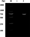
Case presentation: A 5-month old infant with acute myeloid leukemia was admitted due to recurrent skin masses for more than 4 months. The infant had lip swelling, which prevented him from closing the mouth and membranes were present in his mouth and there were also oral ulcers and erosions. Ten maggots were found in the mouth and one in the ear canal with pus flowing out and were confirmed as the third stage larvae of Sarcophaga ruficornis by morphological examination and a comparison of sequence of cytochrome oxidase subunit 1 (COX1) gene. After removal of the maggots and chemotherapy, the infant 's condition was gradually improved.
Conclusions: To the best of our our knowledge, this is the first neonatal oral myiasis case reported in China and its diagnosis requires a high index of suspicion. Microscopy combined with specific DNA sequence analysis is an effective technological tool to provide rapid diagnoses of the larva specimen and cases of rare diseases, as illustrated in the current case.
Keywords: Molecular identification; Neonatal oral myiasis; Nosocomial.
© 2021. The Author(s).
Conflict of interest statement
The authors declare that they have no competing interests.
Figures


Similar articles
-
A Case of Nasopharyngeal Myiasis Caused by Sarcophaga sp.Turkiye Parazitol Derg. 2023 Jun 29;47(2):124-126. doi: 10.4274/tpd.galenos.2023.86547. Turkiye Parazitol Derg. 2023. PMID: 37249117 English.
-
Myiasis of the Tracheostomy Wound Caused by Sarcophaga (Liopygia) argyrostoma (Diptera: Sarcophagidae): Molecular Identification Based on the Mitochondrial Cytochrome c Oxidase I Gene.J Med Entomol. 2015 Nov;52(6):1357-60. doi: 10.1093/jme/tjv108. Epub 2015 Aug 19. J Med Entomol. 2015. PMID: 26336248
-
What's in a child's ear? A case of otomyiasis by Sarcophaga argyrostoma (Diptera, Sarcophagidae).Parasitol Int. 2022 Apr;87:102537. doi: 10.1016/j.parint.2022.102537. Epub 2022 Jan 5. Parasitol Int. 2022. PMID: 34995772
-
Aural myiasis caused by flesh fly larva, Sarcophaga haemorrhoidalis.J Otolaryngol. 1994 Jun;23(3):204-5. J Otolaryngol. 1994. PMID: 8064961 Review.
-
Myiasis of the ear: a review with entomological aspects for the otolaryngologist.Ann Otol Rhinol Laryngol. 2015 May;124(5):345-50. doi: 10.1177/0003489414557021. Epub 2014 Oct 30. Ann Otol Rhinol Laryngol. 2015. PMID: 25358614 Review.
Cited by
-
Oral Myiasis in an Immunocompromised Adult Undergoing Chemotherapy: A Rare Case and Comprehensive Treatment Protocol.Cureus. 2023 Jul 27;15(7):e42555. doi: 10.7759/cureus.42555. eCollection 2023 Jul. Cureus. 2023. PMID: 37637591 Free PMC article.
-
Scalp myiasis associated with soft tissue sarcoma lesion: a case report and review of relevant literature.BMC Infect Dis. 2024 Jan 5;24(1):51. doi: 10.1186/s12879-023-08957-8. BMC Infect Dis. 2024. PMID: 38183025 Free PMC article. Review.
-
Ophthalmomyiasis in a preterm neonate resulting in blindness: A case report from Botswana.Front Pediatr. 2022 Sep 29;10:955212. doi: 10.3389/fped.2022.955212. eCollection 2022. Front Pediatr. 2022. PMID: 36245720 Free PMC article.
References
-
- Hope FW. On insects and their larvae occasionally found in the human body. Trans R Entomol Soc Lond. 1840;2:256–271.
-
- Sharma J, Mamatha GP, Acharya R. Primary oral myiasis: a case report. Med Oral Patol Oral Cir Bucal. 2008;13(11):E714–6. - PubMed
Publication types
MeSH terms
Grants and funding
- 81572023/National Natural Science Foundation of China
- 81371836/National Natural Science Foundation of China
- 82072303/National Natural Science Foundation of China
- NPRC-2019-194-30/National Parasitic Resources Center of China
- grant no. 2019B030316025/Science and Technology Planning Project of Guangdong Province
- 2019A1515011541/Natural Science Foundation of Guangdong Province
- 2020TTM007/Open Foundation of Key Laboratory of Tropical Translational Medicine of Ministry of Education, Hainan Medical University
- B12003/111 Project
- ZDYF2020120/Key Research and Development Program of Hainan Province
- ZDKJ202003/Major Science and Technology Project of Hainan Province
LinkOut - more resources
Full Text Sources

