. 2021 Nov;31(11):1169-1178.
doi: 10.1016/j.nmd.2021.07.016. Epub 2021 Jul 28.

Muscular dystrophy-dystroglycanopathy in a family of Labrador retrievers with a LARGE1 mutation

Affiliations

Muscular dystrophy-dystroglycanopathy in a family of Labrador retrievers with a LARGE1 mutation

G Diane Shelton et al. Neuromuscul Disord. 2021 Nov.

Abstract

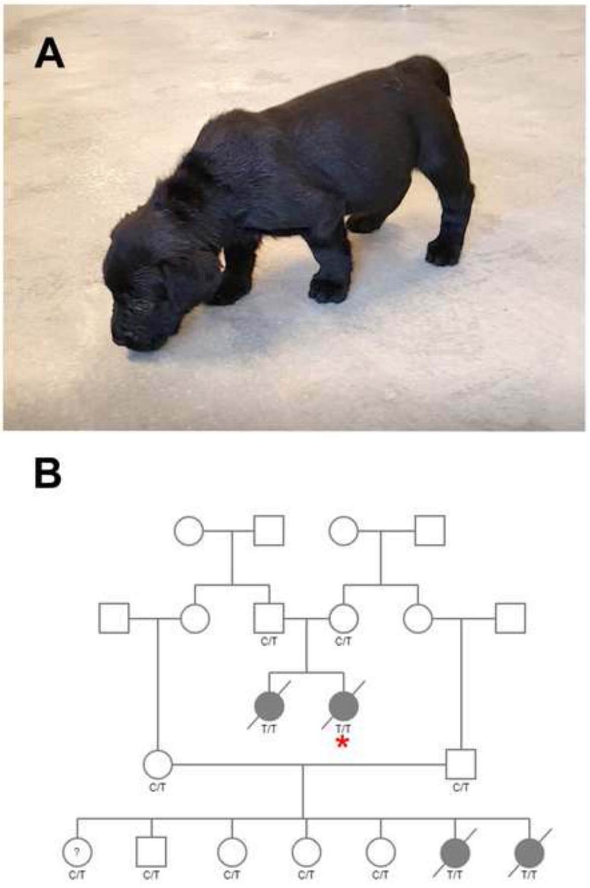
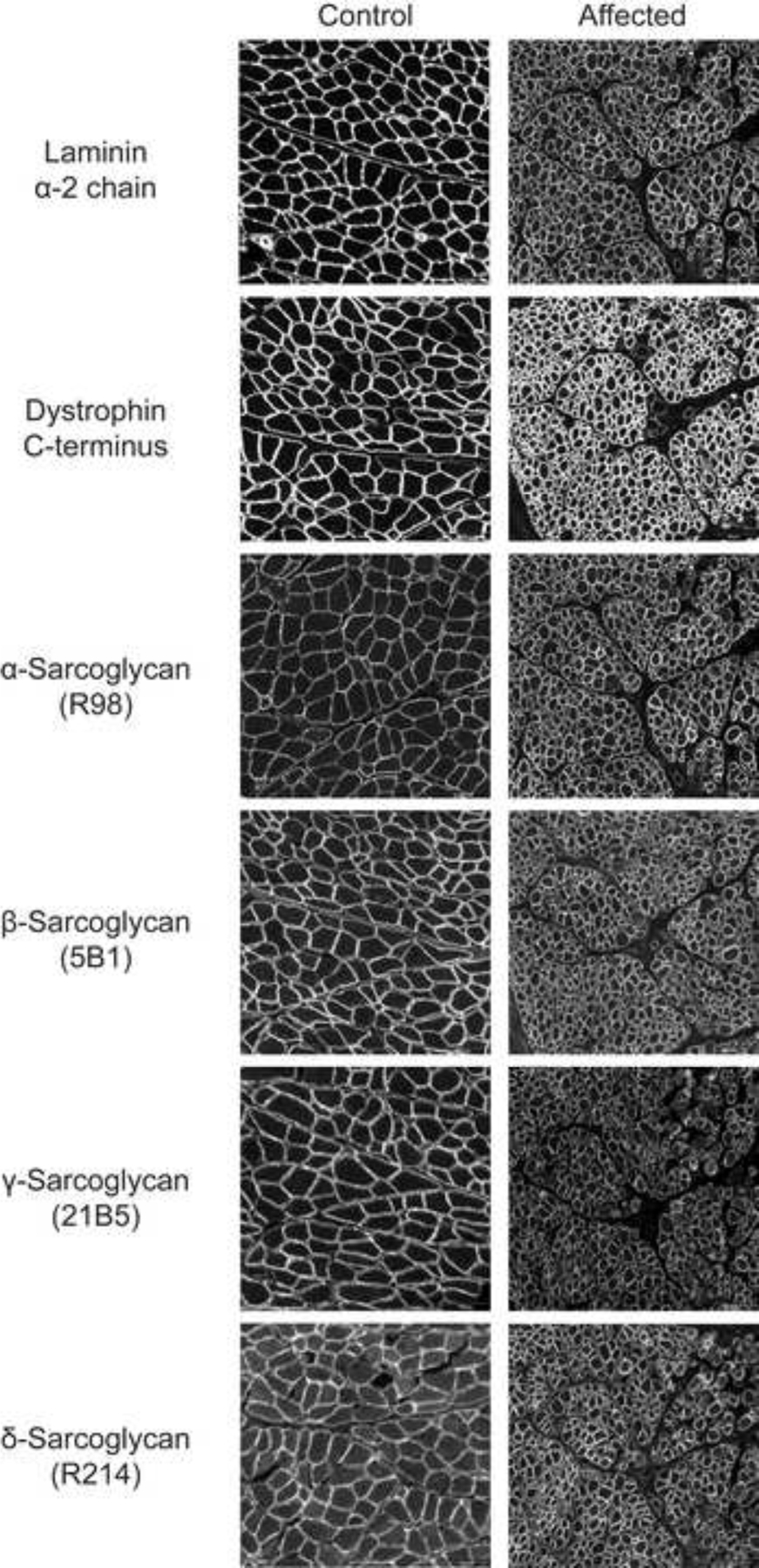
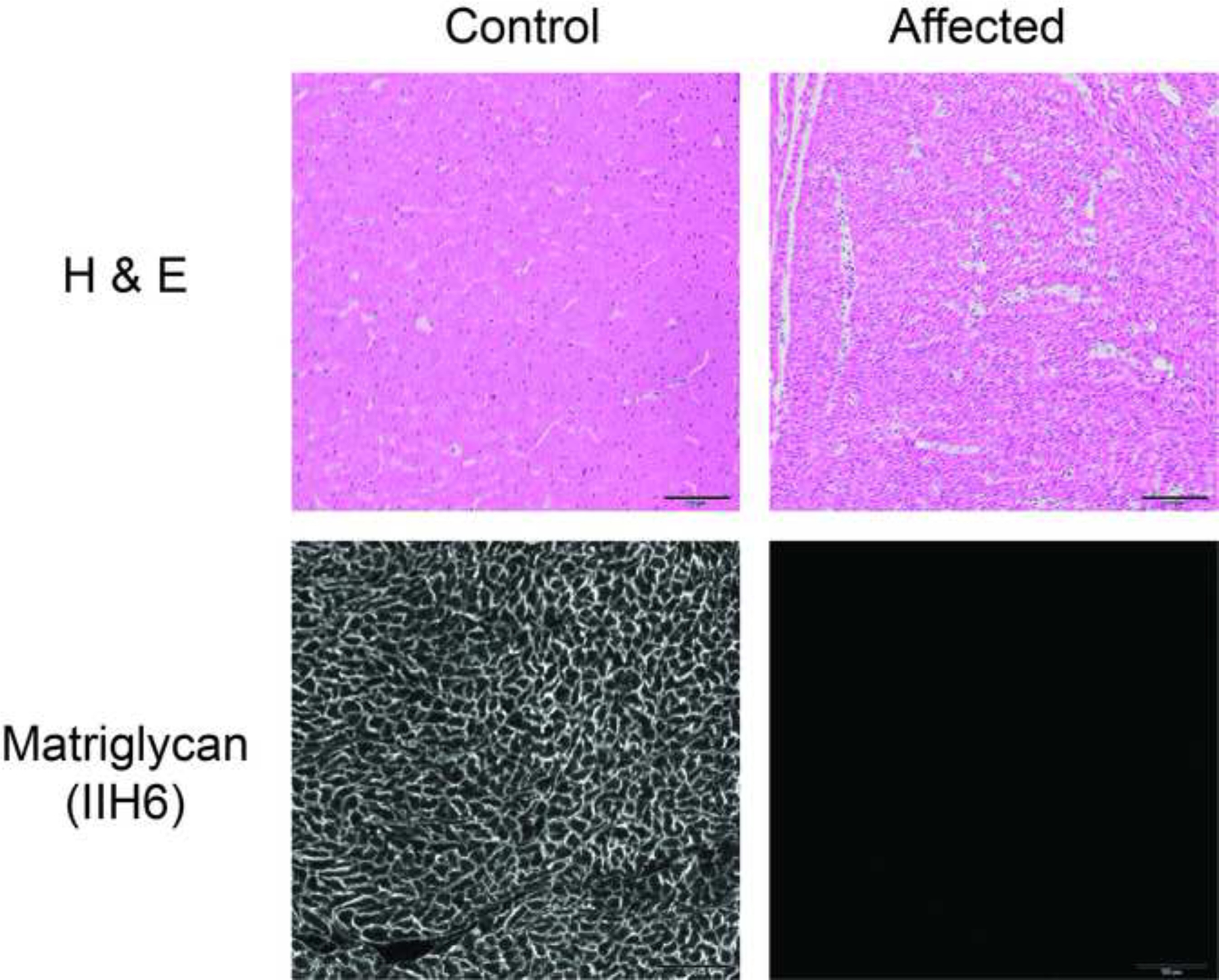
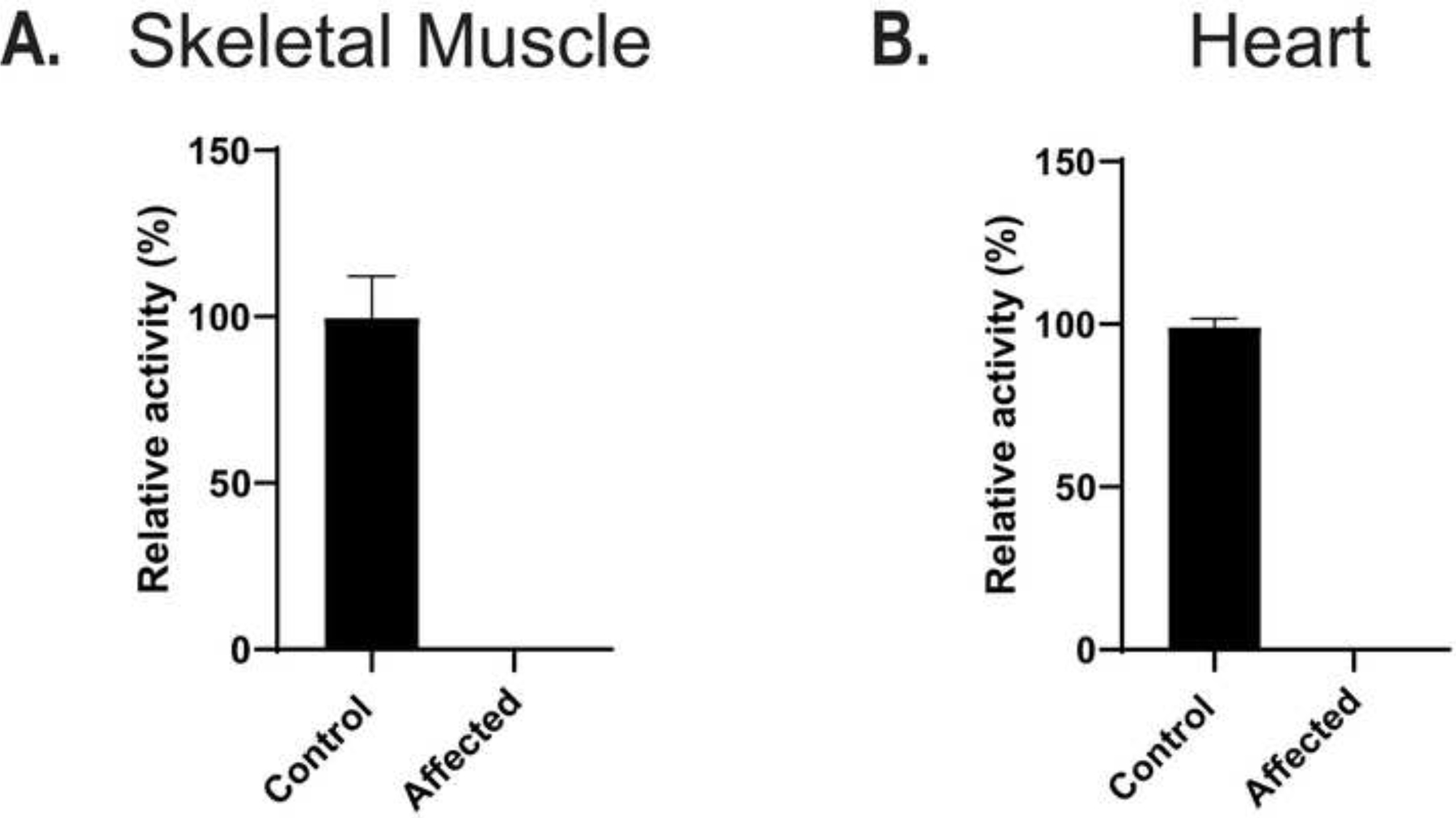
Alpha-dystroglycan (αDG) is a highly glycosylated cell surface protein with a significant role in cell-to-extracellular matrix interactions in muscle. αDG interaction with extracellular ligands relies on the activity of the LARGE1 glycosyltransferase that synthesizes and extends the heteropolysaccharide matriglycan. Abnormalities in αDG glycosylation and formation of matriglycan are the pathogenic mechanisms for the dystroglycanopathies, a group of congenital muscular dystrophies. Muscle biopsies were evaluated from related 6-week-old Labrador retriever puppies with poor suckling, small stature compared to normal litter mates, bow-legged stance and markedly elevated creatine kinase activities. A dystrophic phenotype with marked degeneration and regeneration, multifocal mononuclear cell infiltration and endomysial fibrosis was identified on muscle cryosections. Single nucleotide polymorphism (SNP) array genotyping data on the family members identified three regions of homozygosity in 4 cases relative to 8 controls. Analysis of whole genome sequence data from one of the cases identified a stop codon mutation in the LARGE1 gene that truncates 40% of the protein. Immunofluorescent staining and western blotting demonstrated the absence of matriglycan in skeletal muscle and heart from affected dogs. Compared to control, LARGE enzyme activity was not detected. This is the first report of a dystroglycanopathy in dogs.

Keywords: Dog; Glycosylation; Myopathy; α-dystroglycan.


Conflict of interest statement

Declaration of Competing Interest The authors declare that they have no known competing financial interests or personal relationships that could have appeared to influence the work reported in this paper.

Figures

Figure 1.
Figure 1.
Affected Pup and Pedigree. A) 6-week-old Labrador retriever pup with poor suckling, small stature, and bow-legged stance. B) One family of Labrador retrievers formed the basis for this study. Males are squares and females’ circles. Cases are solid symbols, unaffected are open. ?=phenotype unknown. A diagonal strikethrough indicates deceased pups. Genotypes for the LARGE1 variant are provided for all dogs with available samples. TT = homozygous mutant, C/T = carrier, CC = clear. The case with WGS data is indicated with an asterisk.
Figure 2.
Figure 2.
Histopathology and Immunofluorescence Staining of Skeletal Muscle. Stainings are shown from a control dog and an affected Labrador retriever pup. A). With the H&E stain, a dystrophic phenotype was present in the muscle biopsy from the affected pup. B). Using the IIH6 monoclonal antibody to detect matriglycan on αDG, abnormal glycosylation consistent with a diagnosis of a dystroglycanopathy was identified. Positive staining was observed using the AP83 polyclonal antibody against βDG. Scale bars = 100 μM for all images.
Figure 3.
Figure 3.
Additional Immunofluorescence Staining of Skeletal Muscle. Stainings with antibodies against laminin α−2 chain, dystrophin C-terminus, and α-, β-, γ-, and δ-sarcoglycans in the affected pup were like that of control muscle. Scale bars = 100 μM for all images.
Figure 4.
Figure 4.
Histopathology and Immunofluorescence Staining of Heart Muscle. H&E stainings are shown for a control dog and an affected Labrador retriever pup. Except for the small muscle fiber size due to the young age of the affected pup, no pathological changes were identified compared to the control dog muscle. Using the IIH6 monoclonal antibody to detect matriglycan on αDG, abnormal glycosylation consistent with a diagnosis of a dystroglycanopathy was identified. Scale bars = 100 μm for all images.
Figure 5.
Figure 5.
Histopathology and Immunohistochemical Staining of Brain Frontal Cortex. H&E and Luxol fast blue stains of the frontal cortex are shown from a control dog and an affected Labrador retriever pup. No pathological evidence of dysplasia or abnormal growth patterns were identified in the affected pup compared to the control dog. Using the IIH6 monoclonal antibody to detect matriglycan on αDG, reddish brown staining identified matriglycan on neurons (arrowheads) and their associated axons (arrow) of control brain but not on similar structures from the affected pup. Unidentified cell processes are labeled around blood vessels. Scale bars = 100 μm for all images.
Figure 6.
Figure 6.
Western blotting of Skeletal and Heart Muscle. Glycoproteins were enriched from the biceps femoris muscle using wheat-germ agglutinin (WGA)-agarose. Immunoblotting (three replicates) was performed on control and affected Labrador retriever skeletal and heart muscle with A,D, IIH6 antibody for matriglycan; B,E, antibody AF6868, which recognizes core αDG and βDG; and C,F, laminin overlay to detect laminin binding respectively.
Figure 7.
Figure 7.
LARGE enzyme activity. Lysates (20 μL) from control dog skeletal (Panel A, n=3) and heart (Panel B, n=3) muscle and LARGE mutant skeletal (Panel A, n=3) and heart (Panel B, n=3) muscle (affected) were assayed for enzyme activity for LARGE. Relative activity (%) with respect to control and standard deviation is for triplicate experiments. The activity was normalized by amount of βDG signal from a blot of control and LARGE mutant lane using monoclonal 7D11 which has an epitope to the c-terminus of βDG.

References

    1. Nickolls AR, Bonnemann CG. The roles of dystroglycan in the nervous system: insights from animal models of muscular dystrophy. Dis Model Mech 2018;11:(12):dmm035931. doi: 10.1242/dmm.035931. - DOI - PMC - PubMed
    1. Michele DE, Barresi R, Kanagawa M, Saito F, Cohn RD, Satz JS et al. Post-translational disruption of dystroglycan-ligand interactions in congenital muscular dystrophies. Nature 2002;418:417–22. doi: 10.1038/nature00837. - DOI - PubMed
    1. Longman C, Brockington M, Torelli S, Jimenez-Mallebrera C, Kennedy C, Khalil N et al. Mutations in the human LARGE gene cause CMD1D, a novel form of congenital muscular dystrophy with severe mental retardation and abnormal glycosylation of alpha-dystroglycan. Hum Mol Genet 2003;12:2853–61. doi: 10.1093/hmg/ddg307. - DOI - PubMed
    1. Grewal PK, McLaughlan JM, Moore CJ, Browning CA, Hewitt JE. Characterization of the LARGE family of putative glycosyltransferases associated with dystroglycanopathies. Glycobiology 2005;15:912–23. doi: 10.1093/glycob/cwi094. - DOI - PubMed
    1. Muntoni F, Torelli S, Brockington M. Muscular dystrophies due to glycosylation defects. Neurotherapeutics 2008;5:627–32. doi: 10.1016/j.nurt.2008.08.005. - DOI - PMC - PubMed

Publication types