. 2021 Oct 15;11(1):16866.
doi: 10.1038/s41598-021-96351-2.

Human induced pluripotent stem cell-derived platelets loaded with lapatinib effectively target HER2+ breast cancer metastasis to the brain

Affiliations

Human induced pluripotent stem cell-derived platelets loaded with lapatinib effectively target HER2+ breast cancer metastasis to the brain

Arunoday Bhan et al. Sci Rep. .

Retraction in

Abstract

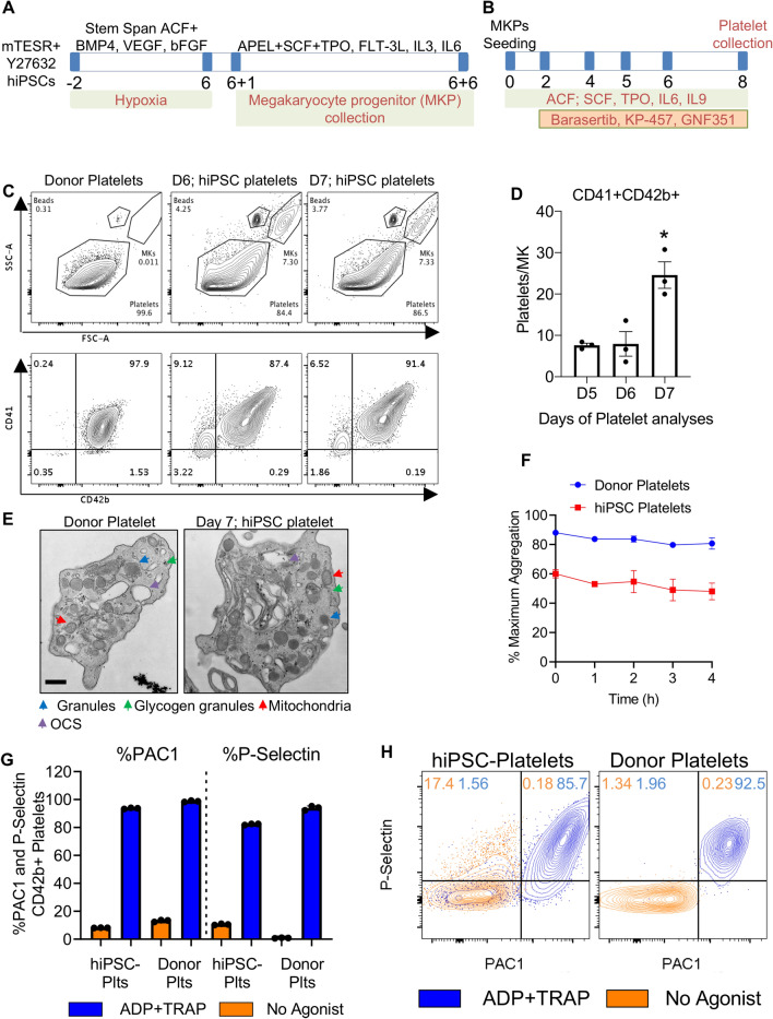
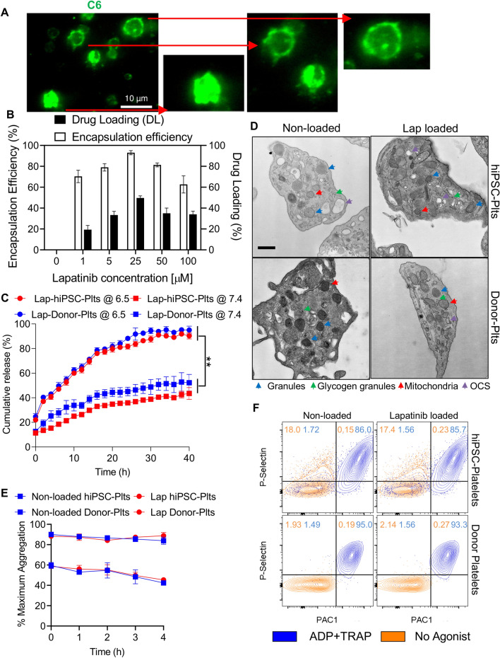
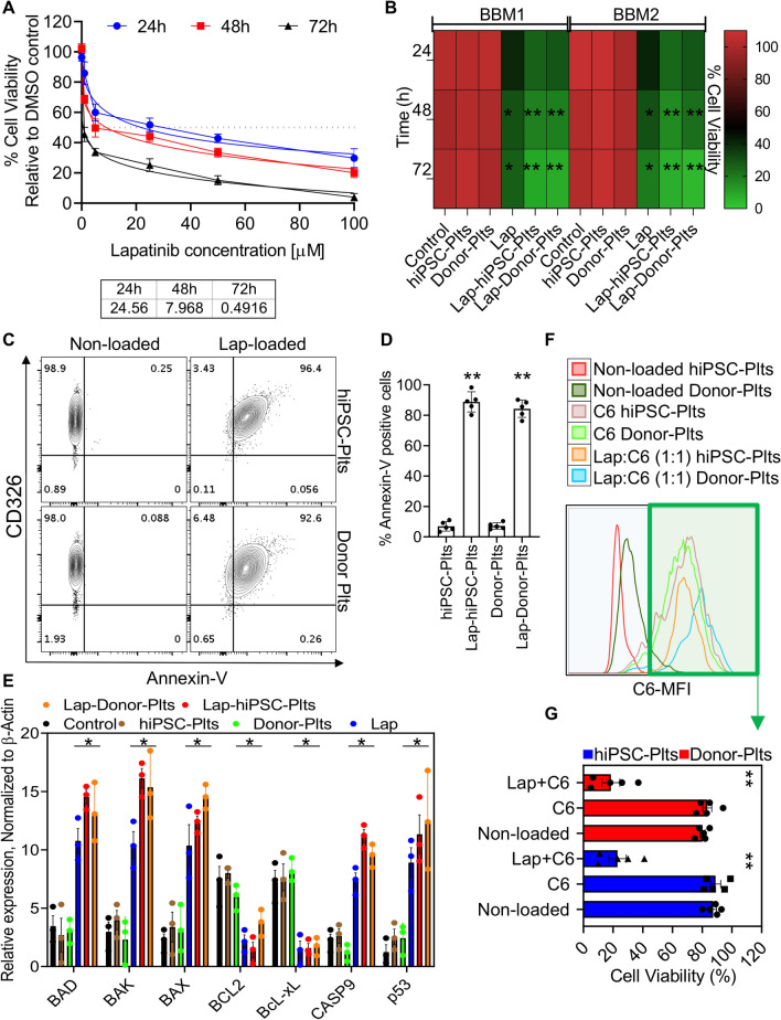
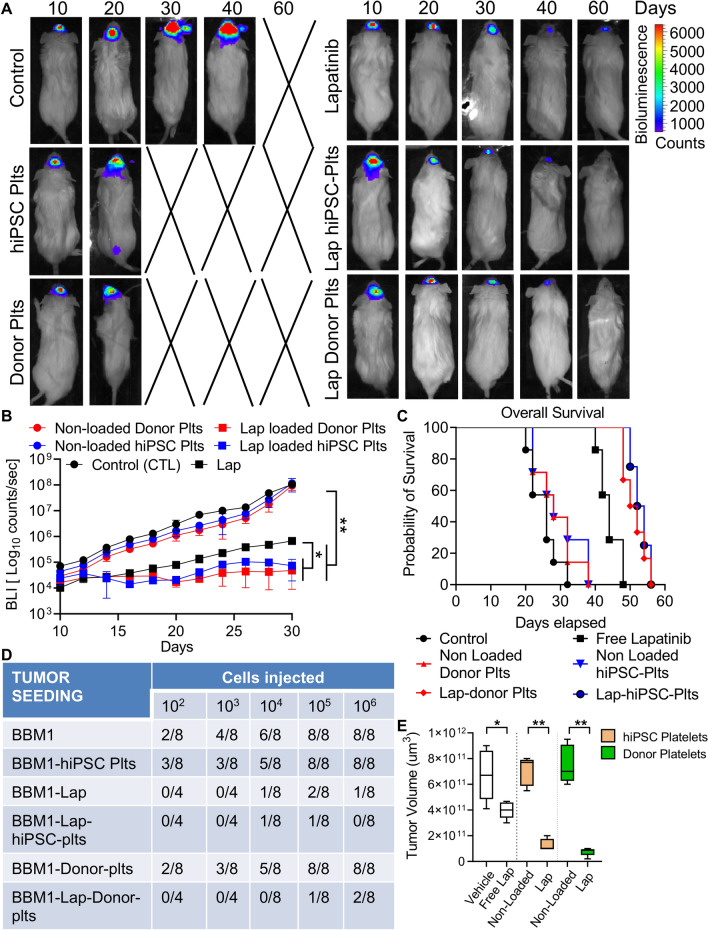
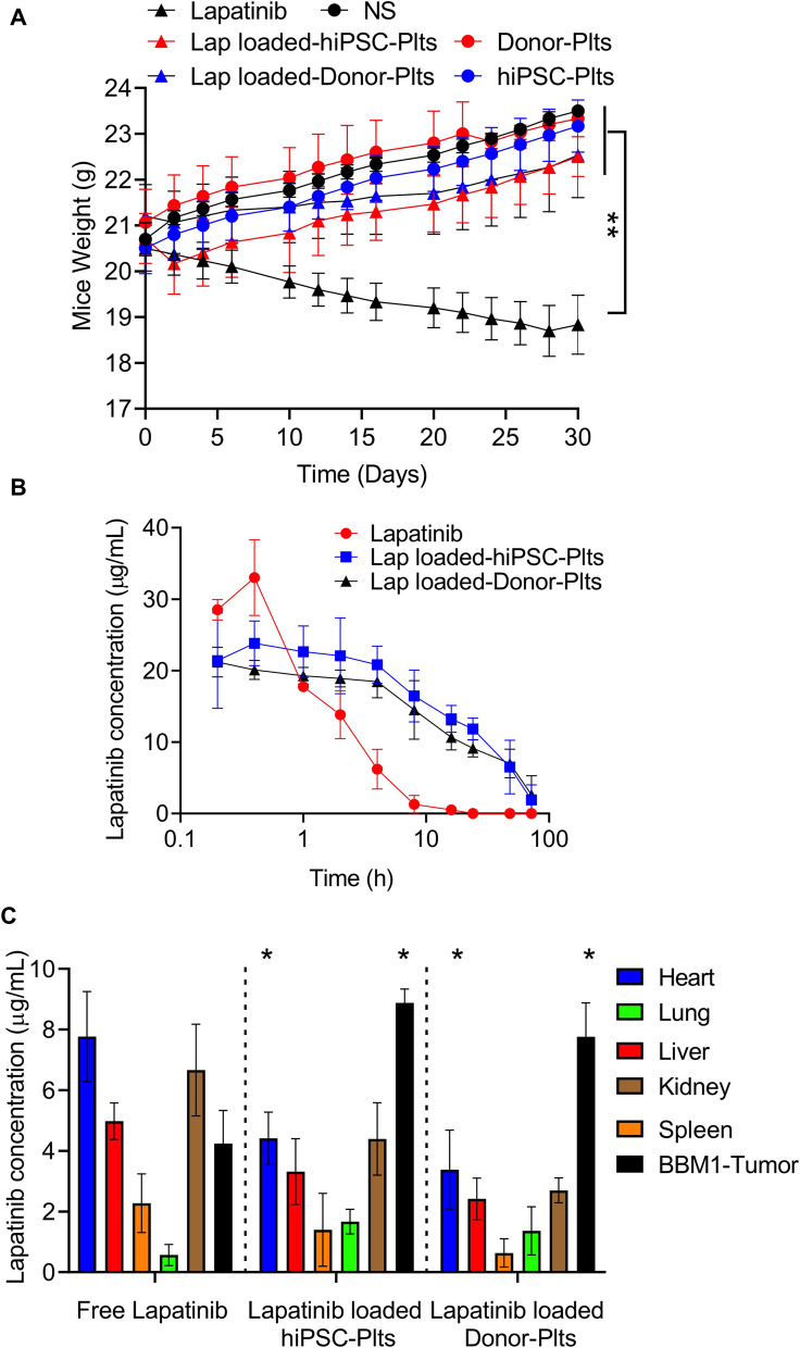
Prognosis of patients with HER2+ breast-to-brain-metastasis (BBM) is dismal even after current standard-of-care treatments, including surgical resection, whole-brain radiation, and systemic chemotherapy. Radiation and systemic chemotherapies can also induce cytotoxicity, leading to significant side effects. Studies indicate that donor-derived platelets can serve as immune-compatible drug carriers that interact with and deliver drugs to cancer cells with fewer side effects, making them a promising therapeutic option with enhanced antitumor activity. Moreover, human induced pluripotent stem cells (hiPSCs) provide a potentially renewable source of clinical-grade transfusable platelets that can be drug-loaded to complement the supply of donor-derived platelets. Here, we describe methods for ex vivo generation of megakaryocytes (MKs) and functional platelets from hiPSCs (hiPSC-platelets) in a scalable fashion. We then loaded hiPSC-platelets with lapatinib and infused them into BBM tumor-bearing NOD/SCID mouse models. Such treatment significantly increased intracellular lapatinib accumulation in BBMs in vivo, potentially via tumor cell-induced activation/aggregation. Lapatinib-loaded hiPSC-platelets exhibited normal morphology and function and released lapatinib pH-dependently. Importantly, lapatinib delivery to BBM cells via hiPSC-platelets inhibited tumor growth and prolonged survival of tumor-bearing mice. Overall, use of lapatinib-loaded hiPSC-platelets effectively reduced adverse effects of free lapatinib and enhanced its therapeutic efficacy, suggesting that they represent a novel means to deliver chemotherapeutic drugs as treatment for BBM.


Conflict of interest statement

Rahul Jandial is a principal investigator on a grant to City of Hope National Medical Center from Department of Defense. City of Hope National medical center has pending patent application related to findings in this manuscript. The authors Arunoday Bhan, and Rahul Jandial are named as inventors in these applications.

Figures

Figure 1
Figure 1
Differentiation of human hiPSC cell lines into megakaryocytes and functional characterization of hiPSC-platelets. (A) Schematic showing the stepwise differentiation of hiPSCs into immature CD41+ megakaryocytes. (B) Schematic showing the stepwise maturation of hiPSC-derived immature CD41+ megakaryocytes and terminal differentiation to produce pro-platelets (hiPSC-platelets). (C) Flow cytometry-based size profile of adult human donor-derived platelets and Day 6 and Day 7 hiPSC-platelets (top), and the percentage of CD41a+CD42b+ donor-derived platelets and hiPSC-platelets (bottom). (D) Time-dependent increase in the CD41a+CD42b+ platelet-generating ability of hiPSC-derived megakaryocytes from Day 5 to Day 7 (mean ± SD, n = 3). * indicates p < 0.001 compared to Day 5 hiPSC-platelets. (E) Transmission electron micrographs of adult human donor-derived platelets and Day 7 hiPSC-platelets. Colored arrows indicate organelles: blue, granules; green, glycogen granules; red, mitochondria; and violet, OCS (open canalicular system). Scale bar = 400 nm. (F) LTA-based aggregation assays of donor-derived platelets and hiPSC-platelets stimulated with 20 µM ADP and 20 µM TRAP, showing time-dependent variability. (mean ± SD, n = 3). (G) Surface PAC1 and P-Sel activation of donor-derived platelets and hiPSC-platelets by 20 µM ADP and 20 µM TRAP, measured via flow cytometry (Fortessa). Orange bars represent unstimulated platelets, and blue bars represent platelets activated upon exposure to ADP and TRAP (mean ± SD, n = 3). (H) Representative flow cytometry (Fortessa) plots showing surface PAC1 and P-Sel activation of donor-derived platelets and hiPSC-platelets by 20 µM ADP and 20 µM TRAP. Orange contour plots represent unstimulated platelets, and blue contour plots represent platelets activated upon exposure to ADP and TRAP.
Figure 2
Figure 2
Functionality characterization and comparison of lapatinib-loaded hiPSC-platelets to non-loaded hiPSC-platelets. (A) Fluorescence image of hiPSC-platelets under an oil immersion lens (×100), demonstrating the in vitro cellular uptake of fluorescent C6 (green). Scale bars = 10 µm. Inset zoomed in images of individual C6-loaded hiPSC-platelets are shown on the right. (B) Encapsulation efficiency and drug loading capability of lapatinib-loaded hiPSC-platelets with different concentrations of lapatinib (means ± SD, n = 3). ** indicates p < 0.01. (C) Analysis of lapatinib release kinetics from hiPSC- and donor-derived platelets in PBS at pH values of 6.5 and 7.4 at 37 °C over 40 h. (D) Transmission electron micrograph of non-loaded hiPSC and donor-derived platelets and platelets loaded with 25 µM lapatinib. Colored arrows indicate organelles: blue, granules; green, glycogen granules; red, mitochondria; and violet, OCS. Scale bar = 400 nm. (E) LTA-based aggregation assays of non-loaded and 25 µM lapatinib-loaded hiPSC- and donor-derived platelets stimulated with 20 µM ADP and 20 µM TRAP, showing time-dependent variability (mean ± SD, n = 3). (F) Representative flow cytometry (Fortessa) plots showing the surface PAC1 and P-Sel activation of non-loaded and 25 µM lapatinib-loaded hiPSC- and donor-derived platelets by 20 µM ADP and 20 µM TRAP. Orange contour plots represent unstimulated platelets, and blue contour plots represent platelets activated upon exposure to ADP and TRAP.
Figure 3
Figure 3
Toxicity studies in BBM cells. (A) The effect of lapatinib-loaded hiPSC-platelets against BBM cells at 24, 48, and 72 h, with various concentrations of lapatinib loaded into the hiPSC-platelets. The inset table (bottom) shows the IC50 (µM) of lapatinib-loaded hiPSC-platelets against BBM cells at 24, 48, and 72 h (mean ± SD, n = 3). (B) Cytotoxic effects of non-loaded hiPSC- and donor-derived platelets, free lapatinib, and lapatinib-loaded hiPSC- and donor-derived platelets on BBM1 and BBM2 cells at 24, 48, and 72 h. ** indicates p < 0.001. (C) Representative flow cytometry plots demonstrating the surface expression of Annexin-V on CD326 + BBM cells exposed to non-loaded and lapatinib-loaded hiPSC- and donor-derived platelets. (D) Surface Annexin-V quantification of BBM cells exposed to non-loaded and lapatinib-loaded hiPSC- and donor-derived platelets (mean ± SD, n = 3). ** indicates p < 0.001. (E) mRNA expression of apoptosis-associated genes in BBM cells under various treatment conditions, evaluated by TaqMan RT-qPCR. * indicates p < 0.001. (F) Representative flow cytometry plot showing BBM cells exposed to non-loaded, C6-loaded, and 1:1 lapatinib + C6-loaded hiPSC- and donor-derived platelets for 48 h. (G) CCK8 viability assay results showing the percentage of viable BBM cells after exposure to non-loaded, C6-loaded, and 1:1 lapatinib + C6-loaded hiPSC- and donor-derived platelets for 48 h (mean ± SD, n = 3). ** indicates p < 0.001.
Figure 4
Figure 4
Therapeutic efficacy of lapatinib-loaded hiPSC-platelets in vivo.(A) Representative BLI images of xenografted BBM1-derived tumor-bearing female NOD/SCID mice treated with vehicle (control), free lapatinib, and non-loaded and lapatinib-loaded hiPSC- and donor-derived platelets every 3 days. (B) Bioluminescence was quantified for BBM1-derived tumor-bearing female NOD/SCID mice throughout the experiment, as indicated. Mice were injected with vehicle (control), free lapatinib, and non-loaded and lapatinib-loaded hiPSC- and donor-derived platelets every 3 days. * indicates p < 0.05, ** indicates p < 0.001. Each group contains n = 7 mice. (C) Overall survival of variously treated BBM1-derived tumor-bearing female NOD/SCID mice. Control vs. non-loaded hiPSC- and donor-derived platelets, ns (non-significant); control vs. lapatinib, p < 0.05; control vs. lapatinib-loaded hiPSC- and donor-derived platelets, p < 0.001. (D) Tumor-seeding capability of BBM1 cells in female NOD/SCID mice pre-treated with vehicle (control), and non-loaded and lapatinib-loaded hiPSC and donor-derived platelets. (E) Tumor volume of BBM-derived tumor-bearing NOD/SCID mice treated with vehicle, free lapatinib, and non-loaded and lapatinib-loaded hiPSC- and donor-derived platelets, measured at 40 days post-implant (n = 7 per group). Control vs. lapatinib, p < 0.05; control vs. free lapatinib, * p < 0.05; non-loaded platelets vs. lapatinib-loaded platelets, ** p < 0.001.
Figure 5
Figure 5
In vivo distribution of lapatinib-loaded hiPSC-platelets. (A) Body weights of variously treated BBM1-derived tumor-bearing female NOD/SCID mice (n = 7) over a 30-day period. ** indicates p < 0.001. (B) Plasma concentrations of lapatinib, measured by LC–MS/MS after injection with free lapatinib or lapatinib-loaded hiPSC- and donor-derived platelets in NOD/SCID mice (n = 7). Lapatinib-loaded hiPSC- and donor-derived platelets vs. free lapatinib, * p < 0.001. (C) Lapatinib concentrations in various tissues of female NOD/SCID mice (n = 7), collected 4 h after infusion with free lapatinib or lapatinib-loaded hiPSC- and donor-derived platelets. * indicates p < 0.001.

Comment in

  • Findings of Research Misconduct.
    [No authors listed] [No authors listed] Fed Regist. 2024 Sep 17;89(180):76121-76123. Fed Regist. 2024. PMID: 39360268 Free PMC article. No abstract available.

References

    1. Zimmer AS, Van Swearingen AED, Anders CK. HER2-positive breast cancer brain metastasis: A new and exciting landscape. Cancer Rep. 2020 doi: 10.1002/cnr2.1274. - DOI - PMC - PubMed
    1. Wolff AC, et al. American Society of Clinical Oncology/College of American Pathologists guideline recommendations for human epidermal growth factor receptor 2 testing in breast cancer. Arch. Pathol. Lab. Med. 2007;131:18–43. doi: 10.1043/1543-2165(2007)131[18:ASOCCO]2.0.CO;2. - DOI - PubMed
    1. Gupta GP, Massague J. Cancer metastasis: Building a framework. Cell. 2006;127:679–695. doi: 10.1016/j.cell.2006.11.001. - DOI - PubMed
    1. Dillekås H, Rogers MS, Straume O. Are 90% of deaths from cancer caused by metastases? Cancer Med. 2019;8:5574–5576. doi: 10.1002/cam4.2474. - DOI - PMC - PubMed
    1. Lin NU, Winer EP. Brain metastases: The HER2 paradigm. Clin. Cancer Res. 2007;13:1648–1655. doi: 10.1158/1078-0432.CCR-06-2478. - DOI - PubMed

Publication types