

Prognostic impact of lingual lymph node metastasis in patients with squamous cell carcinoma of the tongue: a retrospective study
- PMID: 34654881
- PMCID: PMC8520004
- DOI: 10.1038/s41598-021-99925-2
Prognostic impact of lingual lymph node metastasis in patients with squamous cell carcinoma of the tongue: a retrospective study
Abstract
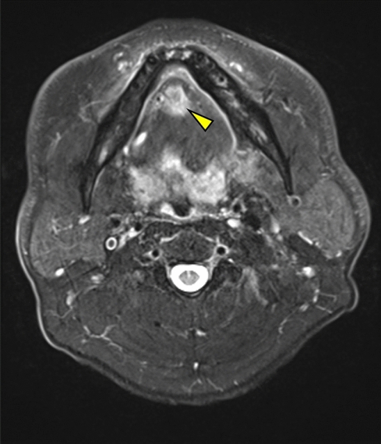
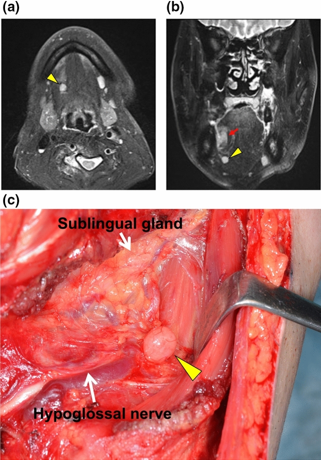
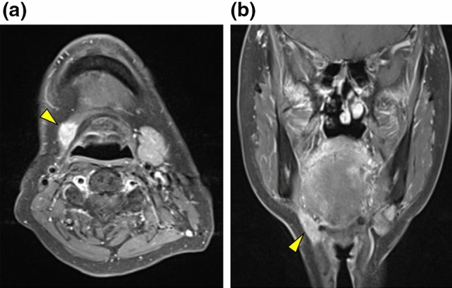
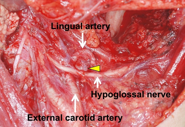
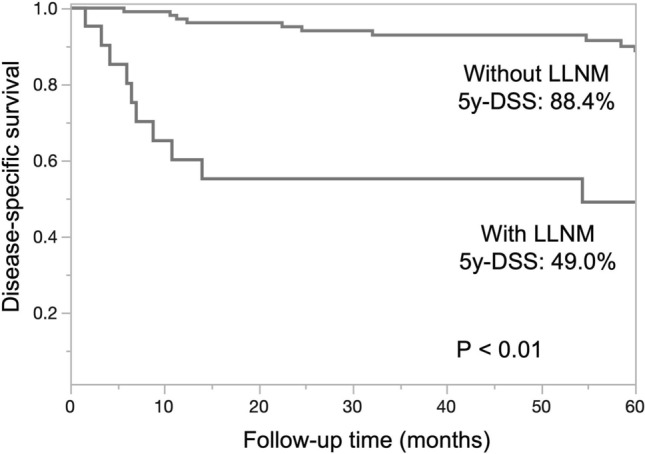
Squamous cell carcinoma (SCC) of the tongue rarely metastasizes to the lingual lymph nodes (LLNs), which are inconstant nodes and often situated outside the areas of basic tongue tumor surgery. The current study evaluated the clinicopathological features and prognostic impact of LLN metastasis (LLNM), compared to that of cervical lymph node metastasis, in patients with tongue SCC. A total of 608 patients underwent radical surgery for tongue SCC at our department between January 2001 and December 2016. During neck dissection, we scrutinized and resected lateral LLNs, when present. Of the 128 patients with lymph node metastasis, 107 had cervical lymph node metastasis and 21 had both cervical lymph node metastasis and LLNM. Univariate analysis demonstrated that LLNM was significantly associated with the adverse features of cervical lymph node metastasis. The 5-year disease-specific survival (5y-DSS) was significantly lower in patients with LLNMs than in those without LLNMs (49.0% vs. 88.4%, P < 0.01). Moreover, Cox proportional hazards model analyses revealed that cervical lymph node metastasis at level IV or V and LLNM were independent prognostic factors for 5y-DSS. LLNM has a strong negative impact on survival in patients with tongue SCC. An advanced status of cervical lymph node metastasis may predict LLNM.
© 2021. The Author(s).
Conflict of interest statement
The authors declare no competing interests.
Figures
Similar articles
-
Value of lingual lymph node metastasis in patients with squamous cell carcinoma of the tongue.Laryngoscope. 2019 Nov;129(11):2527-2530. doi: 10.1002/lary.27927. Epub 2019 Mar 12. Laryngoscope. 2019. PMID: 30861130
-
Lingual lymph nodes in patients with squamous cell carcinoma of the tongue and the floor of the mouth.Head Neck. 2018 Nov;40(11):2383-2388. doi: 10.1002/hed.25340. Epub 2018 Jul 26. Head Neck. 2018. PMID: 30051610
-
Tumor budding correlates with occult cervical lymph node metastasis and poor prognosis in clinical early-stage tongue squamous cell carcinoma.J Oral Pathol Med. 2015 Apr;44(4):266-72. doi: 10.1111/jop.12242. Epub 2014 Aug 28. J Oral Pathol Med. 2015. PMID: 25169851
-
Papillary Thyroid Carcinoma Incidentally Found in Cervical Lymph Nodes During Neck Dissection for Patients With Tongue Squamous Cell Carcinoma: A 3-Case Report and Literature Review.J Oral Maxillofac Surg. 2018 Nov;76(11):2454.e1-2454.e6. doi: 10.1016/j.joms.2018.07.013. Epub 2018 Jul 20. J Oral Maxillofac Surg. 2018. PMID: 30107162 Review.
-
Depth of invasion as a predictive factor for cervical lymph node metastasis in tongue carcinoma.Head Neck. 1997 May;19(3):205-10. doi: 10.1002/(sici)1097-0347(199705)19:3<205::aid-hed7>3.0.co;2-6. Head Neck. 1997. PMID: 9142520 Review.
Cited by
-
MicroRNA-18a regulates the metastatic properties of oral squamous cell carcinoma cells via HIF-1α expression.BMC Oral Health. 2022 Sep 5;22(1):378. doi: 10.1186/s12903-022-02425-6. BMC Oral Health. 2022. PMID: 36064348 Free PMC article.
-
Survival Impact of Lingual Lymph Node Metastases in Tongue Cancer: An Analysis Regarding Anatomical Subsites.In Vivo. 2023 May-Jun;37(3):1328-1333. doi: 10.21873/invivo.13213. In Vivo. 2023. PMID: 37103065 Free PMC article.
-
High expression of Sam68 contributes to metastasis by regulating vimentin expression and a motile phenotype in oral squamous cell carcinoma.Oncol Rep. 2022 Oct;48(4):183. doi: 10.3892/or.2022.8398. Epub 2022 Sep 9. Oncol Rep. 2022. PMID: 36082807 Free PMC article.
References
-
- El-Naggar AK, Chan JKC, Grandis JR, Takata T, Slootweg PJ. WHO Classification of Head and Neck Tumours. 4. IARC; 2017.
MeSH terms
LinkOut - more resources
Full Text Sources
Research Materials
