Review
. 2021 Dec;78(23):7199-7217.
doi: 10.1007/s00018-021-03947-y. Epub 2021 Oct 15.

RIPK3 signaling and its role in the pathogenesis of cancers

Affiliations
Review

RIPK3 signaling and its role in the pathogenesis of cancers

Shanhui Liu et al. Cell Mol Life Sci. 2021 Dec.

Abstract

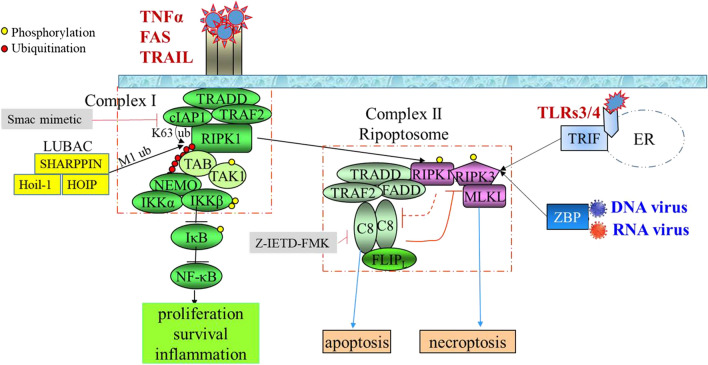
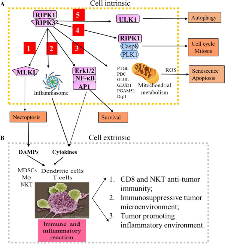
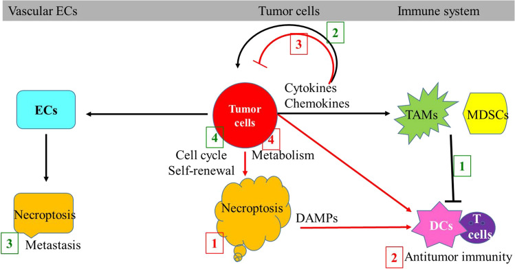
RIPK3 (receptor-interacting protein kinase 3) is a serine/threonine-protein kinase. As a key component of necrosomes, RIPK3 is an essential mediator of inflammatory factors (such as TNFα-tumor necrosis factor α) and infection-induced necroptosis, a programmed necrosis. In addition, RIPK3 signaling is also involved in the regulation of apoptosis, cytokine/chemokine production, mitochondrial metabolism, autophagy, and cell proliferation by interacting with and/or phosphorylating the critical regulators of the corresponding signaling pathways. Similar to apoptosis, RIPK3-signaling-mediated necroptosis is inactivated in most types of cancers, suggesting RIPK3 might play a critical suppressive role in the pathogenesis of cancers. However, in some inflammatory types of cancers, such as pancreatic cancers and colorectal cancers, RIPK3 signaling might promote cancer development by stimulating proliferation signaling in tumor cells and inducing an immunosuppressive response in the tumor environment. In this review, we summarize recent research progress in the regulators of RIPK3 signaling, and discuss the function of this pathway in the regulation of mixed lineage kinase domain-like (MLKL)-mediated necroptosis and MLKL-independent cellular behaviors. In addition, we deliberate the potential roles of RIPK3 signaling in the pathogenesis of different types of cancers and discuss the potential strategies for targeting this pathway in cancer therapy.

Keywords: Cancer pathogenesis; MLKL necroptosis; MLKL-independent signaling; RIPK3 signaling.


Conflict of interest statement

The authors declare that they have no competing financial or professional interests.

Figures

Fig. 1
Fig. 1
RIPK3 gene and protein structure. A Two promoters, proximal and distal, have been identified in the RIPK3 gene. Transcription factors SP1 and SOX17 bind to the proximal promoter to up-regulate RIPK3 expression, whereas CHD4, in the chromatin-remodeling complex NuRD, recognizes the distal promoter and represses RIPK3 expression by deacetylating H3K27. In addition, a HIF1 binding site was identified at 1 kb upstream of the TSS. In hypoxic conditions, HIF1α binds to this site and represses RIPK3 expression. Furthermore, a CpG island was identified approximately 240–600 bp downstream of the TSS. Epigenetic regulator UHRF1 represses RIPK3 expression by recruiting DNMT1 and maintaining the methylation state of this CpG island. B RIPK3 protein is composed of a kinase domain (KD) at the N terminus and a RHIM domain at the C terminus connected by an intermediate domain (IMD). The caspase 8 cleavage site, three NESs and several key phosphorylation and ubiquitination sites are indicated
Fig. 2
Fig. 2
RIPK3-mediated necroptotic pathway. TNF family members stimulate RIPK3 activation through inducing RIPK1 activation and RIPK1–RIPK3 interaction, whereas bacterial and viral infections activate RIPK3 through stimulating either TLR3/4 signal-triggered TRIF-RIPK3 interaction or pathogen DNA/RNA-triggered ZBP1-RIPK3 interaction. Upon TNFα stimulation, RIPK3-mediated necroptosis can be experimentally induced by Smac-mimetic + Caspase 8 inhibitor combination treatment. Smac-mimetic treatment releases RIPK1 from complex I and promotes the formation of complex II. Caspase 8 inhibitor treatment enhances RIP-mediated necroptosis by preventing the degradation of RIPK1 and RIPK3. C8 and C10 represent caspase 8 and caspase 10, respectively. ER endoplasmic reticulum
Fig. 3
Fig. 3
RIPK3 signaling stimulates inflammation and immunity. A In addition to stimulating MLKL-mediated necroptosis (1), RIPK3 signaling also induces: cytokine production by activating ERK/NF-kB/AP1-mediated transcription and inflammasome-mediated pro-cytokine processing (2); mitochondrial metabolism and senescence/apoptosis by phosphorylating several mitochondrial proteins (3); cell cycle and mitosis by regulating the RIPK1/caspase 8/PLK1 mitosis-associated ripoptosome (4); and autophagy by phosphorylating ULK1 (5). Thus RIPK3 activation can cause at least 5 types of cell-intrinsic effects. B In addition, RIPK3 activation also causes amplified damage in local tissues and significant systematic symptoms which are cell extrinsic effects. The necroptotic cells induce immune and inflammatory reactions in tissues by secreting cytokines to recruit both innate and adaptive immune cells, and releasing DAMPs to activate immune cells
Fig. 4
Fig. 4
RIPK3 signaling has both tumor-promoting and tumor-repressive activities. RIPK3 signaling represses cancer development by (1) killing the cancer cells through necroptosis; (2) inducing DC/T cell-mediated antitumor immunity; (3) secreting tumor-suppressive cytokines; and (4) inducing cancer-restrictive mitochondrial metabolism and ROS production (the numbers are indicated in red). RIPK3 signaling promotes cancer development by: (1) inducing immune-suppressive myeloid cells; (2) producing tumor-promoting cytokines; (3) inducing death in vascular endothelial cells; and (4) regulating cell cycle and/or self-renewal signaling (the numbers are indicated in green)
Fig. 5
Fig. 5
Potential strategies to target RIPK3 signaling for cancer treatment. A In apoptosis-resistant cancers, RIPK3 signaling activators overcome apoptosis resistance by inducing necroptosis and potentially stimulating anti-tumor immunity. B In RIPK3-negative cancers, epigenetic drugs and kinase inhibitors reactivate the tumor repressive activity of RIPK3 by inducing RIPK3 expression. C In RIPK3-positive cancers, RIPK1 and RIPK3 inhibitors might repress cancer development and progression by inhibiting the tumor-promoting activity of RIPK3

References

    1. Mulay SR, Desai J, Kumar SV, Eberhard JN, Thomasova D, Romoli S, Grigorescu M, Kulkarni OP, Popper B, Vielhauer V, et al. Cytotoxicity of crystals involves RIPK3-MLKL-mediated necroptosis. Nat Commun. 2016;7:10274. doi: 10.1038/ncomms10274. - DOI - PMC - PubMed
    1. Rickard JA, O'Donnell JA, Evans JM, Lalaoui N, Poh AR, Rogers T, Vince JE, Lawlor KE, Ninnis RL, Anderton H, et al. RIPK1 regulates RIPK3-MLKL-driven systemic inflammation and emergency hematopoiesis. Cell. 2014;157(5):1175–1188. doi: 10.1016/j.cell.2014.04.019. - DOI - PubMed
    1. Daniels BP, Kofman SB, Smith JR, Norris GT, Snyder AG, Kolb JP, Gao X, Locasale JW, Martinez J, Gale M, Jr, et al. The nucleotide sensor ZBP1 and kinase RIPK3 induce the enzyme IRG1 to promote an antiviral metabolic state in neurons. Immunity. 2019;50(1):64–76e64. doi: 10.1016/j.immuni.2018.11.017. - DOI - PMC - PubMed
    1. Kaiser WJ, Sridharan H, Huang C, Mandal P, Upton JW, Gough PJ, Sehon CA, Marquis RW, Bertin J, Mocarski ES. Toll-like receptor 3-mediated necrosis via TRIF, RIP3, and MLKL. J Biol Chem. 2013;288(43):31268–31279. doi: 10.1074/jbc.M113.462341. - DOI - PMC - PubMed
    1. Moriwaki K, Chan FK. RIP3: a molecular switch for necrosis and inflammation. Genes Dev. 2013;27(15):1640–1649. doi: 10.1101/gad.223321.113. - DOI - PMC - PubMed

MeSH terms