. 2021 Oct 16;55(5):605-617.
doi: 10.33594/000000438.

Extracellular Histones Bind Vascular Glycosaminoglycans and Inhibit the Anti-Inflammatory Function of Antithrombin

Affiliations

Extracellular Histones Bind Vascular Glycosaminoglycans and Inhibit the Anti-Inflammatory Function of Antithrombin

Indranil Biswas et al. Cell Physiol Biochem. .

Abstract

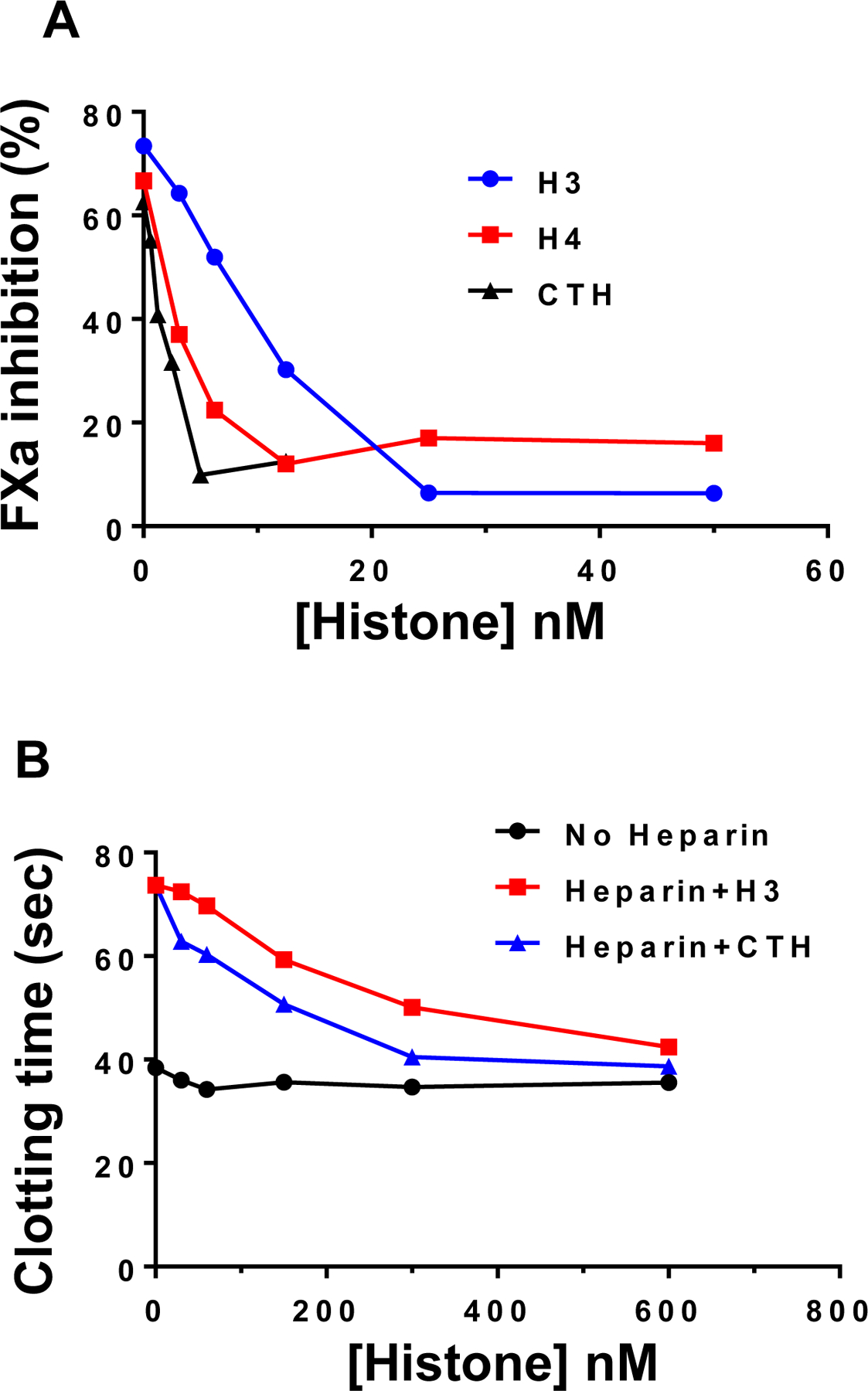
Background/aims: Binding of histones to molecular pattern recognition receptors on endothelial cells and leukocytes provokes proinflammatory responses and promotes activation of coagulation. Histones also bind therapeutic heparins, thereby neutralizing their anticoagulant functions. The aim of this study was to test the hypothesis that histones can interact with the antithrombin (AT)-binding vascular glycosaminoglycans (GAGs) to induce inflammation and inhibit the anti-inflammatory function of AT.

Methods: We evaluated the heparin-binding function of histones by an AT-dependent protease-inhibition assay. Furthermore, we treated endothelial cells with histones in the absence and presence of AT and monitored cellular phenotypes employing established signaling assays.

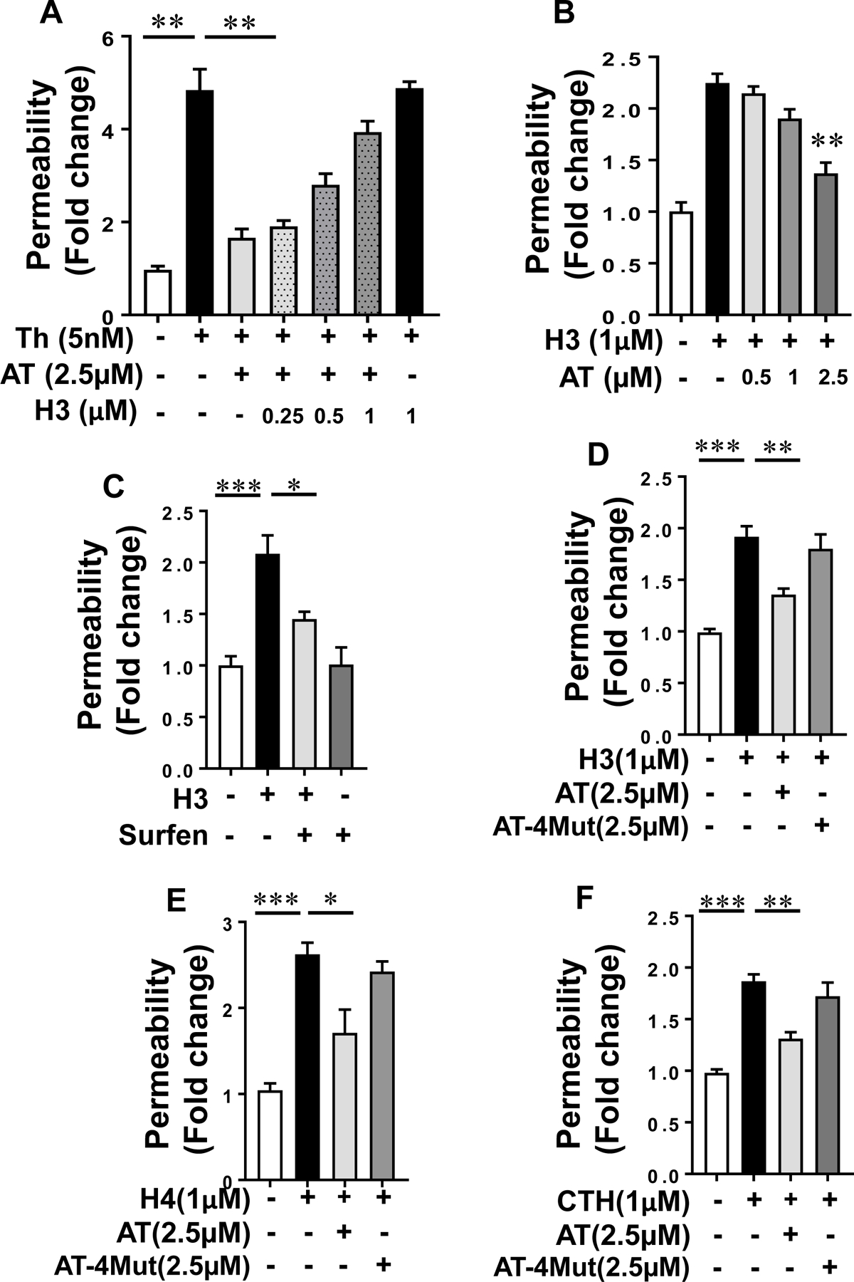
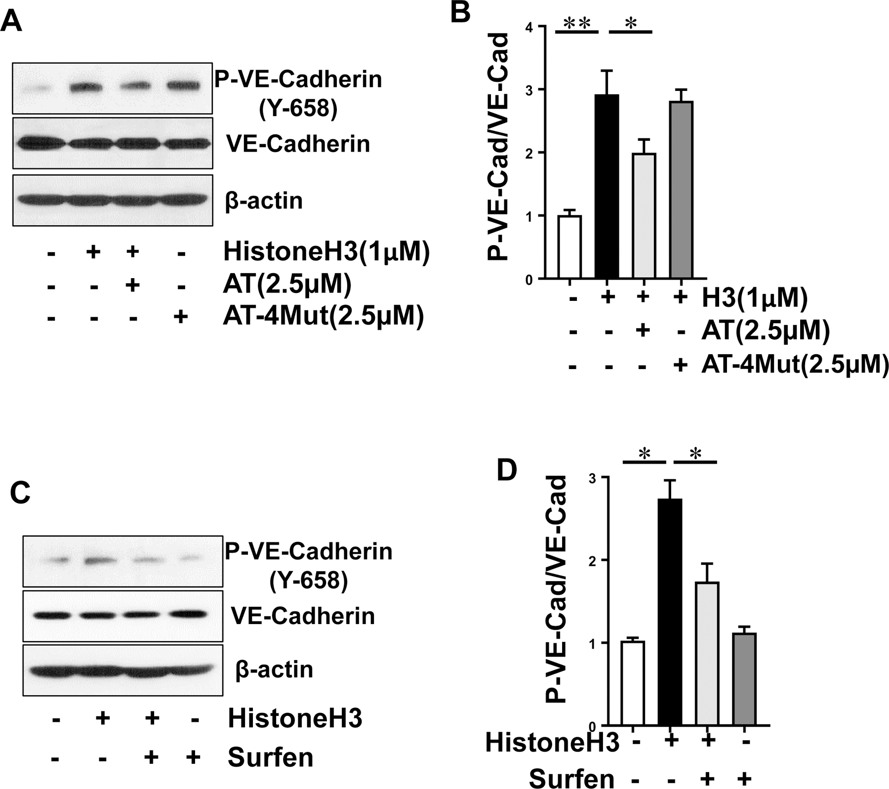
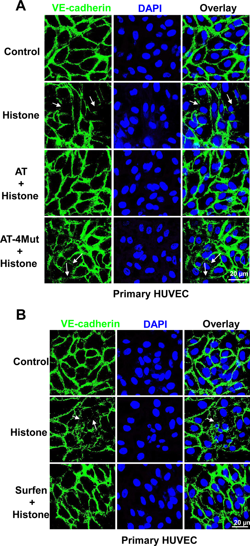
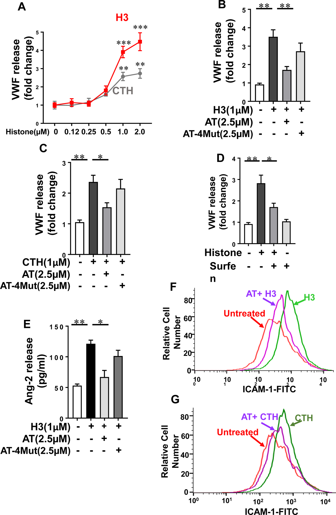
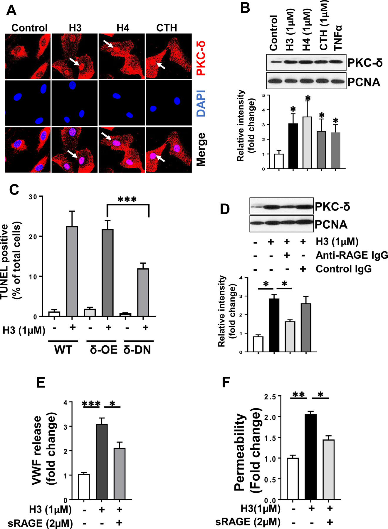
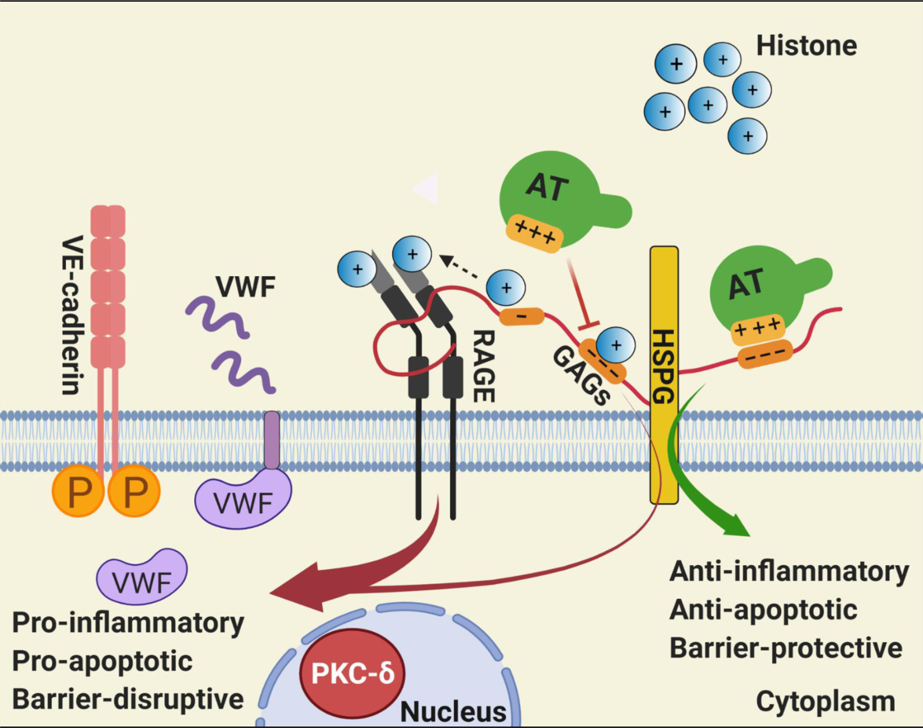
Results: Histones neutralized AT-dependent anticoagulant function of heparin in both purified protease-inhibition and plasma-based assays. Histones also disrupted endothelial cell barrier-permeability function by a GAG-dependent mechanism as evidenced by the GAG-antagonist, surfen, abrogating their disruptive effects. Further studies revealed histones and AT compete for overlapping binding-sites on GAGs, thus increasing concentrations of one protein abrogated effects of the other. Histones elicited proapoptotic effects by inducing nuclear localization of PKC-δ in endothelial cells and barrier-disruptive effects by destabilizing VE-cadherin, which were inhibited by AT, but not by a D-helix mutant of AT incapable of interacting with GAGs. Finally, histones induced release of Weibel-Palade body contents, VWF and angiopoietin-2, and promoted expression of cell adhesion molecules on endothelial cells, which were all downregulated by AT but not by D-helix mutant of AT.

Conclusion: We conclude that histones and AT compete for overlapping binding sites on vascular GAGs to modulate coagulation and inflammation.

Keywords: Histones; Antithrombin; Glycosaminoglycans; Signaling.


Conflict of interest statement

The authors declare no conflict of interests exists.

Figures

Fig. 1-
Fig. 1-
Competitive effect of histones on inhibiting the cofactor function of heparin. (A) Competitive effect of increasing concentrations of histones on inhibiting the cofactor function of heparin in accelerating the AT inhibition of factor Xa (FXa) was monitored as described in methods. (B) The competitive effect of increasing concentrations of histone H3 and CTH on neutralization of the anticoagulant effect of heparin was monitored in an aPTT assay using citrated normal plasma as described in methods.
Fig. 2-
Fig. 2-
Histone H3 inhibits the endothelial barrier protective function of AT. (A) Confluent HTERT-HUVECs were simultaneously incubated with a fixed concentration of AT (2.5 µM) and increasing concentration of histone H3 for 4h followed by measuring cell permeability in response to thrombin by spectrophotometric measurement of the flux of Evans blue-bound albumin across functional endothelial cell monolayer as described in methods. (B) The same as (A) except that the fold change in cell permeability was measured by simultaneous incubation of a fixed concentration of histone H3 with increasing concentration of AT. (C) The cell permeability induced by histone H3 (1 µM for 4h) was monitored in the absence and presence of the GAG-antagonist surfen (10 µM). (D) Protective effect of WT-AT and its D-helix mutant (AT-4Mut) on histone H3-mediated endothelial cell permeability was measured by influx of albumin-bound Evans blue across functional endothelial cell monolayer. (E) The same as (D) except that the effect of WT-AT and AT-4Mut on histone H4-mediated permeability was monitored. (F) The same as (D) except that the effect of WT-AT and AT-4Mut on calf thymus histone (CTH)-mediated cell permeability was monitored. Neither AT-WT nor AT-4Mut had an effect on cell permeability in the absence of histones (not shown). All results are shown as means ± SD of three different experiments. * p < 0.05, ** p < 0.01, *** p < 0.001.
Fig. 3-
Fig. 3-
Analysis of VE-cadherin phosphorylation by histone H3 in the absence and presence of AT. (A) Confluent HTERT-HUVECs were incubated with histone H3 for 15 min in the absence or presence of WT-AT and AT-4Mut and phosphorylated (Y-658) VE-cadherin, total VE-cadherin and β-actin levels in cell lysates were measured by Western blot. Data is representative of three independent repeats. (B) Densitometric analysis of the panel A Western blot. (C) The same as (A) except that histone H3-mediated phosphorylation of VE-cadherin was monitored in the absence or presence of surfen (10 µM). Data is representative of three independent repeats. (D) Densitometric analysis of the panel C Western blot. All results are shown as means ± SD of three different experiments. * p < 0.05, ** p < 0.01.
Fig. 4-
Fig. 4-
Immunofluorescence analysis of histone-mediated VE-cadherin disruption in endothelial cells. (A) Primary HUVECs were incubated with Histone H3 (1 µM) for 1h in the absence or presence of WT-AT and AT-4Mut (2.5 µM each). Cells were then fixed, permeabilized and incubated with rabbit anti-VE-cadherin antibody and Alexa Fluor 488-conjugated goat anti-rabbit IgG. The nucleus was stained with DAPI. Immunofluorescence images were obtained with confocal microscopy. (B) The same as (A) except that primary HUVECs were incubated with histone H3 for 1h in the absence or presence of surfen (10 µM). Arrows indicate loss of VE-cadherin at junctions.
Fig. 5-
Fig. 5-
Histone-mediated WPB exocytosis and ICAM-1 expression in endothelial cells. (A) HTERT-HUVECs were incubated with increasing concentrations of histone H3 (red) or calf thymus histone (CTH, grey) for 4h and VWF release was measured by a sandwich ELISA. (B) The same as (A) except that cells were incubated with histone H3 for 4h in the absence or presence of WT-AT or AT-4Mut and VWF release was measured by a sandwich ELISA. (C) The same as (B) except that cells were incubated with calf thymus histone (CTH). (D) The same as (B) except that cells were incubated with histone H3 in the absence or presence of surfen and VWF release was measured by a sandwich ELISA. (E) The same as (B) except that cells were incubated with histone H3 in the absence or presence of WT-AT or AT-4Mut and Ang-2 release was measured by a sandwich ELISA. (F) HTERT-HUVECs were incubated with histone H3 (1 µM) for 4h in the absence or presence of WT-AT (2.5 µM) followed by measuring expression of surface level of ICAM-1 by flow cytometry. The data is representative of three independent experiments. (G) The same as (F) except that CTH-mediated expression of ICAM-1 in the presence of WT-AT was measured by flow cytometry.
Fig. 6-
Fig. 6-
RAGE signaling is required for histone-mediated nuclear localization of PKC-δ in endothelial cells. (A) HTERT-HUVECs were treated with histones (H3, H4, CTH) for 1h and PKC-δ nuclear localization was analyzed by confocal microscopy. PKC-δ was stained with rabbit monoclonal antibody followed by Alexa Fluor 555-conjugated goat anti-rabbit IgG. DAPI was used to stain the nucleus. (B) The same as (A) except that nuclear PKC-δ in histone-treated cells was monitored through by Western-blotting. TNFα was used as a positive control and PCNA was used as a loading control. Densitometric analysis of the data in the Western-blot is shown below the panel. (C) Wild-type (WT), PKC-δ overexpressing (PKC-δ-OE) and PKC-δ dominant negative (PKC-δ-DN) endothelial (EA.hy926) cells were treated with histones for 16h and cell death was analyzed by a TUNEL assay. TUNEL-positive cells were counted using Nikon C2 Confocal Microscope for at least 10 randomly selected fields from 3 independent samples. (D) HTERT-HUVECs were incubated with histone H3 (1 µM) for 1h in the absence and presence of anti-RAGE antibody (20µg/ml) followed by the analysis of PKC-δ in the nuclear fractions by Western-blotting. Densitometric analysis of the Western-blotting data is shown below the panel. (E) Confluent HTERT-HUVECs were incubated with histone H3 (1 µM) for 4h in the absence and presence of sRAGE (2 µM) followed by measuring the cell permeability by spectrophotometric measurement of the flux of Evans blue-bound albumin across functional endothelial cell monolayer. (F) The same as (E) except that histone H3-mediated VWF release from endothelial cells was measured by an ELISA. Results in all panels are shown as means ± SD of three different experiments. * p < 0.05, ** p < 0.01, *** p < 0.001.
Fig. 7-
Fig. 7-
Hypothetical model of GAG-dependent AT and histone signaling in endothelial cells. Binding of positively charged residues of D-helix of AT on distinct GAGs covalently linked to heparan sulfate proteoglycans (HSPG) culminates in anti-inflammatory, anti-apoptotic and barrier-protective effects. Histones can bind to the same and/or overlapping AT-binding sites on GAGs, thereby excluding AT from binding GAGs. GAGs are known to bind RAGE, activate the receptor by oligomerization and also present histones to RAGE [29], thereby promoting the pro-inflammatory, pro-apoptotic and barrier-disruptive signaling functions of nuclear proteins. The GAG-dependent RAGE signaling by histones induces VWF release from endothelial cells. The pro-apoptotic activity of histones is associated with RAGE-dependent nuclear localization of PKC-δ in endothelial cells. The GAG-dependent histone signaling through RAGE also leads to phosphorylation of VE-cadherin and destabilization of endothelial cell junctions. See the text for further details. Figure was prepared by software provided by Biorender.com.

References

    1. Campos EI, Reinberg D. Histones: annotating chromatin. Annu Rev Genet 2009;43:559–599. - PubMed
    1. Chen R, Kang R, Fan XG, Tang D. Release and activity of histone in diseases. Cell Death Dis 2014;5:e1370. - PMC - PubMed
    1. Xu J, Zhang X, Pelayo R, Monestier M, Ammollo CT, Semeraro F, Taylor FB, Esmon NL, Lupu F, Esmon CT. Extracellular histones are major mediators of death in sepsis. Nat Med 2009;15:1318–1321. - PMC - PubMed
    1. Gould TJ, Lysov Z, Liaw PC. Extracellular DNA and histones: double-edged swords in immunothrombosis. J Thromb Haemost 2015;Suppl 1:S82–91. - PubMed
    1. Brinkmann V, Reichard U, Goosmann C, Fauler B, Uhlemann Y, Weiss DS, Weinrauch Y, Zychlinsky A. Neutrophil extracellular traps kill bacteria. Science 2004;303:1532–5153. - PubMed